Review
. 2021 Feb 2;30(159):200149.
doi: 10.1183/16000617.0149-2020. Print 2021 Mar 31.

The role of mucin 1 in respiratory diseases

Affiliations
Review

The role of mucin 1 in respiratory diseases

Beatriz Ballester et al. Eur Respir Rev. .

Abstract

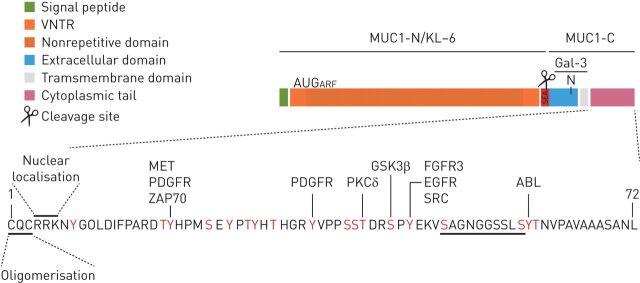
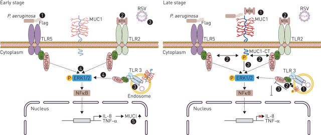
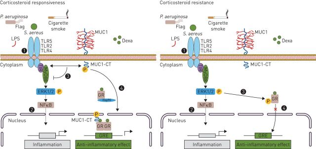
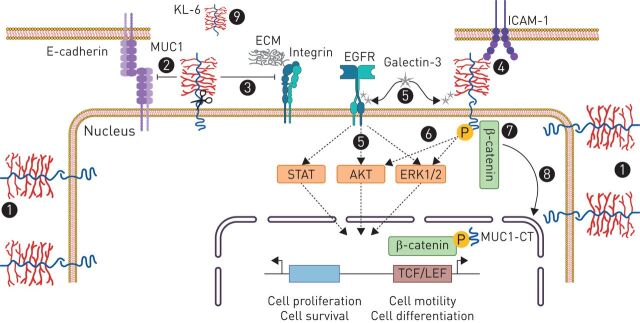
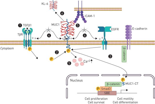
Recent evidence has demonstrated that mucin 1 (MUC1) is involved in many pathological processes that occur in the lung. MUC1 is a transmembrane protein mainly expressed by epithelial and hematopoietic cells. It has a receptor-like structure, which can sense the external environment and activate intracellular signal transduction pathways through its cytoplasmic domain. The extracellular domain of MUC1 can be released to the external environment, thus acting as a decoy barrier to mucosal pathogens, as well as serving as a serum biomarker for the diagnosis and prognosis of several respiratory diseases such as lung cancer and interstitial lung diseases. Furthermore, bioactivated MUC1-cytoplasmic tail (CT) has been shown to act as an anti-inflammatory molecule in several airway infections and mediates the expression of anti-inflammatory genes in lung diseases such as chronic rhinosinusitis, chronic obstructive pulmonary disease and severe asthma. Bioactivated MUC1-CT has also been reported to interact with several effectors linked to cellular transformation, contributing to the progression of respiratory diseases such as lung cancer and pulmonary fibrosis. In this review, we summarise the current knowledge of MUC1 as a promising biomarker and drug target for lung disease.


Conflict of interest statement

Conflict of interest: B. Ballester has nothing to disclose. Conflict of interest: J. Milara Payá has nothing to disclose. Conflict of interest: J. Cortijo Gimeno has nothing to disclose.

Figures

FIGURE 1
FIGURE 1
Structure of mucin 1 (MUC-1). MUC1 protein comprises an N-terminal subunit (also called KL-6) constituted by a signal peptide, a VNTR domain composed of 25–125 repeats of 20 amino acids and the SEA domain. MUC1-C-terminal subunit has a 58-amino acid extracellular domain, a single-pass 28-amino acid transmembrane domain and a 72-amino acid CT. The MUC1 extracellular domain can be shed into the lumen by auto-proteolytic cleavage in the SEA domain or by the action of metalloproteinase 14 in the region following the SEA domain. The MUC1-C extracellular domain is glycosylated on Asn-36 and then serves as a binding site for the profibrotic galectin 3 ligand. MUC1-CT serves as a substrate for phosphorylation (18 documented and putative tyrosine and serine/threonine potential phosphorylation sites) in response to activation of several growth factor receptors and/or kinases. The CQC motif is necessary for MUC1-C oligomerisation and the RRK motif is necessary for MUC1-C binding to importin β and targeting to the nucleus. β-catenin-binding site is also localised to MUC1-CT. An AUG codon downstream to the MUC1 initiation codon has been described to initiate an ARF thereby generating a novel protein, MUC1-ARF. ABL: tyrosine-protein kinase ABL1; ARF: alternate reading frame; CT: cytoplasmic tail; FGFR: fibroblast growth factor receptor; Gal-3: galectin-3; GSK3β: glycogen synthase kinase 3 beta; KL-6: Krebs von den Lungen-6; MET: tyrosine-protein kinase Met; MUC1-C: mucin 1 C-terminal; MUC1-N: mucin 1 N-terminal; PDGFR: platelet-derived growth factor receptor; PKCδ: delta isoform of protein kinase C; SEA: sperm protein, enterokinase and agrin; VNTR: variable number tandem repeat; ZAP70: zeta chain of T-cell receptor associated protein kinase 70.
FIGURE 2
FIGURE 2
The role of MUC1 in airway infection/inflammation. During early stage of infection, primary human airway epithelial cells sense Pseudomonas aeruginosa via TLR5 (1), Haemophilus influenzae (NTHi) via TLR2 (2), and RSV via TLR3 (3), which is expressed in endosomes. After recognition, MyD88/TRIF-dependent secretion of TNF-α and IL-8 is produced (4). Following it, IL-8 and TNF-α lead to an increased expression of MUC1 at the apical surface of lung epithelial cells (5). During the late-stage of infection, P. aeruginosa/NTHi/RSV-induced MUC1-CT tyrosine phosphorylation (1) leads to MUC1-CT interaction with TLR5/TLR2/TLR3 (2), leading to TLR signalling downregulation (3) and inhibition of inflammation (4). CT: cytoplasmic tail; ERK: extracellular signal-regulated kinase; NFκB: nuclear factor-κB; IL: interleukin; MUC1: mucin 1; MyD88: myeloid differentiation primary response gene 88; RSV: respiratory syncytial virus; TIR: Toll/interleukin-1 receptor; TLR: Toll-like receptor; TNF: tumour necrosis factor; TRIF: TIR (Toll/interleukin-1 receptor)-domain-containing adapter-inducing interferon-β.
FIGURE 3
FIGURE 3
The role of MUC1 in the corticosteroid mediated anti-inflammatory response in CRS, severe asthma and COPD. The TLR recognition of airway pathogens such as Pseudomonas aeruginosa or Staphylococcus aureus, as well as, cigarette smoke (1) triggers a MyD88-mediated inflammatory response (2). Among corticosteroid responder patients, corticosteroids increase MUC1 expression. The MUC1-CT interacts with TLR, inhibiting TLR inflammatory signalling (3). Dexamethasone (DEXA) treatment induces the formation of a protein complex between MUC1-CT and GRα, which helps GRα to translocate into the nucleus to exert its anti-inflammatory effects (4). Corticosteroid resistance patients show lower expression of MUC1. Therefore, it is produced an overactivation of TLR signalling (1) and subsequent inflammation (2), which leads to GR-Ser226 phosphorylation through ERK 1/2 hyper-phosphorylation (3). Therefore, MUC1-CT downregulation and GR hyperphosphorylation inhibit the corticosteroids-induced GRα nuclear translocation and GR anti-inflammatory mediated effects (4). CRS: chronic rhinosinusitis; CT: cytoplasmic tail; ERK: extracellular signal-regulated kinase; NFκB: nuclear factor-κB; GR: glucocorticoid receptor; GRE: glucocorticoid response element; Hsp: heat shock protein; IL: interleukin; LPS: lipopolysaccharide; MUC1: mucin 1; MUC1-CT: MUC1 cytoplasmic domain; MyD88: myeloid differentiation primary response gene 88; TIR: Toll/interleukin-1 receptor; TLR: Toll-like receptor; TRIF: TIR-domain-containing adapter-inducing interferon-β.
FIGURE 4
FIGURE 4
The role of MUC1 in lung cancer. (1) MUC1 is overexpressed and depolarised in lung cancer. Therefore, MUC1 interactions with potential binding partners are increased under these circumstances, thus increasing the invasive and metastatic capability of tumour cells by different mechanisms: (2) reducing cell–cell adhesion through disturbing E-cadherin interactions between adjacent cells, (3) reducing cell–ECM adhesion through inhibition of integrin-mediated cell adhesion to ECM components, and (4) interacting with adhesion molecules such as the endothelial ICAM-1. (5) MUC1-C-terminal subunit interacts with galectin-3, increasing MUC1 association with the EGFR and leading to EGFR activation and signalling, which mediates epithelial cell proliferation and survival. (6) Galectin-3 binding to MUC1-C also phosphorylates MUC1-CT, activating mitogen-activated protein kinase and PI3K/AKT signalling pathways, which lead to cell proliferation and motility. (7) β-Catenin preferentially binds to the phosphorylated intracellular tail of MUC1, thus also contributing to the decrease of cell–cell adhesions through E-cadherin. (8) MUC1-CT/β-catenin complex translocates into the nucleus to regulate genes involved in cell proliferation and differentiation. (9) MUC1-N (KL6) is shed from the cell surface and has demonstrated to be a prognostic and treatment responsiveness serum biomarker in lung cancer. AKT: protein kinase B; ECM: extracellular matrix; EGFR: epidermal growth factor receptor; ERK: extracellular signal-regulated kinase; ICAM: intracellular adhesion molecule; KL-6: Krebs von den Lungen-6; LEF: lymphoid enhancer factor; MUC1: mucin 1; MUC1-CT: MUC1 cytoplasmic domain; TCF: T-cell factor protein.
FIGURE 5
FIGURE 5
The role of MUC1 in IPF. Transforming growth factor β1 (TGF-β1) receptor (TβR) activation induces Smad3 phosphorylation (1), which phosphorylates MUC1-CT (2). (3) Bioactivated MUC1-CT has demonstrated to increase the active form of β-catenin, forming a MUC1-CT/phospho-Smad3/active-β-catenin nuclear protein complex that promotes fibrotic processes such as epithelial to mesenchymal transition, fibroblast to mesenchymal transition (FMT), fibroblast proliferation and alveolar type II and fibroblast senescence. (4) MUC1-CT is also activated by galectin 3, which has demonstrated to directly interact with MUC1-C, functioning as a bridge between MUC1-C and the epidermal growth factor receptor (EGFR), as well as, other cell surface receptors such as TβR. (5) MUC1-N-terminal binds to intracellular adhesion molecule-1 (ICAM-1) and the MUC1/ICAM-1 association results in a rapid increase of intracellular calcium in MUC1-expressing cells, inducing cytoskeletal changes and motility. (6) MUC1-N (KL6) is shed from the cell surface and has demonstrated to be a diagnosis and prognostic serum biomarker in idiopathic pulmonary fibrosis KL-6: Krebs von den Lungen-6; LEF: lymphoid enhancer factor; MUC1: mucin 1; SBE: Smad binding element; MUC1-CT: MUC1 cytoplasmic domain.

Similar articles

Cited by

References

    1. Hattrup CL, Gendler SJ. Structure and function of the cell surface (tethered) mucins. Annu Rev Physiol 2008; 70: 431–457. doi:10.1146/annurev.physiol.70.113006.100659 - DOI - PubMed
    1. Pemberton LF, Rughetti A, Taylor-Papadimitriou J, et al. . The epithelial mucin MUC1 contains at least two discrete signals specifying membrane localization in cells. J Biol Chem 1996; 271: 2332–2340. doi:10.1074/jbc.271.4.2332 - DOI - PubMed
    1. Bafna S, Kaur S, Batra SK. Membrane-bound mucins: the mechanistic basis for alterations in the growth and survival of cancer cells. Oncogene 2010; 29: 2893–2904. doi:10.1038/onc.2010.87 - DOI - PMC - PubMed
    1. Kufe D. Oncogenic function of the MUC1 receptor subunit in gene regulation. Oncogene 2010; 29: 5663–5666. doi:10.1038/onc.2010.334 - DOI - PMC - PubMed
    1. Milara J, Peiro T, Armengot M, et al. . Mucin 1 downregulation associates with corticosteroid resistance in chronic rhinosinusitis with nasal polyps. J Allergy Clin Immunol 2015; 135: 470–476. doi:10.1016/j.jaci.2014.07.011 - DOI - PubMed