. 2020 Aug;584(7821):443-449.
doi: 10.1038/s41586-020-2548-6. Epub 2020 Jul 15.

Potently neutralizing and protective human antibodies against SARS-CoV-2

Affiliations

Potently neutralizing and protective human antibodies against SARS-CoV-2

Seth J Zost et al. Nature. 2020 Aug.

Abstract

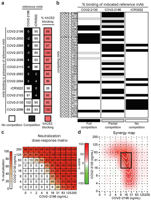
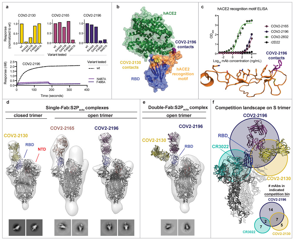
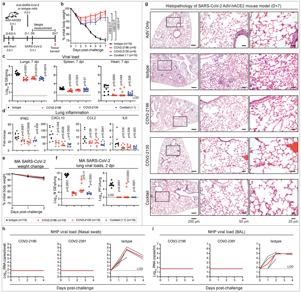
The ongoing pandemic of coronavirus disease 2019 (COVID-19), which is caused by severe acute respiratory syndrome coronavirus 2 (SARS-CoV-2), is a major threat to global health1 and the medical countermeasures available so far are limited2,3. Moreover, we currently lack a thorough understanding of the mechanisms of humoral immunity to SARS-CoV-24. Here we analyse a large panel of human monoclonal antibodies that target the spike (S) glycoprotein5, and identify several that exhibit potent neutralizing activity and fully block the receptor-binding domain of the S protein (SRBD) from interacting with human angiotensin-converting enzyme 2 (ACE2). Using competition-binding, structural and functional studies, we show that the monoclonal antibodies can be clustered into classes that recognize distinct epitopes on the SRBD, as well as distinct conformational states of the S trimer. Two potently neutralizing monoclonal antibodies, COV2-2196 and COV2-2130, which recognize non-overlapping sites, bound simultaneously to the S protein and neutralized wild-type SARS-CoV-2 virus in a synergistic manner. In two mouse models of SARS-CoV-2 infection, passive transfer of COV2-2196, COV2-2130 or a combination of both of these antibodies protected mice from weight loss and reduced the viral burden and levels of inflammation in the lungs. In addition, passive transfer of either of two of the most potent ACE2-blocking monoclonal antibodies (COV2-2196 or COV2-2381) as monotherapy protected rhesus macaques from SARS-CoV-2 infection. These results identify protective epitopes on the SRBD and provide a structure-based framework for rational vaccine design and the selection of robust immunotherapeutic agents.


Figures

Extended Data Figure 1.
Extended Data Figure 1.. SARS-CoV-2 neutralization curves for mAb panel.
Neutralization of authentic SARS-CoV-2 by human mAbs. Mean ± SD of technical duplicates is shown. Data represent one of two or more independent experiments
Extended Data Figure 2.
Extended Data Figure 2.. Inhibition curves for mAb inhibition of S2Pecto binding to hACE2.
Blocking of hACE2 binding to S2Pecto by anti-SARS-CoV-2 neutralizing human mAbs. Mean ± SD of triplicates of one experiment is shown. Antibodies rCR3022 and r2D22 served as controls.
Extended Data Figure 3.
Extended Data Figure 3.. ELISA binding of anti-SARS-CoV-2 neutralizing human mAbs to trimeric SRBD, S2Pecto, or SARS-CoV S2Pecto antigen.
Mean ± SD of triplicates and representative of two experiments are shown. Antibodies rCR3022 and r2D22 served as controls.
Extended Data Figure 4.
Extended Data Figure 4.. Serum or plasma mAbs competition binding
a. Inhibition of hACE2 binding to S2Pecto by serum or plasma of four SARS-CoV-2 immune subjects or one non-immune control in ELISA using SARS-CoV-2 S2Pecto. mAbs were isolated from subjects 3 and 4 as described previously. Mean ± SD of triplicates of one experiment is shown. Dotted line indicates full inhibition (100%) of hACE2 by 500 ng/mL of control mAb COV2-2196 or COV2-2130. b. Inhibition of mAb COV2-2130 (left) or COV2-2196 (right) binding to S2Pecto by serum or plasma of four SARS-CoV-2 immune subjects or one non-immune control in ELISA using SARS-CoV-2 S2Pecto. Mean ± SD of triplicates and representative of two experiments are shown. Dotted line indicates percent of self-competition of mAb COV2-2196 and-2130 on the SARS-CoV-2 S2Pecto antigen.
Extended Data Figure 5.
Extended Data Figure 5.. Mapping of mAb critical contact residues by alanine and arginine mutagenesis and biolayer interferometry.
a. Bar graphs show response values for mAb binding to wt or mutant SRBD constructs normalized to wt. Asterisks denote residues where increased dissociation of mAb was observed, likely indicating the residue is proximal to mAb epitope. Full response curves for mAb association and dissociation with wt or mutant SRBD constructs are also shown. b. Structure of the RBD highlighting the critical contact residues for several mAbs and their location on the structure.
Extended Data Figure 6.
Extended Data Figure 6.. Sequence features of human mAbs used in animal studies and mAb pharmacokinetics following infusion in NHPs
a. Sequence features of human mAbs tested in animal models. Inferred variable genes are indicated and CDR3 amino acids are shown for heavy and light chains. b. NHPs received one 50 mg/kg dose of mAb COV2-2196, COV2-2381, or isotype control mAb (n = 4 animals per group) intravenously on day −3 and then were challenged intranasally and intratracheally with SARS-CoV-2 at day 0. Concentration of human mAbs was determined at indicated timepoints. Each curve shows an individual animal. Data represent a single experiment.
Figure 1.
Figure 1.. Functional characteristics of neutralizing SARS-CoV-2 mAbs.
a. Heatmap of mAb neutralization activity, hACE2 blocking activity, and binding to either trimeric S2Pecto protein or monomeric SRBD. MAbs are ordered by neutralization potency, and dashed lines indicate the 12 antibodies with a neutralization IC50 value lower than 150 ng/mL. IC50 values are visualized for viral neutralization and hACE2 blocking, while EC50 values are visualized for binding. The cross-reactive SARS-CoV SRBD mAb rCR3022 is shown as a positive control, while the anti-dengue mAb r2D22 is shown as a negative control. Data are representative of at least 2 independent experiments performed in technical duplicate. No inhibition or no binding indicates an IC50 or EC50 value of >10,000 ng/mL, respectively. b-d. Correlation of hACE2 blocking, S2Pecto trimer binding, or SRBD binding of mAbs with their neutralization activity. e. Correlation of hACE2 blocking and S2Pecto trimer binding. R2 values are shown for linear regression analysis of log-transformed values. Purple circles indicate mAbs with a neutralization IC50 value lower than 150 ng/mL. f. Neutralization curves for COV2-2196 and COV2-2130 against authentic SARS-CoV-2 virus. Calculated IC50 values are denoted on the graph. Error bars denote the standard deviation of each point. Data are representative of at least 2 independent experiments, each performed in technical duplicate. g. Neutralization curves for COV2-2196 and COV2-2130 in a pseudovirus neutralization assay. Error bars denote the standard deviation of each point. Values shown are technical duplicates from a single experiment. Calculated IC50 values from a minimum of 6 experiments are denoted on the graph. h. hACE2 blocking curves for COV2-2196, COV2-2130, and the non-blocking SARS-CoV mAb rCR3022 in the hACE2 blocking ELISA. Calculated IC50 values are denoted on the graph.. Mean ± SD of technical triplicates are shown from a representative experiment repeated twice. i. ELISA binding of COV2-2196, COV2-2130, and rCR3022 to trimeric S2Pecto. Calculated EC50 values are denoted on the graph. Mean ± SD of technical triplicates are shown from a representative experiment repeated twice.
Figure 2.
Figure 2.. Epitope mapping of mAbs by competition-binding analysis and synergistic neutralization by a pair of mAbs.
a. Left: mAb binding to RBD in presence of reference mAbs COV2-2196 and rCR3022. Values in squares are % binding of the mAb in the presence of the competing mAb relative to a mock-competition control. Black squares denote full competition (<33% relative binding), while white squares denote no competition (>67% relative binding). Right: biolayer interferometry-based competition binding assay measuring the ability of mAbs to prevent binding of hACE2. Values denote % blocking of hACE2 by mAb. Red color denotes high blocking activity. b. Competition of neutralizing mAb panel with reference mAbs COV2-2130, COV2-2196, or rCR3022. Binding of reference mAbs to trimeric S2Pecto was measured in the presence of saturating competitor mAb in a competition ELISA and normalized to binding in the presence of the anti-dengue mAb r2D22. Black denotes full competition (<25% binding of reference mAb), grey denotes partial competition (25-60% binding of reference mAb), and white denotes no competition (>60% binding of reference mAb). c. Neutralization dose-response matrix of wild-type SARS-CoV-2 by COV2-2196 and COV2-2130. Axes denote the concentration of each mAb with % neutralization shown in each square. Data is from a representative experiment that was performed in technical triplicate and repeated twice. A white-to-red heatmap denotes 0% neutralization to 100% neutralization, respectively. d. Synergy map calculated based on the SARS-CoV-2 neutralization in c. Red color denotes areas where synergistic neutralization was observed, and a black box denotes the area of maximal synergy between the two mAbs.
Figure 3.
Figure 3.. Epitope identification and structural characterization of mAbs.
a. Identification of critical contact residues by alanine and arginine mutagenesis. Top: binding of COV2-2130 (gold), COV2-2165 (maroon) or COV2-2196 (dark purple) to wild-type (wt) or mutant SRBD constructs normalized to wt. Bottom: representative binding curves for COV2-2196 to wt or SRBD constructs with mutated critical contact residues. b. Co-crystal structure of SARS-CoV-2 RBD (blue) and hACE2 (green) (PDB 6M0J). The hACE2 recognition motif is colored orange. Critical contact residues are shown for COV2-2130 (gold spheres) and COV2-2196 (purple spheres). c. ELISA binding of mAbs to the hACE2 recognition motif. r2D22 is shown as a negative control. Mean ± SD of technical triplicates are shown from a single experiment repeated twice. Bottom: structure of hACE2 recognition motif in orange with COV2-2196 critical contact residues shown in purple. d. Single-Fab:S2Pecto trimer complexes visualized by negative-stain electron microscopy for COV2-2130 (gold), COV2-2165 (maroon), or COV2-2196 (dark purple). The RBD is shown in blue and the S N-terminal domain (NTD) is shown in red. Electron density is shown in grey. Trimer state (open or closed) is denoted for each complex. Representative 2D class averages for each complex are shown at the bottom (box size is 128 pixels, with 3.06 Å/pixel). Data were collected in a single experiment with detailed collection statistics in Supplemental Table 2. e. COV2-2130 and COV2-2196 Fabs in complex with S2Pecto trimer. Colors and data collection are as in (d). Representative 2D class averages for the complexes are shown at the bottom with scales as in (d). f. Competition-binding analysis visualized on S2Pecto trimer. The CR3022 crystal structure was docked into the double-Fab:S2Pecto trimer model. CR3022 is shown in cyan. Bottom: a quantitative Venn diagram notes the number of mAbs in each competition group.
Figure 4.
Figure 4.. Prophylactic efficacy of neutralizing human mAbs against SARS-CoV-2 infection in mouse and NHP models in vivo.
a. SARS-CoV-2 challenge model. Mice were treated with anti-Ifnar1 mAb, transduced with AdV-hACE2, and 200 μg of mAbs COV2-2196, −2130, or combination (1:1 ratio) or isotype control mAb were passively transferred. One day later, SARS-CoV-2 was inoculated via the i.n. route. Tissues were harvested at 7 dpi for analysis (c, d). b. Body weight change of mice in (a) with comparison to isotype control using a repeated measurements two-way ANOVA with Tukey’s post-test. Mean ± SEM of each experimental group is shown. Numbers of animals (n) for each experimental group are shown. Viral burden in the lung, spleen and heart (c) and cytokine and chemokine gene expression (d) were measured by RT-qPCR assay. Comparisons were performed using a Kruskal-Wallis ANOVA with Dunn’s post-test. e. MA-SARS-CoV-2 challenge model. Mice were treated with indicated mAb and then infected intranasally with MA-SARS-CoV-2. Body weight change of mice is shown. Mean ± SEM of each experimental group is shown. f. Viral burden in the lung was measured at 2 dpi by RT-qPCR (left) or plaque assay (right) from (e); comparisons were made using a Kruskal-Wallis ANOVA with Dunn’s post-test. g. Hematoxylin and eosin staining of lung sections from mice that were treated and challenged as in (a). Images show low- (left), medium- (middle), and high-power magnification (right). Each image is representative of two separate experiments (n = 3 to 5 mice per group). h-i. SARS-CoV-2 NHP challenge model. Animals received one 50 mg/kg dose of mAb COV2-2196 (n=4 NHPs per group) or mAb COV2-2381 (n = 4 NHPs per group) or isotype mAb (n = 4 NHPs per group) served as a contemporaneous control intravenously on day −3 and then challenged intranasally and intratracheally with SARS-CoV-2 in three days. Subgenomic viral RNA levels were assessed in nasal swabs (h) and the bronchioalveolar lavage (i) at multiple timepoints following challenge. Each black curve shows an individual animal, with red lines indicating the median values of animals within each treatment group. Data represent a single experiment. Dashed lines indicate assay limits of detection.
Figure 5.
Figure 5.. Therapeutic efficacy of neutralizing human mAbs against SARS-CoV-2 infection.
a. Mice were inoculated via the i.n. route with MA-SARS-CoV-2 and 12 hrs later given the indicated mAb treatments by i.p. injection. Viral burden in the lungs was measured by plaque assay. Number of mice per group (n) is indicated, and data represent one experiment. b. Mice were treated with anti-Ifnar1 mAb and transduced with AdV-hACE2. Mice were then inoculated via the i.n. route with SARS-CoV-2 and given the indicated mAb treatments by i.p. injection 12 hrs later. Two experiments were performed with 3 to 5 mice per group. Viral burden in the lungs was measured by plaque assay. Controls for plaque neutralization assay performance were included: lung homogenates from individual (n = 3) isotype-control-mAb-treated mice were mixed 1:1 (v:v) with lung homogenates from individual naïve untreated mice or mAb cocktail-treated mice. The latter mixture ensures that neutralization of infection did not occur ex vivo after tissue homogenization. For (a) and (b) measurements from individual mice and median titer are shown, and each group was compared to the isotype-control-treated group using a Kruskal-Wallis ANOVA with Dunn’s post-test. c. Cytokine and chemokine gene expression was measured by qPCR analysis from the lungs harvested as in (b). Measurements from individual mice and median values are shown. Groups were compared using the two-sided Mann-Whitney U test. Number of mice per group (n) is indicated. Two experiments were performed with 3 to 5 mice per group.

Update of

Similar articles

  • Isolation of potent SARS-CoV-2 neutralizing antibodies and protection from disease in a small animal model.
    Rogers TF, Zhao F, Huang D, Beutler N, Burns A, He WT, Limbo O, Smith C, Song G, Woehl J, Yang L, Abbott RK, Callaghan S, Garcia E, Hurtado J, Parren M, Peng L, Ramirez S, Ricketts J, Ricciardi MJ, Rawlings SA, Wu NC, Yuan M, Smith DM, Nemazee D, Teijaro JR, Voss JE, Wilson IA, Andrabi R, Briney B, Landais E, Sok D, Jardine JG, Burton DR. Rogers TF, et al. Science. 2020 Aug 21;369(6506):956-963. doi: 10.1126/science.abc7520. Epub 2020 Jun 15. Science. 2020. PMID: 32540903 Free PMC article.
  • Structures of Human Antibodies Bound to SARS-CoV-2 Spike Reveal Common Epitopes and Recurrent Features of Antibodies.
    Barnes CO, West AP Jr, Huey-Tubman KE, Hoffmann MAG, Sharaf NG, Hoffman PR, Koranda N, Gristick HB, Gaebler C, Muecksch F, Lorenzi JCC, Finkin S, Hägglöf T, Hurley A, Millard KG, Weisblum Y, Schmidt F, Hatziioannou T, Bieniasz PD, Caskey M, Robbiani DF, Nussenzweig MC, Bjorkman PJ. Barnes CO, et al. Cell. 2020 Aug 20;182(4):828-842.e16. doi: 10.1016/j.cell.2020.06.025. Epub 2020 Jun 24. Cell. 2020. PMID: 32645326 Free PMC article.
  • Potent neutralizing antibodies from COVID-19 patients define multiple targets of vulnerability.
    Brouwer PJM, Caniels TG, van der Straten K, Snitselaar JL, Aldon Y, Bangaru S, Torres JL, Okba NMA, Claireaux M, Kerster G, Bentlage AEH, van Haaren MM, Guerra D, Burger JA, Schermer EE, Verheul KD, van der Velde N, van der Kooi A, van Schooten J, van Breemen MJ, Bijl TPL, Sliepen K, Aartse A, Derking R, Bontjer I, Kootstra NA, Wiersinga WJ, Vidarsson G, Haagmans BL, Ward AB, de Bree GJ, Sanders RW, van Gils MJ. Brouwer PJM, et al. Science. 2020 Aug 7;369(6504):643-650. doi: 10.1126/science.abc5902. Epub 2020 Jun 15. Science. 2020. PMID: 32540902 Free PMC article.
  • Antibody tests for identification of current and past infection with SARS-CoV-2.
    Fox T, Geppert J, Dinnes J, Scandrett K, Bigio J, Sulis G, Hettiarachchi D, Mathangasinghe Y, Weeratunga P, Wickramasinghe D, Bergman H, Buckley BS, Probyn K, Sguassero Y, Davenport C, Cunningham J, Dittrich S, Emperador D, Hooft L, Leeflang MM, McInnes MD, Spijker R, Struyf T, Van den Bruel A, Verbakel JY, Takwoingi Y, Taylor-Phillips S, Deeks JJ; Cochrane COVID-19 Diagnostic Test Accuracy Group. Fox T, et al. Cochrane Database Syst Rev. 2022 Nov 17;11(11):CD013652. doi: 10.1002/14651858.CD013652.pub2. Cochrane Database Syst Rev. 2022. PMID: 36394900 Free PMC article. Review.
  • COVID-19 Vaccines: "Warp Speed" Needs Mind Melds, Not Warped Minds.
    Moore JP, Klasse PJ. Moore JP, et al. J Virol. 2020 Aug 17;94(17):e01083-20. doi: 10.1128/JVI.01083-20. Print 2020 Aug 17. J Virol. 2020. PMID: 32591466 Free PMC article. Review.

Cited by

References

    1. Zhou P, et al. A pneumonia outbreak associated with a new coronavirus of probable bat origin. Nature 579, 270–273 (2020). - PMC - PubMed
    1. Zhu N, et al. A Novel Coronavirus from Patients with Pneumonia in China, 2019. N Engl J Med 382, 727–733 (2020). - PMC - PubMed
    1. Tse LV, Meganck RM, Graham RL & Baric RS The Current and Future State of Vaccines, Antivirals and Gene Therapies Against Emerging Coronaviruses. Front Microbiol 11, 658 (2020). - PMC - PubMed
    1. Siracusano G, Pastori C & Lopalco L Humoral Immune Responses in COVID-19 Patients: A Window on the State of the Art. Front Immunol 11, 1049 (2020). - PMC - PubMed
    1. Zost SJ, et al. Rapid isolation and profiling of a diverse panel of human monoclonal antibodies targeting the SARS-CoV-2 spike protein. bioRxiv 10.1101/2020.05.12.091462 (2020). - DOI - PMC - PubMed

Publication types

MeSH terms