Lipid Landscape of the Human Retina and Supporting Tissues Revealed by High-Resolution Imaging Mass Spectrometry
- PMID: 32628476
- PMCID: PMC8161663
- DOI: 10.1021/jasms.0c00119
Lipid Landscape of the Human Retina and Supporting Tissues Revealed by High-Resolution Imaging Mass Spectrometry
Abstract
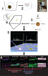
The human retina provides vision at light levels ranging from starlight to sunlight. Its supporting tissues regulate plasma-delivered lipophilic essentials for vision, including retinoids. The macula is an anatomic specialization for high-acuity and color vision that is also vulnerable to prevalent blinding diseases. The retina's exquisite architecture comprises numerous cell types that are aligned horizontally, yielding structurally distinct cell, synaptic, and vascular layers that are visible in histology and in diagnostic clinical imaging. MALDI imaging mass spectrometry (IMS) is now capable of uniting low micrometer spatial resolution with high levels of chemical specificity. In this study, a multimodal imaging approach fortified with accurate multi-image registration was used to localize lipids in human retina tissue at laminar, cellular, and subcellular levels. Multimodal imaging results indicate differences in distributions and abundances of lipid species across and within single cell types. Of note are distinct localizations of signals within specific layers of the macula. For example, phosphatidylethanolamine and phosphatidylinositol lipids were localized to central RPE cells, whereas specific plasmalogen lipids were localized to cells of the perifoveal RPE and Henle fiber layer. Subcellular compartments of photoreceptors were distinguished by PE(20:0_22:5) in the outer nuclear layer, PE(18:0_22:6) in outer and inner segments, and cardiolipin CL(70:5) in the mitochondria-rich inner segments. Several lipids, differing by a single double bond, have markedly different distributions between the central fovea and the ganglion cell and inner nuclear layers. A lipid atlas, initiated in this study, can serve as a reference database for future examination of diseased tissues.
Keywords: age-related macular degeneration; lipid atlas; macula; photoreceptors; retinal pigment epithelium.
Conflict of interest statement
The authors declare no competing financial interest.
Figures


Similar articles
-
Spatial organization of lipids in the human retina and optic nerve by MALDI imaging mass spectrometry.J Lipid Res. 2014 Mar;55(3):504-15. doi: 10.1194/jlr.M044990. Epub 2013 Dec 23. J Lipid Res. 2014. PMID: 24367044 Free PMC article.
-
High resolution MALDI imaging mass spectrometry of retinal tissue lipids.J Am Soc Mass Spectrom. 2014 Aug;25(8):1394-403. doi: 10.1007/s13361-014-0883-2. Epub 2014 May 13. J Am Soc Mass Spectrom. 2014. PMID: 24819461 Free PMC article.
-
Matrix-assisted laser desorption/ionization quadrupole ion trap time-of-flight (MALDI-QIT-TOF)-based imaging mass spectrometry reveals a layered distribution of phospholipid molecular species in the mouse retina.Rapid Commun Mass Spectrom. 2008 Nov;22(21):3415-26. doi: 10.1002/rcm.3751. Rapid Commun Mass Spectrom. 2008. PMID: 18837478
-
Surface analysis of lipids by mass spectrometry: more than just imaging.Prog Lipid Res. 2013 Oct;52(4):329-53. doi: 10.1016/j.plipres.2013.04.005. Epub 2013 Apr 24. Prog Lipid Res. 2013. PMID: 23623802 Review.
-
Imaging mass spectrometry of the visual system: Advancing the molecular understanding of retina degenerations.Proteomics Clin Appl. 2016 Apr;10(4):391-402. doi: 10.1002/prca.201500103. Epub 2016 Jan 28. Proteomics Clin Appl. 2016. PMID: 26586164 Review.
Cited by
-
Untargeted Metabolomic Study of Patients with Wet Age-Related Macular Degeneration in Aqueous Humor.Clin Interv Aging. 2024 Sep 27;19:1571-1580. doi: 10.2147/CIA.S475920. eCollection 2024. Clin Interv Aging. 2024. PMID: 39359698 Free PMC article.
-
Novel technical developments in mass spectrometry imaging in 2020: A mini review.Anal Sci Adv. 2021 Mar 16;2(3-4):225-237. doi: 10.1002/ansa.202000176. eCollection 2021 Apr. Anal Sci Adv. 2021. PMID: 38716449 Free PMC article. Review.
-
Age-Related Macular Degeneration, a Mathematically Tractable Disease.Invest Ophthalmol Vis Sci. 2024 Mar 5;65(3):4. doi: 10.1167/iovs.65.3.4. Invest Ophthalmol Vis Sci. 2024. PMID: 38466281 Free PMC article. Review.
-
Lysolipids are prominent in subretinal drusenoid deposits, a high-risk phenotype in age-related macular degeneration.Front Ophthalmol (Lausanne). 2023;3:1258734. doi: 10.3389/fopht.2023.1258734. Epub 2023 Nov 24. Front Ophthalmol (Lausanne). 2023. PMID: 38186747 Free PMC article.
-
Spectral Analysis of Human Retinal Pigment Epithelium Cells in Healthy and AMD Eyes.Invest Ophthalmol Vis Sci. 2024 Jan 2;65(1):10. doi: 10.1167/iovs.65.1.10. Invest Ophthalmol Vis Sci. 2024. PMID: 38170540 Free PMC article.
References
-
- Harkewicz R; Du H; Tong Z; Alkuraya H; Bedell M; Sun W; Wang X; Hsu YH; Esteve-Rudd J; Hughes G; Su Z; Zhang M; Lopes VS; Molday RS; Williams DS; Dennis EA; Zhang K Essential role of ELOVL4 protein in very long chain fatty acid synthesis and retinal function. J. Biol. Chem. 2012, 287 (14), 11469–80. - PMC - PubMed
MeSH terms
Substances
Grants and funding
LinkOut - more resources
Full Text Sources

