Multicenter Study
. 2020 Mar 17;9(6):e014120.
doi: 10.1161/JAHA.119.014120. Epub 2020 Mar 15.

Helicobacter pylori Infection Impairs Endothelial Function Through an Exosome-Mediated Mechanism

Affiliations
Multicenter Study

Helicobacter pylori Infection Impairs Endothelial Function Through an Exosome-Mediated Mechanism

Xiujuan Xia et al. J Am Heart Assoc. .

Abstract

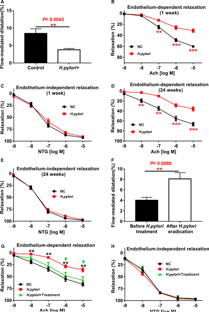
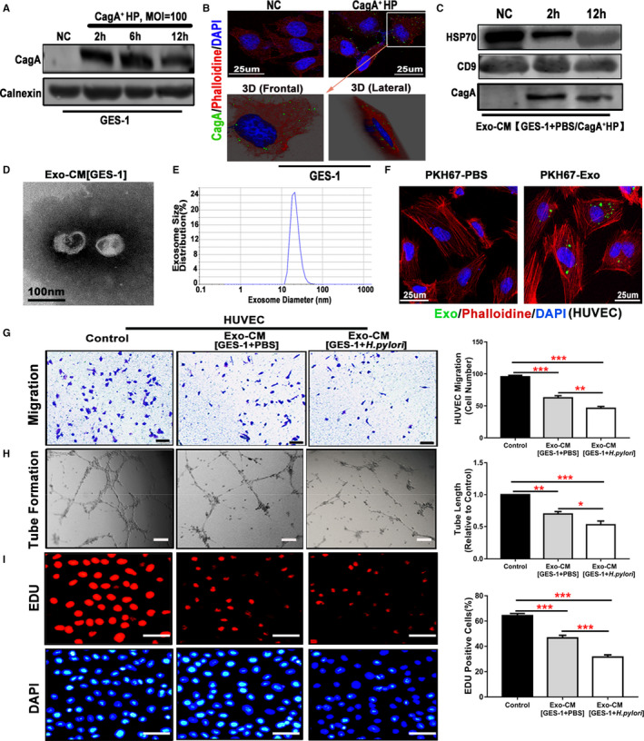
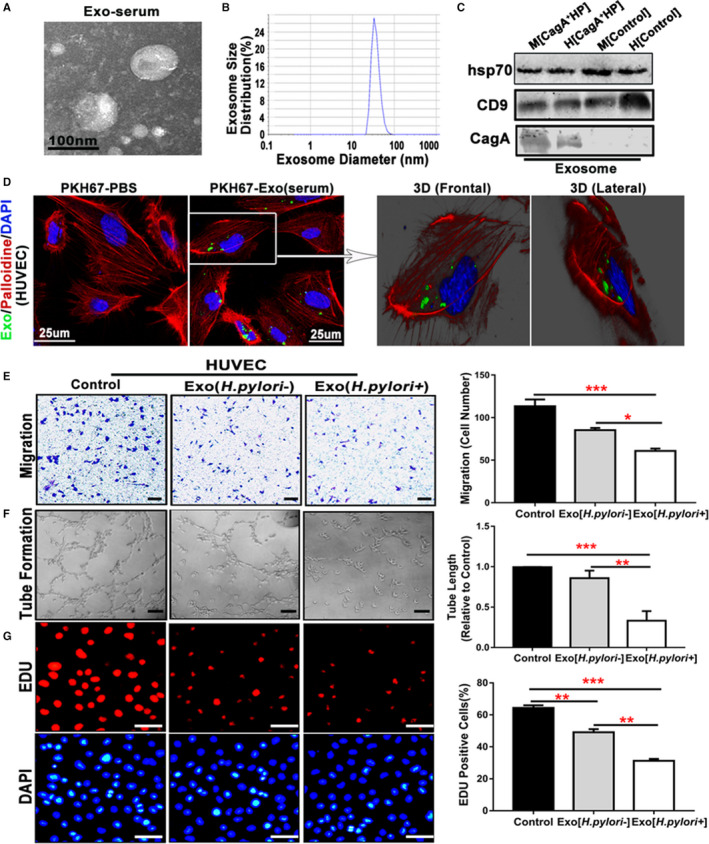
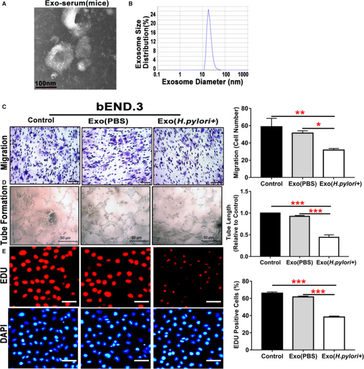
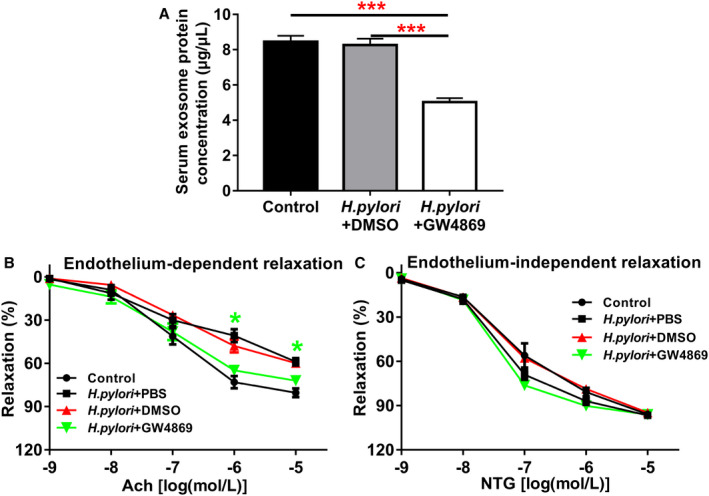
Background Epidemiological studies have suggested an association between Helicobacter pylori (H pylori) infection and atherosclerosis through undefined mechanisms. Endothelial dysfunction is critical to the development of atherosclerosis and related cardiovascular diseases. The present study was designed to test the hypothesis that H pylori infection impaires endothelial function through exosome-mediated mechanisms. Methods and Results Young male and female patients (18-35 years old) with and without H pylori infection were recruited to minimize the chance of potential risk factors for endothelial dysfunction for the study. Endothelium-dependent flow-mediated vasodilatation of the brachial artery was evaluated in the patients and control subjects. Mouse infection models with CagA+H pylori from a gastric ulcer patient were created to determine if H pylori infection-induced endothelial dysfunction could be reproduced in animal models. H pylori infection significantly decreased endothelium-dependent flow-mediated vasodilatation in young patients and significantly attenuated acetylcholine-induced endothelium-dependent aortic relaxation without change in nitroglycerin-induced endothelium-independent vascular relaxation in mice. H pylori eradication significantly improved endothelium-dependent vasodilation in both patients and mice with H pylori infection. Exosomes from conditioned media of human gastric epithelial cells cultured with CagA+H pylori or serum exosomes from patients and mice with H pylori infection significantly decreased endothelial functions with decreased migration, tube formation, and proliferation in vitro. Inhibition of exosome secretion with GW4869 effectively preserved endothelial function in mice with H pylori infection. Conclusions H pylori infection impaired endothelial function in patients and mice through exosome-medicated mechanisms. The findings indicated that H pylori infection might be a novel risk factor for cardiovascular diseases.

Keywords: Helicobacter pylori; cardiovascular risk factor; endothelial dysfunction; exosomes.


Figures

Figure 1
Figure 1
Helicobacter pylori infection significantly impaired endothelium‐dependent flow‐mediated dilatation (FMD) in human subjects and endothelium‐dependent vascular relaxation in mice. Patients with H pylori infection and healthy control subjects were evaluated for endothelium‐dependent FMD of the brachial artery with ultrasound. The diagnosis of H pylori infection was confirmed with gastric endoscopic biopsy and 13C‐urea breath test for each patient. Patients with H pylori infection (n=18 patients) displayed a significant reduction in their endothelium‐dependent FMD compared with the controls (n=13 subjects). A, **P<0.01 by t test. Mice infected with CagA+ H pylori for 1 week significantly decreased acetylcholine (Ach)‐induced endothelium‐dependent relaxation of thoracic aorta (B) without change in NTG‐induced endothelium‐independent vasorelaxation (C) after submaximal contraction with phenylephrine (PE) (10−6M). **P<0.01, ***P<0.001 by 1‐way ANOVA with Bonferroni test or Kruskal‐Wallis test with Dunn post hoc test, n=10 mice for each group. The impaired Ach‐induced endothelium‐dependent vasorelaxation persisted for as long as the infection was present for at least 24 weeks (D) without change in NTG‐induced endothelium‐independent vasorelaxation (E). **P<0.01, ***P<0.001 by 1‐way ANOVA with Bonferroni test or Kruskal‐Wallis test with Dunn post hoc test, n=10 mice for each group. Eradication of H pylori infection with anti–H pylori therapy effectively restored the endothelium‐dependent FMD in patients with H pylori infection (n=10 patients with anti–H pylori therapy) (F) (**P<0.01 by t test with Welch correction), and significantly improved Ach‐induced endothelium‐dependent vasorelaxation in mice with 12 weeks of chronic CagA+ H pylori infection (G), # P<0.05 (compared with CagA+ H pylori +Treatment mice); **P<0.01 (compared with NC mice) by one‐way ANOVA with Bonferroni's test or Kruskal‐Wallis test with Dunn's post hoc test, without change in NTG‐induced endothelium‐independent vasorelaxation (H) (n=8 mice for each group in G and H). Ach indicates acetylcholine; NC, normal control; NTG: nitroglycerin. Data were presented as mean±SE.
Figure 2
Figure 2
Exosomes from human gastric epithelial cells GES‐1 cultured with CagA+Helicobacter pylori significantly inhibited endothelial cell function in vitro. Western blotting analysis (A) and immunofluorescence staining with a 3D confocal microscope (B) demonstrated that the unique H pylori virulence factor CagA entered into GES‐1 after culture with CagA+ H pylori. Exosomes isolated from the conditioned media of GES‐1 cultured with CagA+ H pylori displayed typical features for exosomes including characteristic biomarkers HSP70 and CD9 by Western blotting (C), morphologies on transmission electron microscopy (D), and size using a Zetasizer Nano ZS instrument (E). Western blotting analysis confirmed the presence of CagA protein in the exosomes from the conditioned media of GES‐1 cultured with CagA+ H pylori but not in the ones from GES‐1–conditioned media without CagA+ H pylori (C). PKH67‐labeled GES‐1–derived exosomes (green) were incubated with HUVECs (30 μg protein/5×104 cells), and a significant amount of PKH67‐labeled exosomes were detected inside the HUVECs as visualized using a 3D confocal microscope (F), confirming that the exosomes entered into the cells. Treatment of HUVECs with CagA protein‐containing exosomes (50 μg/mL) from GES‐1–conditioned media for 24 hours significantly inhibited the function of HUVECs with decreased migration (G, scale bars=200 μm), tube formation (H, scale bars=200 μm), and proliferation (I, scale bars=50 μm). CagA+ HP indicates CagA+ H pylori; Exo‐CM, exosomes derived from conditioned medium; GES‐1, human gastric epithelial cells; HUVEC, human umbilical vein endothelial cell; MOI, multiplicity of infection; NC, normal control. *P<0.05, **P<0.01, ***P<0.001 by 1‐way ANOVA with Bonferroni test or Kruskal‐Wallis test with Dunn post hoc test. Data were presented as mean±SE, n=3 independent experiments (experiment was repeated 3 times for every measurement).
Figure 3
Figure 3
Serum exosomes from patients with CagA+Helicobacter pylori infection inhibited endothelial function in vitro. Exosomes isolated from patients with CagA+ H pylori infection exhibited typical exosome characteristics with unique morphologies as shown on transmission electron microscopy (A), size distribution as demonstrated using a Zetasizer Nano ZS (Malvern Instruments, Malvern, Worcestershire, UK) instrument (B), and specific biomarkers (HSP70 and CD9) using Western blot analysis (C). Western blotting also showed that the serum exosomes from mice or patients with CagA+ H pylori infection contained specific CagA protein (C). PKH67‐labeled human serum–derived exosomes (green) were incubated with human umbilical vein endothelial cells (HUVECs) (30 μg protein/5×104 cells). A significant amount of PKH67‐labeled exosomes were detected inside the HUVECs as visualized using a 3‐D confocal microscope (D), confirming that the exosomes entered into the cells (HUVECs). Transwell culture showed that exposure to the serum exosomes from patients with CagA+ H pylori infection significantly inhibited the migration of HUVECs (E, scale bar=200 μm) as compared with the controls. Culture with the serum exosomes from patients with CagA+ H pylori infection also significantly attenuated tube formation (F, scale bar=200 μm), and proliferation (using EdU assay) (G, scale bar=50 μm) of HUVECs. Of note, culture with the serum exosomes from healthy control subjects also moderately and yet significantly inhibited endothelial function with decreased migration, tube formation, and proliferation (E through G); however, the serum exosomes from patients with CagA+ H pylori infection exhibited significantly greater inhibitory effects on endothelial functions than the ones from healthy subjects. CagA+ HP indicates CagA+ H pylori; Exo, Exosomes; H, Human; M, Mouse. *P<0.05, **P<0.01, ***P<0.001 by Kruskal‐Wallis test with Dunn post hoc test or 1‐way ANOVA with the Bonferroni test. Data were presented as mean±SE, n=3 independent experiments (experiment was repeated 3 times for every measurement).
Figure 4
Figure 4
Serum exosomes from mice with Helicobacter pylori infection inhibited endothelial function in vitro. Exosomes isolated from mice with CagA+ H pylori infection exhibited typical exosome characteristics with unique morphologies as shown on transmission electron microscopy (A), and size distribution as demonstrated using a Zetasizer Nano ZS (Malvern Instruments, Malvern, Worcestershire, UK) instrument (B). Transwell culture showed that exposure to the serum exosomes from mice with CagA+ H pylori infection significantly inhibited the migration of mouse brain microvascular endothelial cells bEND.3 (C, scale bar=50 μm) as compared with the controls. Culture with the serum exosomes from mice with CagA+ H pylori infection also significantly attenuated tube formation (D, scale bar=50 μm) and proliferation (using EdU assay) (E, scale bar=50 μm) of bEND.3. bEND.3 indicates mouse brain microvascular endothelial cells; Exo (H pylori +), xosomes derived from mice treated with CagA+ H pylori infection; Exo (PBS), exosomes derived from control mice treated with PBS; Exo‐serum, exosomes isolated from mouse serum. *P<0.05, **P<0.01, ***P<0.001 by Kruskal‐Wallis test with Dunn post hoc test or 1‐way ANOVA with Bonferroni test. Data were presented as mean±SE, n=3 independent experiments (experiment was repeated 3 times for every measurement).
Figure 5
Figure 5
Inhibition of exosome secretion by GW4869 significantly improved endothelium‐dependent vascular relaxation in mice with CagA+Helicobacter pylori infection. Treatment with GW4869 significantly decreased the serum exosome level in the mice with CagA+ H pylori infection (A) as reflected by the significantly decreased total exosome protein levels (***P<0.001 by 1‐way ANOVA with Bonferroni test) and significantly improved acetylcholine (Ach)‐induced endothelium‐dependent relaxation (B) of the aorta in the mice with CagA+ H pylori infection without change in nitroglycerin (NTG)‐induced endothelium‐independent relaxation (C). *P<0.05 by 1‐way ANOVA with Bonferroni test (when CagA+ H pylori+GW4869 group compared with CagA+ H pylori+DMSO group). Ach indicates acetylcholine; DMSO, dimethylsulfoxide (solubilizer of GW4869); NTG, nitroglycerin. Data shown were mean±SE. n=8 mice for control group, and n=10 mice for other groups.

Similar articles

Cited by

References

    1. Mentis A, Lehours P, Megraud F. Epidemiology and diagnosis of Helicobacter pylori infection. Helicobacter. 2015;20(suppl 1):1–7. - PubMed
    1. Eusebi LH, Zagari RM, Bazzoli F. Epidemiology of Helicobacter pylori infection. Helicobacter. 2014;19(suppl 1):1–5. - PubMed
    1. Yamaoka Y, Graham DY. Helicobacter pylori virulence and cancer pathogenesis. Future Oncol. 2014;10:1487–1500. - PMC - PubMed
    1. Xu Z, Li J, Wang H, Xu G. Helicobacter pylori infection and atherosclerosis: is there a causal relationship? Eur J Clin Microbiol Infect Dis. 2017;36:2293–2301. - PubMed
    1. Lee M, Baek H, Park JS, Kim S, Kyung C, Baik SJ, Lee BK, Kim JH, Ahn CW, Kim KR, et al. Current Helicobacter pylori infection is significantly associated with subclinical coronary atherosclerosis in healthy subjects: a cross‐sectional study. PLoS One. 2018;13:e193646. - PMC - PubMed

Publication types

MeSH terms