The functional activity of E-cadherin controls tumor cell metastasis at multiple steps
- PMID: 32127478
- PMCID: PMC7084067
- DOI: 10.1073/pnas.1918167117
The functional activity of E-cadherin controls tumor cell metastasis at multiple steps
Abstract
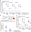
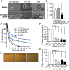
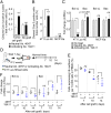
E-cadherin is a tumor suppressor protein, and the loss of its expression in association with the epithelial mesenchymal transition (EMT) occurs frequently during tumor metastasis. However, many metastases continue to express E-cadherin, and a full EMT is not always necessary for metastasis; also, positive roles for E-cadherin expression in metastasis have been reported. We hypothesize instead that changes in the functional activity of E-cadherin expressed on tumor cells in response to environmental factors is an important determinant of the ability of the tumor cells to metastasize. We find that E-cadherin expression persists in metastatic lung nodules and circulating tumor cells (CTCs) in two mouse models of mammary cancer: genetically modified MMTV-PyMT mice and orthotopically grafted 4T1 tumor cells. Importantly, monoclonal antibodies that bind to and activate E-cadherin at the cell surface reduce lung metastasis from endogenous genetically driven tumors and from tumor cell grafts. E-cadherin activation inhibits metastasis at multiple stages, including the accumulation of CTCs from the primary tumor and the extravasation of tumor cells from the vasculature. These activating mAbs increase cell adhesion and reduce cell invasion and migration in both cell culture and three-dimensional spheroids grown from primary tumors. Moreover, activating mAbs increased the frequency of apoptotic cells without affecting proliferation. Although the growth of the primary tumors was unaffected by activating mAbs, CTCs and tumor cells in metastatic nodules exhibited increased apoptosis. Thus, the functional state of E-cadherin is an important determinant of metastatic potential beyond whether the gene is expressed.
Keywords: E-cadherin; E-cadherin-positive tumors; MMTV-PyMT breast cancer model; breast cancer metastasis; circulating tumor cells.
Conflict of interest statement
The authors declare no competing interest.
Figures


Similar articles
-
Core needle biopsy of breast cancer tumors increases distant metastases in a mouse model.Neoplasia. 2014 Nov 20;16(11):950-60. doi: 10.1016/j.neo.2014.09.004. eCollection 2014 Nov. Neoplasia. 2014. PMID: 25425969 Free PMC article.
-
Epithelial requirement for in vitro proliferation and xenograft growth and metastasis of MDA-MB-468 human breast cancer cells: oncogenic rather than tumor-suppressive role of E-cadherin.Breast Cancer Res. 2017 Jul 27;19(1):86. doi: 10.1186/s13058-017-0880-z. Breast Cancer Res. 2017. PMID: 28750639 Free PMC article.
-
Phenotypic Heterogeneity and Metastasis of Breast Cancer Cells.Cancer Res. 2021 Jul 1;81(13):3649-3663. doi: 10.1158/0008-5472.CAN-20-1799. Epub 2021 May 11. Cancer Res. 2021. PMID: 33975882 Free PMC article.
-
Epithelial-mesenchymal transition in tumor metastasis.Mol Oncol. 2017 Jan;11(1):28-39. doi: 10.1002/1878-0261.12017. Epub 2016 Dec 9. Mol Oncol. 2017. PMID: 28085222 Free PMC article. Review.
-
Molecular mechanisms controlling E-cadherin expression in breast cancer.Biochem Biophys Res Commun. 2009 Jun 19;384(1):6-11. doi: 10.1016/j.bbrc.2009.04.051. Epub 2009 Apr 18. Biochem Biophys Res Commun. 2009. PMID: 19379710 Free PMC article. Review.
Cited by
-
Epigenetic Regulation in Oral Squamous Cell Carcinoma Microenvironment: A Comprehensive Review.Cancers (Basel). 2023 Nov 27;15(23):5600. doi: 10.3390/cancers15235600. Cancers (Basel). 2023. PMID: 38067304 Free PMC article. Review.
-
USP10 Regulates ZEB1 Ubiquitination and Protein Stability to Inhibit ZEB1-Mediated Colorectal Cancer Metastasis.Mol Cancer Res. 2023 Jun 1;21(6):578-590. doi: 10.1158/1541-7786.MCR-22-0552. Mol Cancer Res. 2023. PMID: 36940483 Free PMC article.
-
Molecular mechanism of palmitic acid and its derivatives in tumor progression.Front Oncol. 2023 Aug 9;13:1224125. doi: 10.3389/fonc.2023.1224125. eCollection 2023. Front Oncol. 2023. PMID: 37637038 Free PMC article. Review.
-
Predicting T and N Staging of Resectable Gastric Cancer According to Whole Tumor Histogram Analysis About a Non-Cartesian k-Space Acquisition DCE-MRI: A Feasibility Study.Cancer Manag Res. 2021 Oct 18;13:7951-7960. doi: 10.2147/CMAR.S326874. eCollection 2021. Cancer Manag Res. 2021. PMID: 34703316 Free PMC article.
-
The Apoptosis Inhibitor Protein Survivin Is a Critical Cytoprotective Resistor against Silica-Based Nanotoxicity.Nanomaterials (Basel). 2023 Sep 12;13(18):2546. doi: 10.3390/nano13182546. Nanomaterials (Basel). 2023. PMID: 37764575 Free PMC article.
References
-
- Bracke M. E., Van Roy F. M., Mareel M. M., The E-cadherin/catenin complex in invasion and metastasis. Curr. Top. Microbiol. Immunol. 213, 123–161 (1996). - PubMed
-
- Hanahan D., Weinberg R. A., Hallmarks of cancer: The next generation. Cell 144, 646–674 (2011). - PubMed
-
- Kang Y., Massagué J., Epithelial-mesenchymal transitions: Twist in development and metastasis. Cell 118, 277–279 (2004). - PubMed
-
- Bukholm I. K., Nesland J. M., Børresen-Dale A. L., Re-expression of E-cadherin, alpha-catenin and beta-catenin, but not of gamma-catenin, in metastatic tissue from breast cancer patients [seecomments]. J. Pathol. 190, 15–19 (2000). - PubMed
Publication types
MeSH terms
Substances
Grants and funding
LinkOut - more resources
Full Text Sources
Medical
Molecular Biology Databases

