A pneumonia outbreak associated with a new coronavirus of probable bat origin
- PMID: 32015507
- PMCID: PMC7095418
- DOI: 10.1038/s41586-020-2012-7
A pneumonia outbreak associated with a new coronavirus of probable bat origin
Erratum in
-
Addendum: A pneumonia outbreak associated with a new coronavirus of probable bat origin.Nature. 2020 Dec;588(7836):E6. doi: 10.1038/s41586-020-2951-z. Nature. 2020. PMID: 33199918 Free PMC article. No abstract available.
Abstract
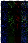
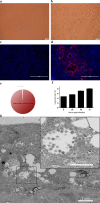
Since the outbreak of severe acute respiratory syndrome (SARS) 18 years ago, a large number of SARS-related coronaviruses (SARSr-CoVs) have been discovered in their natural reservoir host, bats1-4. Previous studies have shown that some bat SARSr-CoVs have the potential to infect humans5-7. Here we report the identification and characterization of a new coronavirus (2019-nCoV), which caused an epidemic of acute respiratory syndrome in humans in Wuhan, China. The epidemic, which started on 12 December 2019, had caused 2,794 laboratory-confirmed infections including 80 deaths by 26 January 2020. Full-length genome sequences were obtained from five patients at an early stage of the outbreak. The sequences are almost identical and share 79.6% sequence identity to SARS-CoV. Furthermore, we show that 2019-nCoV is 96% identical at the whole-genome level to a bat coronavirus. Pairwise protein sequence analysis of seven conserved non-structural proteins domains show that this virus belongs to the species of SARSr-CoV. In addition, 2019-nCoV virus isolated from the bronchoalveolar lavage fluid of a critically ill patient could be neutralized by sera from several patients. Notably, we confirmed that 2019-nCoV uses the same cell entry receptor-angiotensin converting enzyme II (ACE2)-as SARS-CoV.
Conflict of interest statement
The authors declare no competing interests.
Figures


Comment in
-
Potential of large "first generation" human-to-human transmission of 2019-nCoV.J Med Virol. 2020 Apr;92(4):448-454. doi: 10.1002/jmv.25693. Epub 2020 Feb 14. J Med Virol. 2020. PMID: 31997390 Free PMC article.
-
Calling all coronavirus researchers: keep sharing, stay open.Nature. 2020 Feb;578(7793):7. doi: 10.1038/d41586-020-00307-x. Nature. 2020. PMID: 32020126 No abstract available.
-
Evolving status of the 2019 novel coronavirus infection: Proposal of conventional serologic assays for disease diagnosis and infection monitoring.J Med Virol. 2020 May;92(5):464-467. doi: 10.1002/jmv.25702. Epub 2020 Feb 17. J Med Virol. 2020. PMID: 32031264 Free PMC article. No abstract available.
-
Novel coronavirus takes flight from bats?Nat Rev Microbiol. 2020 Apr;18(4):191. doi: 10.1038/s41579-020-0336-9. Nat Rev Microbiol. 2020. PMID: 32051570 Free PMC article.
-
Chloroquine for the 2019 novel coronavirus SARS-CoV-2.Int J Antimicrob Agents. 2020 Mar;55(3):105923. doi: 10.1016/j.ijantimicag.2020.105923. Epub 2020 Feb 15. Int J Antimicrob Agents. 2020. PMID: 32070753 Free PMC article. No abstract available.
-
SARS Coronavirus Redux.Trends Immunol. 2020 Apr;41(4):271-273. doi: 10.1016/j.it.2020.02.007. Epub 2020 Mar 12. Trends Immunol. 2020. PMID: 32173256 Free PMC article.
-
A Unique Protease Cleavage Site Predicted in the Spike Protein of the Novel Pneumonia Coronavirus (2019-nCoV) Potentially Related to Viral Transmissibility.Virol Sin. 2020 Jun;35(3):337-339. doi: 10.1007/s12250-020-00212-7. Epub 2020 Mar 20. Virol Sin. 2020. PMID: 32198713 Free PMC article. No abstract available.
-
Inefficiency of Sera from Mice Treated with Pseudotyped SARS-CoV to Neutralize 2019-nCoV Infection.Virol Sin. 2020 Jun;35(3):340-343. doi: 10.1007/s12250-020-00214-5. Epub 2020 Mar 31. Virol Sin. 2020. PMID: 32236815 Free PMC article. No abstract available.
-
COVID-19: A New Virus, but a Familiar Receptor and Cytokine Release Syndrome.Immunity. 2020 May 19;52(5):731-733. doi: 10.1016/j.immuni.2020.04.003. Epub 2020 Apr 22. Immunity. 2020. PMID: 32325025 Free PMC article.
-
Virus, veritas, vita.Postgrad Med J. 2020 Jul;96(1137):371-372. doi: 10.1136/postgradmedj-2020-137802. Epub 2020 Apr 28. Postgrad Med J. 2020. PMID: 32345756 No abstract available.
-
Calling for a COVID-19 One Health Research Coalition.Lancet. 2020 May 16;395(10236):1543-1544. doi: 10.1016/S0140-6736(20)31028-X. Epub 2020 May 7. Lancet. 2020. PMID: 32386563 Free PMC article. No abstract available.
-
[Asthma and COVID-19: a risk population?].Rev Mal Respir. 2020 Sep;37(7):606-607. doi: 10.1016/j.rmr.2020.05.002. Epub 2020 May 15. Rev Mal Respir. 2020. PMID: 32419737 Free PMC article. French. No abstract available.
-
The role of Instagram in public health education in COVID-19 in Iran.J Clin Anesth. 2020 Oct;65:109887. doi: 10.1016/j.jclinane.2020.109887. Epub 2020 May 20. J Clin Anesth. 2020. PMID: 32454342 Free PMC article. No abstract available.
-
Isolation and Growth Characteristics of SARS-CoV-2 in Vero Cell.Virol Sin. 2020 Jun;35(3):348-350. doi: 10.1007/s12250-020-00241-2. Epub 2020 Jun 19. Virol Sin. 2020. PMID: 32562199 Free PMC article. No abstract available.
-
SARS-CoV-2 Does Not Replicate in Aedes Mosquito Cells nor Present in Field-Caught Mosquitoes from Wuhan.Virol Sin. 2020 Jun;35(3):355-358. doi: 10.1007/s12250-020-00251-0. Epub 2020 Jun 29. Virol Sin. 2020. PMID: 32602045 Free PMC article. No abstract available.
-
The use of SARS-CoV-2-related coronaviruses from bats and pangolins to polarize mutations in SARS-Cov-2.Sci China Life Sci. 2020 Oct;63(10):1608-1611. doi: 10.1007/s11427-020-1764-2. Epub 2020 Jul 1. Sci China Life Sci. 2020. PMID: 32621057 Free PMC article. No abstract available.
-
Indian perspective of remdesivir: A promising COVID-19 drug.Indian J Pharmacol. 2020 May-Jun;52(3):227-228. doi: 10.4103/ijp.IJP_486_20. Epub 2020 Aug 4. Indian J Pharmacol. 2020. PMID: 32874008 Free PMC article. No abstract available.
-
Genomic Characterization of a New Coronavirus from Migratory Birds in Jiangxi Province of China.Virol Sin. 2021 Dec;36(6):1656-1659. doi: 10.1007/s12250-021-00402-x. Epub 2021 Jul 8. Virol Sin. 2021. PMID: 34236588 Free PMC article. No abstract available.
Similar articles
-
Receptor Recognition by the Novel Coronavirus from Wuhan: an Analysis Based on Decade-Long Structural Studies of SARS Coronavirus.J Virol. 2020 Mar 17;94(7):e00127-20. doi: 10.1128/JVI.00127-20. Print 2020 Mar 17. J Virol. 2020. PMID: 31996437 Free PMC article.
-
Genomic characterisation and epidemiology of 2019 novel coronavirus: implications for virus origins and receptor binding.Lancet. 2020 Feb 22;395(10224):565-574. doi: 10.1016/S0140-6736(20)30251-8. Epub 2020 Jan 30. Lancet. 2020. PMID: 32007145 Free PMC article.
-
Isolation and characterization of a bat SARS-like coronavirus that uses the ACE2 receptor.Nature. 2013 Nov 28;503(7477):535-8. doi: 10.1038/nature12711. Epub 2013 Oct 30. Nature. 2013. PMID: 24172901 Free PMC article.
-
The origin, transmission and clinical therapies on coronavirus disease 2019 (COVID-19) outbreak - an update on the status.Mil Med Res. 2020 Mar 13;7(1):11. doi: 10.1186/s40779-020-00240-0. Mil Med Res. 2020. PMID: 32169119 Free PMC article. Review.
-
Angiotensin-converting enzyme 2: The old door for new severe acute respiratory syndrome coronavirus 2 infection.Rev Med Virol. 2020 Sep;30(5):e2122. doi: 10.1002/rmv.2122. Epub 2020 Jun 30. Rev Med Virol. 2020. PMID: 32602627 Free PMC article. Review.
Cited by
-
Characterization of Collaborative Cross mouse founder strain CAST/EiJ as a novel model for lethal COVID-19.Sci Rep. 2024 Oct 24;14(1):25147. doi: 10.1038/s41598-024-77087-1. Sci Rep. 2024. PMID: 39448712 Free PMC article.
-
The dynamics of inflammatory markers in coronavirus disease-2019 (COVID-19) patients: A systematic review and meta-analysis.Clin Epidemiol Glob Health. 2021 Jul-Sep;11:100727. doi: 10.1016/j.cegh.2021.100727. Epub 2021 Mar 20. Clin Epidemiol Glob Health. 2021. PMID: 33778183 Free PMC article. Review.
-
Synthetic Homogeneous Glycoforms of the SARS-CoV-2 Spike Receptor-Binding Domain Reveals Different Binding Profiles of Monoclonal Antibodies.Angew Chem Int Ed Engl. 2021 Jun 1;60(23):12904-12910. doi: 10.1002/anie.202100543. Epub 2021 Apr 8. Angew Chem Int Ed Engl. 2021. PMID: 33709491 Free PMC article.
-
Impact of cytokine storm and systemic inflammation on liver impairment patients infected by SARS-CoV-2: Prospective therapeutic challenges.World J Gastroenterol. 2021 Apr 21;27(15):1531-1552. doi: 10.3748/wjg.v27.i15.1531. World J Gastroenterol. 2021. PMID: 33958841 Free PMC article. Review.
-
Emerging mechanisms of immunocoagulation in sepsis and septic shock.Trends Immunol. 2021 Jun;42(6):508-522. doi: 10.1016/j.it.2021.04.001. Epub 2021 Apr 24. Trends Immunol. 2021. PMID: 33906793 Free PMC article. Review.
References
-
- Li W, et al. Bats are natural reservoirs of SARS-like coronaviruses. Science. 2005;310:676–679. - PubMed
MeSH terms
Substances
LinkOut - more resources
Full Text Sources
Other Literature Sources
Molecular Biology Databases
Miscellaneous

