. 2019 Nov 15;7(1):307.
doi: 10.1186/s40425-019-0796-5.

A new synthetic toll-like receptor 1/2 ligand is an efficient adjuvant for peptide vaccination in a human volunteer

Affiliations

A new synthetic toll-like receptor 1/2 ligand is an efficient adjuvant for peptide vaccination in a human volunteer

Hans-Georg Rammensee et al. J Immunother Cancer. .

Erratum in

Abstract

Background: We previously showed that the bacterial lipopeptide Pam3Cys-Ser-Ser, meanwhile established as a toll-like receptor (TLR) 1/2 ligand, acts as a strong adjuvant for the induction of virus specific CD8+ T cells in mice, when covalently coupled to a synthetic peptide.

Case presentation: We now designed a new water-soluble synthetic Pam3Cys-derivative, named XS15 and characterized it in vitro by a TLR2 NF-κB luciferase reporter assay. Further, the capacity of XS15 to activate immune cells and stimulate peptide-specific CD8+ T and NK cells by 6-sulfo LacNAc+ monocytes was assessed by flow cytometry as well as cytokine induction using immunoassays. The induction of a functional immune response after vaccination of a volunteer with viral peptides was assessed by ELISpot assay and flow cytometry in peripheral blood cells and infiltrating cells at the vaccination site, as well as by immunohistochemistry and imaging. XS15 induced strong ex vivo CD8+ and TH1 CD4+ responses in a human volunteer upon a single injection of XS15 mixed to uncoupled peptides in a water-in-oil emulsion (Montanide™ ISA51 VG). A granuloma formed locally at the injection site containing highly activated functional CD4+ and CD8+ effector memory T cells. The total number of vaccine peptide-specific functional T cells was experimentally assessed and estimated to be 3.0 × 10⁵ in the granuloma and 20.5 × 10⁶ in peripheral blood.

Conclusion: Thus, in one volunteer we show a granuloma forming by peptides combined with an efficient adjuvant in a water-in-oil-emulsion, inducing antigen specific T cells detectable in circulation and at the vaccination site, after one single vaccination only. The ex vivo T cell responses in peripheral blood were detectable for more than one year and could be strongly boosted by a second vaccination. Hence, XS15 is a promising adjuvant candidate for peptide vaccination, in particular for tumor peptide vaccines in a personalized setting.

Keywords: Adjuvant; Immunotherapy; Lipopeptide; TLR1/2 ligand; Vaccines.


Conflict of interest statement

H.G. Rammensee has ownership interest (including patents) in Immatics Biotechnologies GmbH, CureVac AG, and Synimmune GmbH, further he shares the patent for XS15.

M.W. Löffler, D.J. Kowalewski, H. Schuster, S. Stevanović, and S.P. Haen are the inventors of patents for vaccine peptides owned by Immatics. D.J. Kowalewski, L. Backert, and H. Schuster are currently employees of Immatics Biotechnologies. P. Anoop Chandran is an employee of Adaptimmune Therapeutics Ltd. H. Zelba is employed by CeGaT GmbH.

K.H. Wiesmüller shares the patent for XS15 and holds ownership interest in EMC microcollections GmbH.

No competing interests were disclosed by the other authors.

Figures
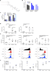
Fig. 1
Fig. 1
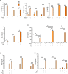
Pam3Cys-GDPKHPKSF (XS15) is a TLR1/2 ligand activating immune cells and stimulating DCs and cytokine release. (a) Structure of Pam3Cys-GDPKHPKSF: Skeletal structural formula of the molecular structure of the lipopeptide Pam3Cys-GDPKHPKSF termed XS15. (b) Dual-luciferase assay on HEK293T cells transfected with TLR2: HEK293T cells were transiently transfected with a human TLR2 plasmid and a NF-κB luciferase reporter plasmid or left untreated (− ctrl.). Culture medium was replaced after 30 h and stimuli added at the stated concentrations. The cells were incubated for 18 h and lysates were prepared and analysed by dual-luciferase assay. Pam3CysSK4 (P3CSK4) and two different lots of XS15 (XS15#1/ XS15#2) were used. (c) HEK-Dual hTLR2 cells, stably expressing a NF-κB/AP-1-inducible secreted embryonic alkaline phosphatase (SEAP) reporter, were incubated for 1 h with TLR1, TLR2 and TLR6 blocking antibodies, isotype control or negative controls (no Abs) (4 μg/ml). Then, cells were stimulated for 24 h with the established TLR2/6 agonist FSL-1 (1 ng/ml), XS15 (10 ng/ml) or left unstimulated (− ctrl.). Supernatants were collected and SEAP levels determined using QUANTI-Blue detection assay. Error bars represent SD. The graph shows the mean + SEM of n = 2 experiments, significance was assessed by two-way ANOVA. (d) Immune cell activation by XS15: Fresh PBMCs were cultured for 40 h in the presence of Phytohemagglutinin-L (PHA) + Pokeweed (PWM) (P + P), Pam3CysSK4 (P3CSK4), XS15 or left untreated (− ctrl.). Activated NK (left panel) and B cells (right panel) were assessed with the marker CD69 following the gating strategy: time gate, single cells (FSC-H/ FSC-A), living cells (Zombie-Aqua/ FSC-A), lymphocytes (FSC-A/ SSC-A); B-cells were defined as CD14neg CD3neg CD19+ cells and NK cells as CD14neg CD3neg CD19neg CD56+ cells. Healthy donors (n = 6), means are shown, significance was assessed by one-way ANOVA. (e) Dendritic cell (DC) stimulation by XS15: DCs were differentiated from blood monocytes and then matured as described in the material and methods section. Gating strategy was: time gate, single cells (FSC-H/ FSC-A), living cells (Zombie Aqua/ FSC-A). Upper panel: scatter plots for healthy donors (n = 6), means are shown significance was assessed by one-way ANOVA. Lower panel: modal histograms and median fluorescences for one representative donor. Medium control without maturation cocktail = − ctrl. Standard maturation cocktail = Mat. (f) Induction of cytokine release by XS15: Anticoagulated whole blood was incubated with XS15 (10 μg/ml) as well as LPS (100 ng/ml) and PHA (2 μg/ml)/ PWM (1 μg/ml) as positive (+ ctrl.) and medium only as negative controls (− ctrl.) and supernatants harvested after 20 h. Multiplexed bead-based sandwich immunoassays were performed using a LUMINEX device with a 42-analyte panel. Exemplary findings obtained in three healthy donors (HD) for IL-8 (left), MCP1 (middle) and MIP-1β (right) are shown with means. HD1 (blue square) designates the vaccinated volunteer characterized in more detail subsequently. Additional results are provided in Additional file 7: Table S1. In case of saturation, the upper limit of quantification (ULOQ) was assigned. p ≤ 0.05*; **p ≤ 0.01; ***p ≤ 0.001
Fig. 2
Fig. 2
Impact of XS15 on cytokine release by slanMo and their capacity to stimulate WT1 peptide-specific CD8+ T cells and NK cells. (a) slanMo were maintained for 6 h to allow spontaneous maturation into DCs. Subsequently, slanMo were cultivated alone (slanMo) or in the presence of XS15 (slanMo + XS15) for additional 18 h. Supernatants were collected and the concentration of (a) TNF (left), IL-1β (middle), IL-6 (right), IL-23 (lower left) analysed by ELISA. (b) slanMo were maintained for 6 h to allow spontaneous maturation into DCs. Subsequently, slanMo were cultivated in the absence (slanMo) or presence of XS15 (slanMo + XS15) for additional 18 h, alternatively, slanMo were incubated with IFNγ for the first 6 h. Thereafter, slanMo were cultivated in the presence of IFNγ alone (slanMo + IFNγ) or together with XS15 (slanMo + IFNγ + XS15) for additional 18 h. Then, IL-12 was analysed by ELISA. The results of three different healthy donors (HD) are presented as mean ± SE of duplicate or triplicate measurements. (c) Effect of XS15 on the capacity of slanMo to stimulate IFNγ release by WT1 peptide-specific CD8+ T cells: slanMo were maintained for 6 h to allow spontaneous maturation. Subsequently, slanMo were coincubated with the specific CD8+ T cell clone CC7 (slanMo + CD8+), in the presence of the WT1 peptide (WT1) and/or XS15. After 42 h, supernatants were collected and IFNγ was quantified by ELISA. The results of three different healthy donors (HD) are presented as mean ± SE of triplicate determinations. (d) Impact of XS15 on the ability of slanMo to stimulate IFNγ secretion by NK cells: slanMo were maintained for 6 h to allow spontaneous maturation. Then, autologous NK cells were cultured either alone (NK) or incubated with XS15 (NK + XS15), cocultured with slanMo alone (NK + slanMo) or additionally incubated with XS15 (NK + slanMo +XS15). After 42 h, supernatants were collected and the concentration of IFNγ was determined by ELISA. The results of three different HD are presented as mean ± SE of triplicate determinations. Asterisks indicate a statistically significant difference (*p ≤ 0.05; **p ≤ 0.01; ***p ≤ 0.001; assessed by Student’s t-test). Exemplary flow cytometry results showing effects of XS15 on slanMo-mediated T cell programming regarding the percentage of IFNγ- and IL-4-producing CD4+ T cells are provided as Additional file 8: Fig. S1
Fig. 3
Fig. 3
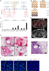
A single vaccination with peptides and XS15 induces a granuloma and local immune cell infiltration with functional T cells. (a) Time line providing an overview on blood and tissues samples as well as analyses described subsequently and interventions performed (i.e. vaccination, 18F-FDG-PET-MR imaging/ granuloma resection). Vaccinated peptides used at each time point are provided in Tables 1 & 2, respectively; Pre (before vaccination); d (day after first vaccination). (b) Induction of functional T cells by XS15 detected in ex vivo ELISpot: PBMCs were isolated from peripheral blood of a volunteer before vaccination (pre-vac), 28 days and 44 days after vaccination. IFNγ response towards the three vaccine peptides (ADV-Hex, FLU-NCAP and EBV-GP350) was determined in two ELISpot assays (Pre-vac + 28d, and 44d). HIV-A*01, HIV-B*08 and Fil-A peptides served as the relevant negative controls. 300,000 cells were seeded per well. Phytohemagglutinin-L (PHA-L) stimulation was used as a positive control (ELISpot plate wells rearranged, and negative controls left out). (c) Respective mean spot counts and SD/100,000 cells per well are shown. (d) Granuloma formation at the vaccination site: 18F-FDG-PET/MR (upper panel) performed on day 43 demonstrated an intense 18F-FDG uptake at the site of the induration (standardized uptake value ((SUV(mean) 4.6; SUV(max) 6.4), but no 18F-FDG-uptake was observed in the draining lymph nodes or in any other secondary lymphoid organs; corresponding MR (lower panel). (e) Immune cell infiltration of the granuloma induced by vaccination: A tissue sample from the granuloma center was processed as formalin-fixed paraffin-embedded (FFPE) tissue and assessed by hematoxylin & eosin (HE) staining (right) and immunohistochemistry (left). T cells (CD8+ and CD4+), B cells (CD20+) as well as macrophages (CD68+) and granulocytes appeared as ordered structures in separated areas resembling lymphoid tissues. Mineral oil deposits (black arrows) were still discernible, surrounded by macrophages, whereas both CD4+ and CD8+ T cells were located closely to the macrophages but separated from the oil patches. Original magnification was × 100. Black scale bars indicate 200 μm. (f) Co-localization of slanMo and CD8+ T cells in the granuloma. Immunofluorescence staining was performed to detect slanMo and CD8+ lymphocytes in the granuloma of the XS15-vaccinated volunteer. As representative examples, images of single CD8+ T cell or slanMo stainings as well as merged images are shown. Original magnification was × 400. White scale bars are 20 μm
Fig. 4
Fig. 4
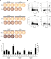
Functionality and antigen-specificity of granuloma infiltrating cells (GICs). GICs were isolated as described in the Material and Methods section and analysed alongside PBMCs isolated from blood drawn on the same day from the same individual. (a) GICs were rested overnight after isolation and the IFNγ response towards the three vaccinated peptides (ADV-Hex, FLU-NCAP and EBV-GP350; Table 1) was determined by IFNγ ELISpot assay. 50,000 cells were seeded per well. HIV-A*01, HIV-B*08 and Fil-A peptides served as the relevant negative controls (rearranged wells). Ex vivo phenotype of GICs is provided as Additional File 8: Fig. S3. (b) PBMCs and GICs were harvested from the ELISpot plate (see panel A) and stained with ADV-Hex APC- and FLU-NCAP-PE- multimers. Percentages of CD8+ multimer-positive and multimer-negative cells within CD4neg are indicated. (c) GICs were stimulated and expanded in vitro using anti-CD3 mAb and IL-2. The cells were then re-stimulated with the indicated peptides or with an equal volume of 10% DMSO for 12 h and the indicated secreted cytokines and surface CD107a expression (degranulation) were quantified by flow cytometry (% of functional cells are given after subtraction of marker-positive cells in the DMSO control well)
Fig. 5
Fig. 5
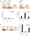
Induction of CMV specific T cells after a single multi-peptide vaccination, and evidence for long lasting memory and boosting. The same volunteer as previously shown was vaccinated with the peptides shown in Table 2, this time with 50 μg of a new batch of XS15. At day 28 after vaccination (Post-vac), PBMCs were assayed by ex vivo ELISpot (a; upper panel and b, 300.000 cells/well), and additionally tested after a short time of in vitro expansion in the presence of the relevant peptides (in vitro stimulation; IVS) (A; lower panels, 250.000 cells/well). Reactivities against the HLA class I and HLA class II peptides are shown in panels (a) and (b), respectively (rearranged wells). In addition, bar charts with respective mean spot counts /100,000 cells + SD (when applicable) are shown. Negative control (− ctrl.) was DMSO or respective HLA-matched peptides (HIV); vac (vaccination)

Similar articles

Cited by

References

    1. Gubin MM, Zhang X, Schuster H, et al. Checkpoint blockade cancer immunotherapy targets tumour-specific mutant antigens. Nature. 2014;515(7528):577–581. doi: 10.1038/nature13988. - DOI - PMC - PubMed
    1. Luksza M, Riaz N, Makarov V, et al. A neoantigen fitness model predicts tumour response to checkpoint blockade immunotherapy. Nature. 2017;551(7681):517–520. doi: 10.1038/nature24473. - DOI - PMC - PubMed
    1. Panda A, Betigeri A, Subramanian K, et al. Identifying a clinically applicable mutational burden threshold as a potential biomarker of response to immune checkpoint therapy in solid tumors. JCO Precis Oncol. 2017;2017. - PMC - PubMed
    1. Rini BI, Stenzl A, Zdrojowy R, et al. IMA901, a multipeptide cancer vaccine, plus sunitinib versus sunitinib alone, as first-line therapy for advanced or metastatic renal cell carcinoma (IMPRINT): a multicentre, open-label, randomised, controlled, phase 3 trial. Lancet Oncol. 2016;17(11):1599–1611. doi: 10.1016/S1470-2045(16)30408-9. - DOI - PubMed
    1. Walter S, Weinschenk T, Stenzl A, et al. Multipeptide immune response to cancer vaccine IMA901 after single-dose cyclophosphamide associates with longer patient survival. Nat Med. 2012;18(8):1254–1261. doi: 10.1038/nm.2883. - DOI - PubMed

Publication types