. 2016 Jul 21;63(2):261-276.
doi: 10.1016/j.molcel.2016.06.015. Epub 2016 Jul 14.

The Molecular Basis for Ubiquitin and Ubiquitin-like Specificities in Bacterial Effector Proteases

Affiliations

The Molecular Basis for Ubiquitin and Ubiquitin-like Specificities in Bacterial Effector Proteases

Jonathan N Pruneda et al. Mol Cell. .

Abstract

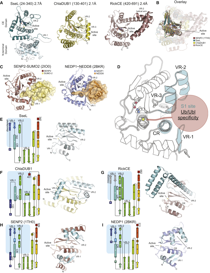
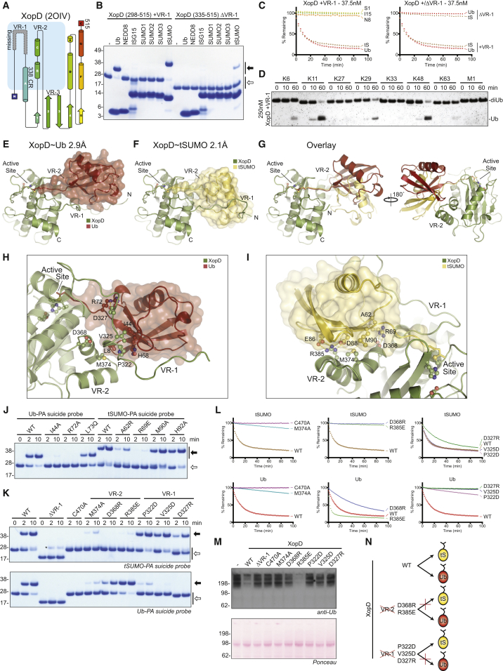
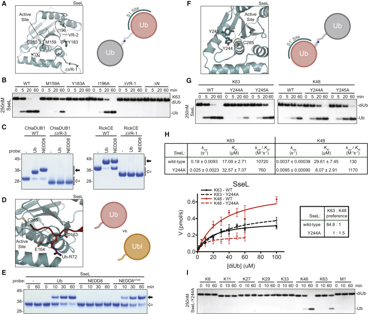
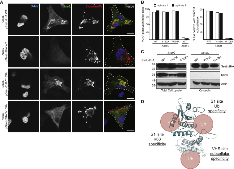
Pathogenic bacteria rely on secreted effector proteins to manipulate host signaling pathways, often in creative ways. CE clan proteases, specific hydrolases for ubiquitin-like modifications (SUMO and NEDD8) in eukaryotes, reportedly serve as bacterial effector proteins with deSUMOylase, deubiquitinase, or, even, acetyltransferase activities. Here, we characterize bacterial CE protease activities, revealing K63-linkage-specific deubiquitinases in human pathogens, such as Salmonella, Escherichia, and Shigella, as well as ubiquitin/ubiquitin-like cross-reactive enzymes in Chlamydia, Rickettsia, and Xanthomonas. Five crystal structures, including ubiquitin/ubiquitin-like complexes, explain substrate specificities and redefine relationships across the CE clan. Importantly, this work identifies novel family members and provides key discoveries among previously reported effectors, such as the unexpected deubiquitinase activity in Xanthomonas XopD, contributed by an unstructured ubiquitin binding region. Furthermore, accessory domains regulate properties such as subcellular localization, as exemplified by a ubiquitin-binding domain in Salmonella Typhimurium SseL. Our work both highlights and explains the functional adaptations observed among diverse CE clan proteins.


Figures

None
Graphical abstract
Figure 1
Figure 1
CE Effector Proteins Demonstrate Mixed Activities (A) Domain annotation of selected bacterial CE effectors. Construct boundaries and accessory domains used in this study are indicated. α, predicted α-helical domain; α/β, predicted α/β fold; TM, transmembrane helix; Pro, proline-rich sequence; GUB, wall-associated receptor kinase galacturonan-binding (GUB_WAK_bind) domain; CC, coiled coil. (B) Purified CE enzymes from bacteria. CA, catalytic Cys-to-Ala mutant. GST-YopJ contains co-purifying degradation products at lower molecular weight. (C) Suicide probe reaction following incubation with the Ub-PA probe for 1 hr at room temperature. Asterisks () mark catalytic cysteine-dependent reactivity. (D) Schematic of the Ub/Ubl substrate cleavage assay. Ub/Ubl modifiers are isopeptide linked to a TAMRA-labeled Lys-Gly peptide, and fluorescence polarization (FP) is used to monitor cleavage over time. N8, NEDD8; S1, SUMO1; I15, ISG15. (E) Normalized FP measured as a function of time following the addition of the listed active CE enzymes to the TAMRA-linked Ub/Ubl reagents. (F) Linkage specificity analysis for bacterial CE DUBs active in the TAMRA assay. A silver-stained SDS-PAGE gel shows diUb hydrolysis over time. See also Figure S1.
Figure 2
Figure 2
Structural Analysis of Bacterial CE Deubiquitinases (A) Cartoon representations of SseL (2.7 Å, teal), ChlaDUB1 (2.1 Å, yellow), and RickCE (2.4 Å, violet) aligned on the catalytic triad (ball and stick). (B) Superposition of the CE core based on the catalytic triad for structures in (A) and human SENP2 (PDB: 1TH0). (C) Representations of the human SENP2-SUMO2 noncovalent complex (PDB: 2IO0, left) and NEDP1∼NEDD8 covalent complex (PDB: 2BKR, right) illustrating a common Ubl binding site. (D) Schematic of the CE fold based on the NEDP1 structure (PDB: 2BKR), highlighting the constant region (CR) and variable regions (VR) that form the Ub/Ubl-binding S1 site (blue). (E–I) Left: topology diagrams for CE structures, with the S1 binding site highlighted (blue box). Structure boundaries are indicated by numbers. Features in dashed outline are either disordered or outside the crystallized constructs, as marked. Right: view of the S1 substrate binding site in CE-fold structures, with variable regions highlighted in cyan and labeled accordingly. See also Table 1 and Figure S2.
Figure 3
Figure 3
Molecular Analysis of XopD Ub/Ubl Specificity (A) Topology diagram, as in Figure 2E, for the XopD crystal structure (PDB: 2OIV). The crystallized construct lacks a potential VR-1 region. (B) XopD constructs with VR-1 (+VR-1: 298–515) or without VR-1 (ΔVR-1: 335–515) were tested against a panel of Ub/Ubl suicide probes for 1 hr at room temperature (propargylamine-derived probes). Open arrow, unmodified; closed arrow, modified. (C) FP assays as in Figure 1D, including a substrate derived from S. lycopersicum SUMO (tS, tomato SUMO). Left: Ub/Ubl specificity of XopD +VR-1. Right: comparison of XopD +VR-1 cleaving the Ub and tSUMO substrates (shown in left) with the ΔVR-1 construct. (D) Linkage specificity analysis as in Figure 1F for XopD +VR-1. (E) Cartoon representation of the 2.9 Å XopD∼Ub covalent complex crystal structure. (F) Cartoon representation of the 2.1 Å XopD∼tSUMO covalent complex crystal structure. (G) Overlay of structures shown in (E) and (F), illustrating differences in Ub/tSUMO conformation and usage of XopD variable regions. For clarity, VR-1 is not shown. (H) Close-up of the XopD-Ub interaction. In the foreground, VR-1 coordinates Ub Arg72 and the Ile44 hydrophobic patch. In the background, VR-2 contacts the Ub Leu8 loop. (I) Close-up of the XopD-tSUMO interaction. VR-2 forms the basis of tSUMO binding and makes a number of polar and salt-bridge interactions surrounding the hydrophobic contact of tSUMO Met90. (J) Series of Ub and tSUMO suicide probe reactions performed on ice, testing interactions observed in the Ub/tSUMO domain, as well as in their C termini (propargylamine-derived probes). Open arrow, unmodified; closed arrow, modified. (K) As in (J), for XopD mutations encompassing VR-1 and VR-2. Open arrow, unmodified; closed arrow, modified. Asterisk, contamination in tSUMO suicide probe. (L) FP-based cleavage assays testing the XopD variants used in (K) against the Ub and tSUMO KG-modified substrates. Wild-type XopD data from (C) are shown for reference. (M) S. lycopersicum protein extract blotted for total Ub (Ubi-1; Novus Biologicals) following a 1 hr room temperature treatment with the XopD variants used in (K) at 1 μM final concentration. (N) Schematic summarizing the activities displayed by wild-type and VR-1/VR-2 mutant XopD variants. See also Table 1 and Figure S3.
Figure 4
Figure 4
Structure-Based Manipulation of CE DUB Activities and Specificities (A) Mutations and truncations designed in the SseL S1 binding site (SseL ΔVR-1: Δ138–147; SseL ΔN: aa 158–340). (B) K63 diUb cleavage assay using mutants from (A). (C) Suicide probe assay monitoring Ub and NEDD8 reactivity following 1 hr incubation at room temperature, for wild-type ChlaDUB1 and RickCE, and constructs containing truncated variable regions (ChlaDUB1 ΔVR-3: Δ250-272; RickCE ΔVR-1: aa 420-691; propargylamine-derived probes). Open arrow, unmodified; closed arrow, modified. (D) Model of the Ub C terminus entering the SseL active site, based on analogous Ubl-bound structures. Putative acidic residues that can coordinate Ub Arg72 are shown. (E) SseL suicide probe assay with Ub, NEDD8, and NEDD8 A72R probes performed at room temperature (chloroethylamine-derived probes). Open arrow, unmodified; closed arrow, modified. (F) Close-up of the conserved hydrophobic S1’ site of SseL. (G) K63 and K48 diUb cleavage assays using S1’ site mutations. (H) Kinetic analysis of K48 and K63 diUb linkage preference for SseL wild-type and Y244A. (I) Linkage specificity analysis for all diUb substrates, as in Figure 1F, for the SseL Y244A S1’ site mutant. See also Figure S4.
Figure 5
Figure 5
Bioinformatic Analysis of the CE Protease Clan Structure-informed dendrogram for the CE clan, with representative members from eukaryotes, bacteria, and viruses. Clustered families are highlighted and labeled according to the archetypal examples. Eukaryotic family members are labeled with numbers corresponding to their SENP nomenclature. Characterized members are highlighted in bold. See also Figure S5.
Figure 6
Figure 6
CE Catalytic Domains Are Fitted with Accessory Domains (A) SseL crystal structure (active-site Cys in a ball-and-stick representation), focusing on the superhelical N-terminal VHS domain. (B) Superposition of the SseL N-terminal domain (cyan) with the VHS domains of STAM1 (PDB: 3LDZ, dark blue), and GGA1 (PDB: 1PY1, orange). (C) 1H,15N-HSQC TROSY spectra of 80 μM 15N-labeled Ub alone (black) and in the presence of 0.25 (20 μM, red) or 0.5 (40 μM, blue) molar equivalents of the SseL VHS domain (see the Supplemental Experimental Procedures). Numbers refer to the assigned Ub residues. (D) Calculated ratio in peak intensity observed between 15N-labeled Ub alone and in the presence of 0.5 molar equivalent SseL VHS domain. A red dashed line marks the level of significance used in (E). Gray bars indicate prolines or missing resonances. (E) Surface representation of the Ub structure (PDB: 1UBQ) with red painted surface corresponding to regions significantly affected by SseL VHS binding. (F) Exposed hydrophobic patch within the SseL VHS domain; W105 was chosen for mutation. (G) 1H,15N-HSQC TROSY spectra of 80 μM 15N-labeled Ub alone (black) and in the presence of 0.5 molar equivalents of SseL (24–340) W105A (40 μM, orange). (H) Time course assays monitoring cleavage of K63- and K48-linked tetraUb chains with the SseL VHS domain Ub-binding mutant. See also Figure S6.
Figure 7
Figure 7
The SseL Accessory VHS Domain Dictates Subcellular Localization (A) Representative images of HeLa cells infected with S. Typhimurium strains expressing 2HA-tagged wild-type or mutant SseL, in the ΔsseL or ΔssaV (SPI-2 T3SS null) background. Cells were immunostained with HA (SseL) and CSA-1 (S. Typhimurium), and DNA was stained with DAPI. White bar, 10 μM. (B) Quantitation of (A). Infections were performed in duplicate (white and black bars). 200 cells from each replicate of each infection were assessed for SseL localization to SCV and Sifs. (C) Cytosolic and total cell lysate fractions of infections performed as in (A), separated by SDS-PAGE, and immunoblotted for HA (SseL), DnaK, and Actin. (D) Model demonstrating the multiple Ub binding modes exhibited by SseL and their effects on various levels of specificity. See also Figure S7.

Similar articles

Cited by

References

    1. Ashida H., Kim M., Sasakawa C. Exploitation of the host ubiquitin system by human bacterial pathogens. Nat. Rev. Microbiol. 2014;12:399–413. - PubMed
    1. Auweter S.D., Yu H.B., Arena E.T., Guttman J.A., Finlay B.B. Oxysterol-binding protein (OSBP) enhances replication of intracellular Salmonella and binds the Salmonella SPI-2 effector SseL via its N-terminus. Microbes Infect. 2012;14:148–154. - PubMed
    1. Balakirev M.Y., Jaquinod M., Haas A.L., Chroboczek J. Deubiquitinating function of adenovirus proteinase. J. Virol. 2002;76:6323–6331. - PMC - PubMed
    1. Basters A., Geurink P.P., El Oualid F., Ketscher L., Casutt M.S., Krause E., Ovaa H., Knobeloch K.P., Fritz G. Molecular characterization of ubiquitin-specific protease 18 reveals substrate specificity for interferon-stimulated gene 15. FEBS J. 2014;281:1918–1928. - PubMed
    1. Byrd C.M., Bolken T.C., Hruby D.E. Molecular dissection of the vaccinia virus I7L core protein proteinase. J. Virol. 2003;77:11279–11283. - PMC - PubMed

MeSH terms