

Dali server update
- PMID: 27131377
- PMCID: PMC4987910
- DOI: 10.1093/nar/gkw357
Dali server update
Abstract
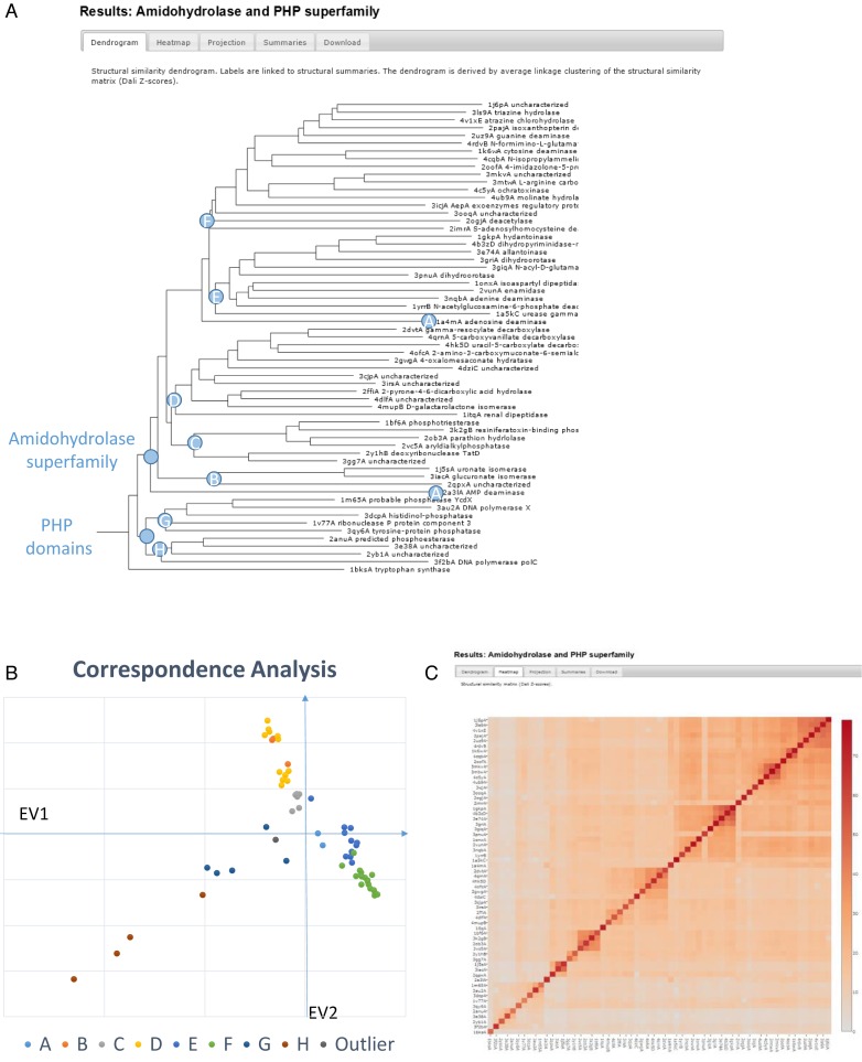
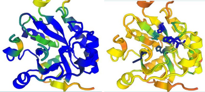
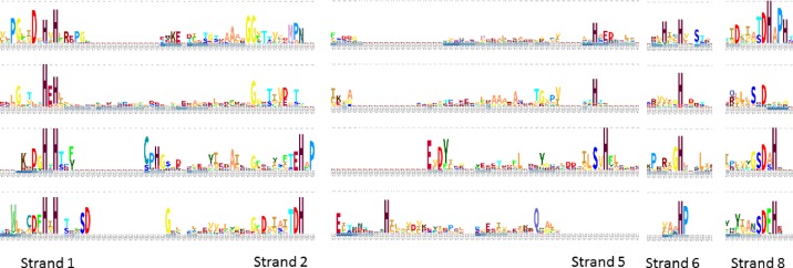
The Dali server (http://ekhidna2.biocenter.helsinki.fi/dali) is a network service for comparing protein structures in 3D. In favourable cases, comparing 3D structures may reveal biologically interesting similarities that are not detectable by comparing sequences. The Dali server has been running in various places for over 20 years and is used routinely by crystallographers on newly solved structures. The latest update of the server provides enhanced analytics for the study of sequence and structure conservation. The server performs three types of structure comparisons: (i) Protein Data Bank (PDB) search compares one query structure against those in the PDB and returns a list of similar structures; (ii) pairwise comparison compares one query structure against a list of structures specified by the user; and (iii) all against all structure comparison returns a structural similarity matrix, a dendrogram and a multidimensional scaling projection of a set of structures specified by the user. Structural superimpositions are visualized using the Java-free WebGL viewer PV. The structural alignment view is enhanced by sequence similarity searches against Uniprot. The combined structure-sequence alignment information is compressed to a stack of aligned sequence logos. In the stack, each structure is structurally aligned to the query protein and represented by a sequence logo.
© The Author(s) 2016. Published by Oxford University Press on behalf of Nucleic Acids Research.
Figures
Similar articles
-
Dali server: conservation mapping in 3D.Nucleic Acids Res. 2010 Jul;38(Web Server issue):W545-9. doi: 10.1093/nar/gkq366. Epub 2010 May 10. Nucleic Acids Res. 2010. PMID: 20457744 Free PMC article.
-
Searching protein structure databases with DaliLite v.3.Bioinformatics. 2008 Dec 1;24(23):2780-1. doi: 10.1093/bioinformatics/btn507. Epub 2008 Sep 25. Bioinformatics. 2008. PMID: 18818215 Free PMC article.
-
Using Dali for structural comparison of proteins.Curr Protoc Bioinformatics. 2006 Jul;Chapter 5:Unit 5.5. doi: 10.1002/0471250953.bi0505s14. Curr Protoc Bioinformatics. 2006. PMID: 18428766
-
Comparison of proteins based on segments structural similarity.Acta Biochim Pol. 2004;51(1):161-72. Acta Biochim Pol. 2004. PMID: 15094837 Review.
-
QSalignWeb: A Server to Predict and Analyze Protein Quaternary Structure.Front Mol Biosci. 2022 Jan 5;8:787510. doi: 10.3389/fmolb.2021.787510. eCollection 2021. Front Mol Biosci. 2022. PMID: 35071324 Free PMC article. Review.
Cited by
-
Phylogenetic, functional and structural characterization of a GH10 xylanase active at extreme conditions of temperature and alkalinity.Comput Struct Biotechnol J. 2021 May 3;19:2676-2686. doi: 10.1016/j.csbj.2021.05.004. eCollection 2021. Comput Struct Biotechnol J. 2021. PMID: 34093984 Free PMC article.
-
Characterisation of a tripartite α-pore forming toxin from Serratia marcescens.Sci Rep. 2021 Mar 19;11(1):6447. doi: 10.1038/s41598-021-85726-0. Sci Rep. 2021. PMID: 33742033 Free PMC article.
-
Crystal structure and mutational analysis of the human TRIM7 B30.2 domain provide insights into the molecular basis of its binding to glycogenin-1.J Biol Chem. 2021 Jan-Jun;296:100772. doi: 10.1016/j.jbc.2021.100772. Epub 2021 May 11. J Biol Chem. 2021. PMID: 33989636 Free PMC article.
-
ppGpp Coordinates Nucleotide and Amino-Acid Synthesis in E. coli During Starvation.Mol Cell. 2020 Oct 1;80(1):29-42.e10. doi: 10.1016/j.molcel.2020.08.005. Epub 2020 Aug 27. Mol Cell. 2020. PMID: 32857952 Free PMC article.
-
Structure and function of bacteriophage CBA120 ORF211 (TSP2), the determinant of phage specificity towards E. coli O157:H7.Sci Rep. 2020 Sep 21;10(1):15402. doi: 10.1038/s41598-020-72373-0. Sci Rep. 2020. PMID: 32958885 Free PMC article.
References
-
- Levitt M., Chothia C. Structural patterns in globular proteins. Nature. 1976;261:5552–558. - PubMed
-
- Richardson J. The anatomy and taxonomy of protein structure. Adv. Protein Chem. 1981;34:167–339. - PubMed
-
- Holm L., Sander C. Globin fold in a bacterial toxin. Nature. 1993;361:309. - PubMed
-
- Holm L., Sander C. Structural similarity between plant endochitinase and lysozymes from animals and phage: an evolutionary connection. FEBS Lett. 1994;340:129–132. - PubMed
-
- Holm L., Murzin A., Sander C. Three sisters, different names: 3alpha,20beta-hydroxysteroid dehydrogenase, dihydropteridine reductase and UDP-galactose 4-epimerase. Nat. Struct. Biol. 1994;1:146–147. - PubMed
Publication types
MeSH terms
Substances
LinkOut - more resources
Full Text Sources
Other Literature Sources
