Review
. 2016 Jun;14(6):385-400.
doi: 10.1038/nrmicro.2016.30. Epub 2016 Apr 25.

Chlamydia cell biology and pathogenesis

Affiliations
Review

Chlamydia cell biology and pathogenesis

Cherilyn Elwell et al. Nat Rev Microbiol. 2016 Jun.

Abstract

Chlamydia spp. are important causes of human disease for which no effective vaccine exists. These obligate intracellular pathogens replicate in a specialized membrane compartment and use a large arsenal of secreted effectors to survive in the hostile intracellular environment of the host. In this Review, we summarize the progress in decoding the interactions between Chlamydia spp. and their hosts that has been made possible by recent technological advances in chlamydial proteomics and genetics. The field is now poised to decipher the molecular mechanisms that underlie the intimate interactions between Chlamydia spp. and their hosts, which will open up many exciting avenues of research for these medically important pathogens.


Figures
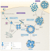
Figure 1
Figure 1. The life cycle of Chlamydia trachomatis
The binding of elementary bodies to host cells is initiated by the formation of a trimolecular bridge between bacterial adhesins, host receptors and host heparan sulfate proteoglycans (HSPGs). Next, pre-synthesized type III secretion system (T3SS) effectors are injected into the host cell, some of which initiate cytoskeletal rearrangements to facilitate internalization and/or initiate mitogenic signalling to establish an anti-apoptotic state. The elementary body is endocytosed into a membrane-bound compartment, known as the inclusion, which rapidly dissociates from the canonical endolysosomal pathway. Bacterial protein synthesis begins, elementary bodies convert to reticulate bodies and newly secreted inclusion membrane proteins (Incs) promote nutrient acquisition by redirecting exocytic vesicles that are in transit from the Golgi apparatus to the plasma membrane. The nascent inclusion is transported, probably by an Inc, along microtubules to the microtubule-organizing centre (MTOC) or centrosome. During mid-cycle, the reticulate bodies replicate exponentially and secrete additional effectors that modulate processes in the host cell. Under conditions of stress, the reticulate bodies enter a persistent state and transition to enlarged aberrant bodies. The bacteria can be reactivated upon the removal of the stress. During the late stages of infection, reticulate bodies secrete late-cycle effectors and synthesize elementary-body-specific effectors before differentiating back to elementary bodies. Elementary bodies exit the host through lysis or extrusion.
Figure 2
Figure 2. Chlamydia–host interactions
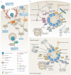
a | Elementary bodies contain pre-synthesized type III secretion system (T3SS) effectors along with their respective chaperones. On contact with host cells, invasion-related effectors are injected through the T3SS to induce cytoskeletal rearrangements and host signalling. In Chlamydia trachomatis, translocated actin-recruiting phosphoprotein (TarP), CT166 and CT694 are secreted first followed by TepP. TarP and TepP are tyrosine phosphorylated by host kinases. Phosphorylated TarP interacts with SRC homology 2 domain-containing transforming protein C1 (SHC1) to activate extracellular signal-regulated kinase 1 (ERK1; also known as MAPK3) and ERK2 (also known as MAPK1) for pro-survival signalling, whereas other phosphorylated TarP residues mediate interactions with two RAC guanine nucleotide exchange factors (GEFs), VAV2 and son of sevenless homologue 1 (SOS1). SOS1 is part of a multiprotein complex with ABL interactor 1 (ABI1) and epidermal growth factor receptor kinase substrate 8 (EPS8) in which ABI1 is thought to mediate the interaction of the complex with phosphorylated TarP, which leads to the activation of RAS-related C3 botulinum toxin substrate 1 (RAC1) and the host actin-related protein 2/3 (ARP2/3) complex. Phosphorylated TarP binds to the p85 subunit of phosphoinositide 3-kinase (PI3K), producing phosphatidylinositol-3,4,5-triphosphate PI(3,4,5)P3, which may activate VAV2. TarP also directly mediates the formation of actin filaments. TarP orthologues in Chlamydia caviae (which do not contain phosphorylation sites) bind to focal adhesion kinase (FAK) through a mammalian leucine–aspartic acid (LD2)-like motif and activate cell division control protein 42 (CDC42)-related actin assembly. CT694 contains a membrane-binding domain and interacts with the AHNAK protein, which links the membrane to the rearrangement of actin. CT166 glycosylates and inactivates RAC1. TarP activates the polymerization of actin, whereas CT694 and CT166 promote the depolymerization of actin. Phosphorylated TepP interacts with CRKI and CRKII to initiate innate immune signalling. b | The chlamydial inclusion is actively remodelled by host proteins and bacterial inclusion membrane proteins (Incs). Incs may regulate fusion with intracellular compartments and modulate membrane dynamics. Several RAB GTPases localize to the inclusion, including RAB4 and RAB11, which are recruited from recycling endosomes soon after entry by CT229 in C. trachomatis and Cpn0585 in Chlamydia pneumoniae, respectively. RAB11 is also recruited from the Golgi apparatus and binds to RAB11 family-interacting protein 2 (RAB11FIP2) to promote the recruitment of RAB14. RAB1 is recruited from the endoplasmic reticulum, whereas RAB6 and RAB10 relocalize from the Golgi apparatus. The RAB effector bicaudal-D homologue 1 (BICD1) may link the inclusion to dynein for transport along microtubules. The phosphatidylinositol-4-phosphate (PI4P)-producing enzymes OCRL1 and phosphatidylinositol-4-kinase type IIα (PI4KIIα) are recruited and may generate PI4P. Sorting nexin 5 (SNX5) and SNX6 are recruited from early endosomes by IncE to remodel the inclusion membrane and potentially inhibit retromer trafficking. RAB39a regulates the interaction between multivesicular bodies and the inclusion. Several soluble N-ethylmaleimide-sensitive factor attachment protein receptors (SNAREs) are recruited, including the Golgi-specific SNAREs vesicle-associated membrane protein 4 (VAMP4), syntaxin 6 (STX6) and GS15. In addition, the endocytic SNAREs, VAMP 3, VAMP 7 and VAMP 8, are recruited by IncA, InaC and inclusion protein acting on microtubules (IPAM), and are thought to act as inhibitory SNAREs (iSNAREs) to block the fusion with lysosomes. c | Chlamydia spp. interact with several subcellular compartments to acquire essential lipids. Sphingomyelin and cholesterol are incorporated into reticulate body membranes by intercepting vesicles from fragmented Golgi mini-stacks and multivesicular bodies. The trafficking of lipid-containing vesicles from Golgi mini-stacks is regulated by the GBF1-dependent activation of ADP-ribosylation factor (ARF) GTPases, by dynein heavy chain (DYN1) GTPase, and by Golgi-associated SNAREs and RABs. RAB39 mediates the interaction between the inclusion and multivesicular bodies. Lipid droplets and peroxisomes are translocated into the inclusion. Lipid droplets may be intercepted by Lda1 or Lda3, or by the Incs: Cap1, CT618, IncG and IncA. FYN kinase signalling from Inc microdomains that contain IncB, CT101, CT222 and CT850, contributes to lipid acquisition, possibly through the positioning of the inclusion at the microtubule-organizing centre (MTOC) or centrosome. Non-vesicular mechanisms of lipid acquisition involve the formation of endoplasmic reticulum–inclusion membrane contact sites (mediated by vesicle-associated membrane proteins (VAPs), the lipid transporter ceramide endoplasmic reticulum transport protein (CERT), and IncD), the recruitment of members of the high-density lipoprotein (HDL) machinery, and ERK signalling. The sphingomyelin biosynthetic enzyme, sphingomyelin synthase 2 (SMS2), may convert ceramide to sphingomyelin directly on the inclusion. ABCA1, ATP-binding cassette transporter 1; APOA1, apolipoprotein A1; CLA1, CD36 and LIMPII analagous 1; cPLA2, cytosolic phospholipase A2; WAVE2, Wiskott–Aldrich syndrome protein family member 2.
Figure 3
Figure 3. Modulation of host cell survival and death
Chlamydial infection promotes host cell proliferation and survival through the activation of phosphoinositide 3-kinase (PI3K) and mitogen-activated protein kinase kinase (MAPKK, also known as MEK)–mitogen-activated protein kinase (MAPK, also known as ERK) signalling cascades by binding to receptor tyrosine kinases or through the secretion of the early effector translocated actin-recruiting phosphoprotein (TarP). Chlamydia pneumoniae sequesters cytoplasmic activation/proliferation-associated protein 2 (CAPRIN2) and glycogen synthase kinase 3β (GSK3β), members of the β-catenin destruction complex, to the inclusion membrane possibly through their interaction with Cpn1027, which leads to increased stabilization of β-catenin and transcriptional activation of survival genes by β-catenin. Infected host cells are resistant to various apoptotic stimuli and apoptosis is blocked both upstream and downstream of the permeabilization of the outer membrane of mitochondria by numerous mechanisms. The upregulation of the anti-apoptotic proteins BAG family molecular chaperone regulator 1 (BAG1) and myeloid leukaemia cell differentiation protein 1 (MCL1) inhibits the ability of BH3-only proteins to destabilize the mitochondrial membrane. The BH3-only protein BCL-2-associated agonist of cell death (BAD) is also sequestered at the inclusion membrane by binding to the host protein 14-3-3β. The degradation of p53 by MDM2-mediated ubiquitylation and sequestration of protein kinase Cγ (PKCγ) also prevents the depolarization of the mitochondrial membrane. Downstream of the release of cytochrome c, Chlamydia spp. are still able to prevent apoptosis through unknown mechanisms. Infection also leads to the upregulation of inhibitors of apoptosis (IAPs), which contributes to the anti-apoptotic phenotype. The chlamydial protein CADD is implicated in the modulation of apoptosis by binding to the death domains of tumour necrosis factor (TNF) family receptors. C. trachomatis can block the activation of caspase 8 through the regulator cellular FLICE-like inhibitory protein (cFLIP) by an unknown mechanism. The text in the red boxes denotes the individual steps in which Chlamydia spp. are proposed to modulate host cell function and the bacterial effector that is involved, if it is known. Casp8, caspase 8; DAG, diacylglycerol; EGFR, epidermal growth factor receptor; EPHA2, ephrin receptor A2; ERK, extracellular signal-regulated kinase; FGFR, fibroblast growth factor receptor; Inc, inclusion membrane protein; PDGFR, platelet derived growth factor receptor.
Figure 4
Figure 4. Modulation of the innate immune response
The recognition of chlamydial infection by pattern-recognition receptors leads to cell-autonomous immunity and the production of pro-inflammatory cytokines. Chlamydial antigens (green boxes) can be recognized by cell surface, endosomal or cytosolic pathogen sensors. Toll-like receptor 4 (TLR4) recognizes lipopolysaccharide (LPS) or the 60 kDa heat shock protein (HSP60), whereas TLR2 recognizes peptidoglycan, macrophage inhibitory protein (MIP) and/or plasmid-regulated ligands. Both TLR2 and TLR4 signalling require the adaptors myeloid differentiation primary response protein 88 (MYD88) and tumour necrosis factor (TNF) receptor-associated factor 6 (TRAF6), and lead to the nuclear translocation of nuclear factor-κB (NF-κB) and the induction of innate immune responses. The activation of the cytosolic sensor STING (stimulator of interferon genes) by the bacterial second messenger cyclic di-AMP (c-di-AMP) or through the host secondary messenger cyclic GMP–AMP (cGAMP) leads to the phosphorylation of interferon regulatory factor 3 (IRF3), nuclear translocation and induction of type I interferon (IFN) genes (encoding IFNα and IFNβ) and IFN-stimulated genes (ISG). The peptidoglycan sensor nucleotide-binding oligomerization domain-containing 1 (NOD1) is activated during chlamydial infection and induces the production of pro-inflammatory cytokines through NF-κB signalling. The activation of the NOD-, LRR- and pyrin domain-containing 3 (NLRP3)–apoptosis-associated speck-like protein containing a CARD (ASC) inflammasome (NLRP3–ASC inflammasome) requires reactive oxygen species (ROS) and K+ efflux. The production of ROS is amplified by the mitochondrial NOD-like receptor X1 (NLRX1), which augments the production of ROS, creating a feed-forward loop. Some innate immune molecules that recognize vacuolar pathogens, such as human guanylate-binding protein 1 (hGBP1) and hGBP2, mouse immunity-related GTPase family M protein 1 (mIRGM1) and mIRGM3, and mouse IRGB10 (mIRGB10), localize to Chlamydia trachomatis inclusions and promote bacterial clearance. Chlamydia muridarum prevents the recognition of inclusions by these receptors. Chlamydial virulence factors (depicted in red) can disrupt or augment the innate immune response. During infection with Chlamydia pneumoniae, an unknown protease cleaves TRAF3, which blocks the phosphorylation of IRF3 and the production of type 1 IFNs. The deubiquitinase Dub1 removes ubiquitin from NF-κB inhibitor-α (IκBα), which stabilizes the p65–p50–IκBα complex and prevents the nuclear translocation of NF-κB. The C. pneumonia protein CP0236 sequesters NF-κB activator 1 (ACT1) to the inclusion membrane. Chlamydial infection also leads to the upregulation of olfactomedin 4 (OLFM4), which potentially blocks NOD1-mediated signalling. cGAS, cGAMP synthase.

Similar articles

Cited by

References

    1. Bachmann NL, Polkinghorne A, Timms P. Chlamydia genomics: providing novel insights into chlamydial biology. Trends Microbiol. 2014;22:464–472. - PubMed
    1. Mandell GL, Bennett JE, Dolin R, editors. Mandell, Douglas, and Bennett's Principles and Practice of Infectious Diseases. 7th edn Vol. 2. Churchill Livingstone/Elsevier; 2010.
    1. Rank RG. In: Intracellular Pathogens 1: Chlamydiales. Tan M, Bavoil PM, editors. Vol. 1. ASM Press; 2012. pp. 285–310.
    1. Malhotra M, Sood S, Mukherjee A, Muralidhar S, Bala M. Genital Chlamydia trachomatis: an update. Indian J. Med. Res. 2013;138:303–316. - PMC - PubMed
    1. de Vrieze NH, de Vries HJ. Lymphogranuloma venereum among men who have sex with men. An epidemiological and clinical review. Expert Rev. Anti Infect. Ther. 2014;12:697–704. - PubMed

MeSH terms

Substances