. 2016 Mar 11:7:10961.
doi: 10.1038/ncomms10961.

CCC- and WASH-mediated endosomal sorting of LDLR is required for normal clearance of circulating LDL

Affiliations

CCC- and WASH-mediated endosomal sorting of LDLR is required for normal clearance of circulating LDL

Paulina Bartuzi et al. Nat Commun. .

Abstract

The low-density lipoprotein receptor (LDLR) plays a pivotal role in clearing atherogenic circulating low-density lipoprotein (LDL) cholesterol. Here we show that the COMMD/CCDC22/CCDC93 (CCC) and the Wiskott-Aldrich syndrome protein and SCAR homologue (WASH) complexes are both crucial for endosomal sorting of LDLR and for its function. We find that patients with X-linked intellectual disability caused by mutations in CCDC22 are hypercholesterolaemic, and that COMMD1-deficient dogs and liver-specific Commd1 knockout mice have elevated plasma LDL cholesterol levels. Furthermore, Commd1 depletion results in mislocalization of LDLR, accompanied by decreased LDL uptake. Increased total plasma cholesterol levels are also seen in hepatic COMMD9-deficient mice. Inactivation of the CCC-associated WASH complex causes LDLR mislocalization, increased lysosomal degradation of LDLR and impaired LDL uptake. Furthermore, a mutation in the WASH component KIAA0196 (strumpellin) is associated with hypercholesterolaemia in humans. Altogether, this study provides valuable insights into the mechanisms regulating cholesterol homeostasis and LDLR trafficking.


Figures

Figure 1
Figure 1. COMMD1-deficient dogs are hypercholesterolaemic.
(a) Western blot analysis of COMMD1, CCDC22 and CCDC93 in livers from an unaffected dog (COMMD1+/+) and an affected dog homozygous for a loss-of-function mutation in COMMD1 (COMMD1−/−). (b) Plasma TC and (c) TG levels of dogs heterozygous (+/−) (n=5) or homozygous (−/−) (n=4) for a COMMD1 mutation (d) FPLC lipoprotein profile of COMMD1+/− (n=5) and COMMD1−/− dogs (n=4). Pooled plasma samples of each experimental group were separated using FPLC gel filtration. Fifty fractions were collected from each separation. TC content was determined and lipoprotein profile was plotted. The results are presented as mean±s.e.m., and significance was calculated relative to the control group by unpaired Student's t-test; *P<0.05.
Figure 2
Figure 2. Ablation of hepatic Commd1 increases plasma total and LDL cholesterol levels.
(a) Plasma TC and (b) TG levels of hepatic Commd1 knockout mice (Commd1ΔHep) and WT mice (n=6–8) fed a chow diet or a HFC (0.2%) diet for 20 weeks. Pooled plasma samples of each experimental group of mice fed either the (c) chow or (d) the HFC diet were separated using FPLC gel filtration. Fifty fractions were collected from each separation. TC content was determined and lipoprotein profile was plotted. Fractions #13–26 containing cholesterol were collected and loaded on an SDS polyacrylamide gel and blotted using antibodies against apoA1 and apoB100 lipoproteins. The chow group (e) and HFC group (f) are shown. (g) Livers of chow-fed mice were homogenized, and 30 μg of protein was subjected to immunoblot analysis. Levels of LDLR, LRP1, ARH, CCDC22, CCDC93 and tubulin were determined. Three representative samples from WT and Commd1ΔHep mice are shown. (h) Relative levels of the proteins shown in g were determined by immunoblot analysis, and densitometry analysis was performed using tubulin as a loading control (n=6–8 per genotype). The results are presented as mean±s.e.m., and significance was calculated relative to the control group by unpaired Student's t-test; ***P<0.001. ARH, autosomal recessive hypercholesterolaemia protein.
Figure 3
Figure 3. LDLR associates with COMMD1 and the WASH complex.
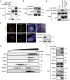
(a) Human embryonic kidney 293T (HEK293T) cells were transfected with constructs expressing Flag-LDLR with either COMMD1-GST or GST alone. Interaction with COMMD1 was detected via pull-down assay using glutathione sepharose beads. (b) HEK293T cells were transfected with Flag-LDLR vector, and interaction with endogenous COMMD1 was detected by immunoprecipitation with rabbit anti-Flag-antibody. (c) Colocalization of LDLR (red) and COMMD1 (green) in Commd1f/f MEFs examined by immunofluorescence staining. Representative images are shown; scale bar, 5μm. LDLR (red) and COMMD1 (green) was stained in COMMD1-deficient MEFs (Commd1−/−) and imaged by confocal fluorescence microscopy. COMMD1 levels in Commd1f/f and in Commd1−/− MEFs determined by immunoblot analysis. (d) Liver of a WT chow-fed mouse was homogenized and loaded on a continuous 10–40% sucrose gradient. Fractions were separated by ultracentrifugation and immunoblotted using antibodies against COMMD1, LDLR, WASH1, FAM21, VPS35 and CCDC22. The figure represents results of three independent experiments. (e) HEK293T cells were transfected with Ha-COMMD1 construct together with GST alone, GST-LDLRct (GST-tagged cytosolic domain of LDLR) or GST-LDLRct Y807A (GST-tagged mutated cytosolic domain of LDLR). Pull-down assay was performed to study the interaction between LDLRct and COMMD1. (f) Lysates of Flag-LDLR-transfected HEK293T cells were used for immunoprecipitation assays. Immunoprecipitates were washed, separated by SDS–polyacrylamide gel electrophoresis and immunoblotted as indicated.
Figure 4
Figure 4. COMMD1 deficiency impairs the function of LDLR.
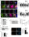
(a) LDLR (green), COMMD1 (red) and VPS35 (pink) were stained in Commd1f/f and Commd1−/− MEFs and imaged by confocal microscopy. Representative images are shown; scale bar, 5μm. (b) Quantification of the colocalization of LDLR with COMMD1, VPS35, EEA1 and LAMP1 was performed by the analysis of 30–40 cells. (c) Total and plasma membrane LDLR levels of Commd1f/f and Commd1−/− MEFs determined by biotinylation assay. Data represent three independent experiments, and (d) the relative levels of LDLR at the cell surface are quantified in all experiments. (e) In vitro LDL and transferrin uptake assay. Dil-labelled LDL (5 μg ml−1) or Alexa-633-labelled transferrin (5 μg ml−1) was added to serum-depleted medium and incubated with MEFs at 4 °C for 1 h and subsequently at 37 °C for 5 min. Dil-labelled LDL and Alexa-633-labelled transferrin uptake was measured by FACS analysis, and the relative uptake in triplicate is shown. The results are presented as mean±s.e.m.; significance was calculated relative to the control group by unpaired Student's t-test; *P<0.05, ***P<0.001.
Figure 5
Figure 5. The WASH complex is essential for endosomal sorting of LDLR.
(a) Cellular localization of LDLR (green), COMMD1 (red) and LAMP1 (pink) in Wash1f/f and Wash1−/− MEFs was determined by immunofluorescence staining. Representative images are shown; scale bar, 5 μm. Relative colocalization of (b) LDLR and (c) COMMD1 with VPS35, EEA1 and LAMP1 was quantified. (d) LDLR, WASH1, VPS35 and COMMD1 levels in Wash1f/f and Wash1−/− MEFs analysed by western blot. (e) Representative images (n=3) of immunoblot analysis of total LDLR levels in Wash1f/f and Wash1−/− MEFs treated with bafilomycin A (100 nM) for 0, 4 and 6 h. (f) Densitometry revealed the relative levels of LDLR in bafilomycin A-treated cells (n=3). (g) Representative images (n=3) of LDLR on the surface of Wash1f/f and Wash1−/− MEFs determined by surface biotinylation assay. (h) Densitometry revealed relative LDLR surface levels (n=3). (i) Wash1f/f and Wash1−/− MEFs were incubated with DiI-LDL for 30 min and imaged by fluorescence microscope. (j) Fluorescence intensity was quantified using ImageJ software and was normalized to the number of DAPI nuclei per image; >30 cells per condition were recorded. The results are presented as mean±s.e.m.; significance was calculated relative to the control group by unpaired Student's t-test; ***P<0.001.
Figure 6
Figure 6. Hypothetical model of CCC- and WASH-mediated endosomal LDLR sorting.
After internalization mediated by ARH, LDLR is sorted in endosomes. LDLR is directed to the lysosome for proteolysis—which is induced by either PCSK9 (refs 59, 60) or IDOL—or retrieved and recycled back to the cell surface. SNX17 enhances LDL endocytosis likely by promoting LDLR retrieval, which might precede CCC and retromer/WASH-mediated endosomal trafficking. Through the interaction of the retromer component VPS35 with FAM21 the WASH complex and the CCC complex are recruited to the endosomes. Subsequently, CCC and WASH form a protein complex with LDLR; WASH facilitates the formation of branched actin patches to define restricted domains of the endosomes from where LDLR is sorted back to the cell surface. NDRG1 is required for the formation of MVB: loss of NDRG1 impairs LDL uptake and LDLR recycling, and causes the accumulation of IDOL-mediated ubiquitinated LDLR in MVB. Further research is needed to assess whether CCC and WASH are functionally related to SNX17, NDRG1 and to the lysosomal degradation pathways of LDLR. ARH, autosomal recessive hypercholesterolaemia protein; MVB, multivesicular body; PCSK9, proprotein convertase subtilisin/kexin type 9; Ub, ubiquitin; WASH, WASH1, FAM21, strumpellin, KIAA1033, CCDC53.

Similar articles

Cited by

References

    1. Gomez T. S. & Billadeau D. D. A FAM21-containing WASH complex regulates retromer-dependent sorting. Dev. Cell 17, 699–711 (2009) . - PMC - PubMed
    1. Harbour M. E., Breusegem S. Y. & Seaman M. N. J. Recruitment of the endosomal WASH complex is mediated by the extended ‘tail' of Fam21 binding to the retromer protein Vps35. Biochem. J. 442, 209–220 (2012) . - PubMed
    1. Seaman M. N. J., Gautreau A. & Billadeau D. D. Retromer-mediated endosomal protein sorting: all WASHed up!. Trends Cell Biol. 23, 522–528 (2013) . - PMC - PubMed
    1. Harbour M. E. et al.. The cargo-selective retromer complex is a recruiting hub for protein complexes that regulate endosomal tubule dynamics. J. Cell. Sci. 123, 3703–3717 (2010) . - PMC - PubMed
    1. Jia D., Gomez T. S., Billadeau D. D. & Rosen M. K. Multiple repeat elements within the FAM21 tail link the WASH actin regulatory complex to the retromer. Mol. Biol. Cell 23, 2352–2361 (2012) . - PMC - PubMed

Publication types

MeSH terms