

Structural basis for the RING-catalyzed synthesis of K63-linked ubiquitin chains
- PMID: 26148049
- PMCID: PMC4529489
- DOI: 10.1038/nsmb.3052
Structural basis for the RING-catalyzed synthesis of K63-linked ubiquitin chains
Abstract
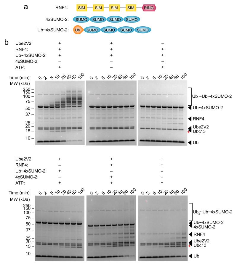
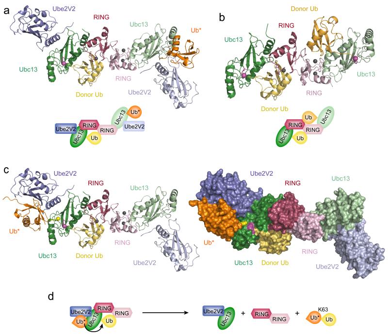
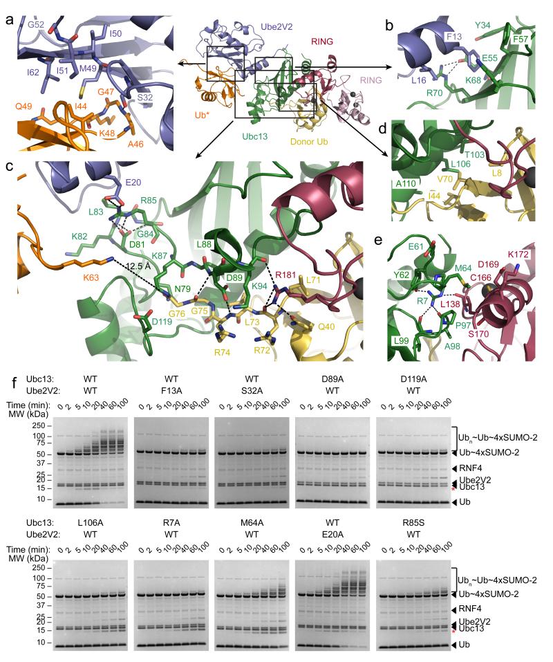
RING E3 ligase-catalyzed formation of K63-linked ubiquitin chains by the Ube2V2-Ubc13 E2 complex is required in many important biological processes. Here we report the structure of the RING-domain dimer of rat RNF4 in complex with a human Ubc13∼Ub conjugate and Ube2V2. The structure has captured Ube2V2 bound to the acceptor (priming) ubiquitin with K63 in a position favorable for attack on the linkage between Ubc13 and the donor (second) ubiquitin held in the active 'folded back' conformation by the RING domain of RNF4. We verified the interfaces identified in the structure by in vitro ubiquitination assays of site-directed mutants. To our knowledge, this represents the first view of synthesis of K63-linked ubiquitin chains in which both substrate ubiquitin and ubiquitin-loaded E2 are juxtaposed to allow E3 ligase-mediated catalysis.
Figures
Similar articles
-
Structure of a RING E3 ligase and ubiquitin-loaded E2 primed for catalysis.Nature. 2012 Sep 6;489(7414):115-20. doi: 10.1038/nature11376. Nature. 2012. PMID: 22842904 Free PMC article.
-
Molecular insights into the function of RING finger (RNF)-containing proteins hRNF8 and hRNF168 in Ubc13/Mms2-dependent ubiquitylation.J Biol Chem. 2012 Jul 6;287(28):23900-10. doi: 10.1074/jbc.M112.359653. Epub 2012 May 15. J Biol Chem. 2012. PMID: 22589545 Free PMC article.
-
The mechanism of OTUB1-mediated inhibition of ubiquitination.Nature. 2012 Feb 22;483(7391):618-22. doi: 10.1038/nature10911. Nature. 2012. PMID: 22367539 Free PMC article.
-
Structural basis of generic versus specific E2-RING E3 interactions in protein ubiquitination.Protein Sci. 2019 Oct;28(10):1758-1770. doi: 10.1002/pro.3690. Epub 2019 Aug 23. Protein Sci. 2019. PMID: 31340062 Free PMC article. Review.
-
Structural Diversity of Ubiquitin E3 Ligase.Molecules. 2021 Nov 4;26(21):6682. doi: 10.3390/molecules26216682. Molecules. 2021. PMID: 34771091 Free PMC article. Review.
Cited by
-
Structural insights into the catalysis and regulation of E3 ubiquitin ligases.Nat Rev Mol Cell Biol. 2016 Oct;17(10):626-42. doi: 10.1038/nrm.2016.91. Epub 2016 Aug 3. Nat Rev Mol Cell Biol. 2016. PMID: 27485899 Free PMC article. Review.
-
Hemi-methylated DNA regulates DNA methylation inheritance through allosteric activation of H3 ubiquitylation by UHRF1.Elife. 2016 Sep 6;5:e17101. doi: 10.7554/eLife.17101. Elife. 2016. PMID: 27595565 Free PMC article.
-
A tri-ionic anchor mechanism drives Ube2N-specific recruitment and K63-chain ubiquitination in TRIM ligases.Nat Commun. 2019 Oct 3;10(1):4502. doi: 10.1038/s41467-019-12388-y. Nat Commun. 2019. PMID: 31582740 Free PMC article.
-
Mechanism of ubiquitin transfer promoted by TRAF6.Proc Natl Acad Sci U S A. 2018 Feb 20;115(8):1783-1788. doi: 10.1073/pnas.1721788115. Epub 2018 Feb 5. Proc Natl Acad Sci U S A. 2018. PMID: 29432170 Free PMC article.
-
MKP-1 modulates ubiquitination/phosphorylation of TLR signaling.Life Sci Alliance. 2021 Sep 27;4(12):e202101137. doi: 10.26508/lsa.202101137. Print 2021 Dec. Life Sci Alliance. 2021. PMID: 34580177 Free PMC article.
References
-
- Scheffner M, Kumar S. Mammalian HECT ubiquitin-protein ligases: Biological and pathophysiological aspects. Biochimica et Biophysica Acta (BBA) - Molecular Cell Research. 2014;1843:61–74. - PubMed
Publication types
MeSH terms
Substances
Associated data
- Actions
- Actions
Grants and funding
LinkOut - more resources
Full Text Sources
Other Literature Sources
Molecular Biology Databases
Miscellaneous
