. 2013:4:2863.
doi: 10.1038/ncomms3863.

Proteasomal degradation of Nck1 but not Nck2 regulates RhoA activation and actin dynamics

Affiliations

Proteasomal degradation of Nck1 but not Nck2 regulates RhoA activation and actin dynamics

Lisa Buvall et al. Nat Commun. 2013.

Abstract

The ubiquitously expressed adapter proteins Nck1/2 interact with a multitude of effector molecules to regulate diverse cellular functions including cytoskeletal dynamics. Here we show that Nck1, but not Nck2, is a substrate of c-Cbl-mediated ubiquitination. We uncover lysine 178 in Nck1 as the evolutionarily conserved ubiquitin acceptor site. We previously reported that synaptopodin, a proline-rich actin-binding protein, induces stress fibres by blocking the Smurf1-mediated ubiquitination of RhoA. We now find that synaptopodin competes with c-Cbl for binding to Nck1, which prevents the ubiquitination of Nck1 by c-Cbl. Gene silencing of c-Cbl restores Nck1 protein abundance and stress fibres in synaptopodin knockdown cells. Similarly, expression of c-Cbl-resistant Nck1(K178R) or Nck2 containing the SH3 domain 2 of Nck1 restores stress fibres in synaptopodin-depleted podocytes through activation of RhoA signalling. These findings reveal proteasomal regulation as a key factor in the distinct and non-redundant effects of Nck on RhoA-mediated actin dynamics.


Conflict of interest statement

Competing financial interests: The authors declare no competing financial interests.

Figures

Figure 1
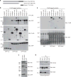
Figure 1. Nck directly binds to synaptopodin
(a) Schematic of synaptopodin isoforms. (b) GFP-Nck1 and GFP-Nck2 co-precipitate with FLAG-Synpo-long, FLAG-Synpo-short and FLAG-Synpo-T from co-transfected HEK293 cells. No interaction is found with GFP-sui serving as negative control. The interaction between FLAG-N-WASP and GFP-Nck1/2 serves as positive control. (c) Western blot analysis of Nck antibody specificity in HEK293 cells transfected with GFP-Nck1, GFP-Nck2 or GFP. (d) Endogenous Co-IP shows that Nck1 and Nck2 interact with synaptopodin in isolated mouse glomeruli. IP with anti-GFP (control) serves as negative control. IP: immunoprecipitation. (e) Direct binding of purified FLAG-Synpo to purified GST-Nck1 and GST-Nck2 but not the GST control. The interaction between purified FLAG-N-WASP and GST-Nck1/2 serves as positive control. Molecular weight markers are in kDa.
Figure 2
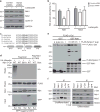
Figure 2. Synaptopodin increases the protein abundance of Nck1 but not Nck2
(a) Synaptopodin is not expressed in undifferentiated (undiff) podocytes and is markedly downregulated in synaptopodin knockdown podocytes (synpo shRNA), when compared with differentiated (diff) and non-silencing shRNA control cells. Similarly, Nck1 protein levels are low in undifferentiated and synaptopodin knockdown podocytes. Nck2 levels are comparable under all conditions. GAPDH serves as a loading control. (b) Quantification of Nck1 and Nck2 protein abundance in the presence or absence of synaptopodin. Statistical analysis was performed by pairwise comparison of undifferentiated versus differentiated podocytes, or scrambled shRNA versus synaptopodin shRNA-infected cells using Student’s t-test. Values are presented as % band intensity of differentiated podocytes ±s.e.m., n = 3, *P<0.05. (c) Transfection of undifferentiated podocytes with GFP-Synpo-long but not GFP-sui (negative control) increases Nck1 but not Nck2 protein abundance. GAPDH serves as loading control. Molecular weight markers are in kDa.
Figure 3
Figure 3. c-Cbl-mediated ubiquitination of Nck1 is blocked by competitive binding of synaptopodin to Nck1
(a) Lactacystin increases Nck1 and cyclin D1 (positive control) steady-state protein levels in synaptopodin knockdown podocytes. Nck2 protein abundance remains unchanged. GAPDH serves as loading control. (b) Quantification of lactacystin induced changes in Nck1 and Nck2 abundance in synaptopodin-depleted podocytes. Values are presented as % of untreated control shRNA-expressing cells ±s.e.m., n = 3, ANOVA, *P<0.05. (c) Nck1 contains a lysine at position 178 that is conserved from Xenopus to human. (d) FLAG-Nck1 is ubiquitinated by wild-type (WT) but not by catalytically inactive (381A) c-Cbl in transfected HEK cells co-expressing GFP-sui, as revealed using anti-HA immunoblotting. GFP-Synpo-T (Syn) but not GFP-sui blocks the ubiquitination of Nck1. Nck1(K178R) is resistant to ubiquitination by c-Cbl. No ubiquitination is detected in the absence of HA-ubiquitin. (e) Immobilized GST-Nck1 directly binds to purified FLAG-c-Cbl. In the presence of increasing amounts of FLAG-Synpo-T, binding of Nck1 to c-Cbl is gradually lost, whereas increased binding of Synpo-T to Nck1 is detected. (f) Endogenous Nck1 and Nck2 co-precipitate with c-Cbl in undifferentiated (non-synaptopodin containing) podocytes. In differentiated (synaptopodin replete) podocytes, Nck1 (and Nck2) binds to synaptopodin at the expense of c-Cbl. No interaction is found between c-Cbl and synaptopodin. Molecular weight markers are in kDa.
Figure 4
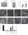
Figure 4. Gene silencing of c-Cbl or overexpression of Nck1 rescues stress fibre formation in synaptopodin-depleted podocytes
(a) Western blot analysis confirms synaptopodin depletion in synaptopodin knockdown (synpo shRNA) podocytes compared with cells expressing a non-silencing control shRNA (con). Western blot for Nck shows comparable expression levels of GFP-Nck1, GFP-Nck1(K178R) and GFP-Nck2 in synaptopodin knockdown cells. (b) Western blots analysis confirms the rescue of Nck1 protein abundance in synaptopodin knockdown podocytes by gene silencing of c-Cbl shRNA. Nck2 levels are comparable under all conditions. GAPDH serves as a loading control. (c) Quantification of Nck1 and Nck2 protein abundance in c-Cbl-depleted synaptopodin shRNA cells. Values are presented as % of control shRNA band intensity±s.e.m., n = 3, ANOVA, **P<0.01. (d) Phalloidin labelling shows the loss of stress fibres in synaptopodin knockdown (synpo shRNA) but not in control shRNA-infected cells. GFP-Nck1 and GFP-Nck1(K178R) but not GFP-Nck2 or GFP alone restore stress fibres. Gene silencing of c-Cbl but not a control shRNA also restore stress fibres in synaptopodin-depleted podocytes. Scale bar 20 μM (e) Quantitative analysis shows a partial rescue of stress fibre formation by GFP-Nck1 and a complete rescue by GFP-Nck1(K178R) or c-Cbl shRNA. Values are presented as % stress fibre-containing cells ±s.e.m., n = 3, ANOVA, ****P<0.0001. Molecular weight markers are in kDa.
Figure 5
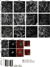
Figure 5. Nck1 promotes stress fibre formation via Rho kinase and formin signalling
(a) Phalloidin labelling shows well-developed stress fibres in podocytes infected with a non-silencing control shRNA and GFP, and in synaptopodin knockdown (synpo shRNA) podocytes infected with GFP-Nck1(K178R) or GFP-RhoA(K6,7R). The Arp2/3 inhibitor CK666 does not disrupt stress fibres. In contrast, the formin inhibitor SMIFH2 or the Rho kinase inhibitor Y27632 cause a loss of stress fibres in control shRNA-infected cells and synaptopodin knockdown podocytes infected with GFP-Nck1(K178R) or GFP-RhoA(K6,7R). Scale bar 20 μM (b) Double labelling for phalloidin and ARP3 confirms the loss of ARP3 in CK666-treated podocytes but not in dimethylsulphoxide-treated control cells without affecting stress fibre integrity. Scale bar 20 μM (c) Quantitative analysis showing a significant loss of stress fibres after SMIFH2 (formin inhibitor) or Y-27632 (Rho kinase inhibitor) treatment. Values are presented as % stress fibre-containing cells ±s.e.m., n = 3, ANOVA, ****P<0.0001.
Figure 6
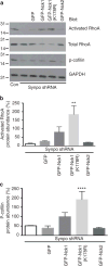
Figure 6. Nck1 increases RhoA activation and cofilin phosphorylation
(a) GFP-Nck1 and more strongly c-Cbl-resistant GFP-Nck1(K178R) but GFP-Nck2 or GFP alone increase the abundance of GTP-bound active RhoA and the downstream target phospho-cofilin. GAPDH serves as loading control. Molecular weight markers are in kDa. (b) Quantitative analysis of RhoA activation in synaptopodin-depleted cells overexpressing c-Cbl-resistant GFP-Nck1 (K178R), **P<0.01. Data are presented as % ±s.e.m. of control shRNA-expressing cells, n = 3. (c) Quantitative analysis of p-cofilin abundance in synaptopodin-depleted cells overexpressing GFP-Nck1 (K178R). Data are presented as % ±s.e.m. of control shRNA-expressing cells, n = 3, ANOVA, ****P<0.0001.
Figure 7
Figure 7. SH3 domain 2 in Nck1 is essential for stress fibre formation
(a) Schematic showing the domain structure of Nck1, Nck2 and four Nck2 constructs containing Nck1 domains. (b) Western blot for Nck shows comparable expression levels for GFP-Nck1(K178R), GFP-Nck2, GFP-Nck2/Nck1 SH3-1, GFP-Nck2/Nck1 SH2, GFP-Nck2/Nck1 SH3 and GFP-Nck2/Nck1 SH2 in synaptopodin knockdown cells. Molecular weight markers are in kDa. (c) Phalloidin labelling shows the induction of stress fibres in synaptopodin-depleted (synpo shRNA) cells expressing c-Cbl-resistant Nck1(K178R) or Nck2/Nck1 SH3-2. Scale bar 20 μM. (d) Quantitative analysis showing a significant increase in stress fibres containing synaptopodin-depleted cells expressing c-Cbl-resistant Nck1(K178R) or Nck2/Nck1 SH3-2. Values are presented as % stress fibre-containing cells ±s.e.m., n = 3, ANOVA ***P<0.001, ****P<0.0001. (e) A model for the co-regulation of RhoA signalling by Nck and synaptopodin. (Left) In the absence of synaptopodin, RhoA and Nck1 are targeted for proteasomal degradation by Smurf1-or c-Cbl, respectively. (Right) Synaptopodin (Synpo) prevents the targeting of RhoA and Nck1 for proteasomal degradation by blocking the binding of Smurf1 to RhoA and the binding of c-Cbl to Nck1, thereby increasing RhoA activation and stress fibre formation. Ub, ubiquitin.

Similar articles

Cited by

References

    1. Buday L, Wunderlich L, Tamas P. The Nck family of adapter proteins: regulators of actin cytoskeleton. Cell Signal. 2002;14:723–731. - PubMed
    1. Lettau M, Pieper J, Janssen O. Nck adapter proteins: functional versatility in T cells. Cell commun Signal. 2009;7:1. - PMC - PubMed
    1. Rohatgi R, Nollau P, Ho HY, Kirschner MW, Mayer BJ. Nck and phosphatidylinositol 4,5-bisphosphate synergistically activate actin polymerization through the N-WASP-Arp2/3 pathway. J Biol Chem. 2001;276:26448–26452. - PubMed
    1. Yamaguchi H, et al. Molecular mechanisms of invadopodium formation: the role of the N-WASP-Arp2/3 complex pathway and cofilin. J Cell Biol. 2005;168:441–452. - PMC - PubMed
    1. Lu W, Katz S, Gupta R, Mayer BJ. Activation of Pak by membrane localization mediated by an SH3 domain from the adaptor protein Nck. Curr Biol. 1997;7:85–94. - PubMed

Publication types

MeSH terms

LinkOut - more resources