. 2013 Feb 7;49(3):558-70.
doi: 10.1016/j.molcel.2012.11.019. Epub 2012 Dec 20.

LSD2/KDM1B and its cofactor NPAC/GLYR1 endow a structural and molecular model for regulation of H3K4 demethylation

Affiliations

LSD2/KDM1B and its cofactor NPAC/GLYR1 endow a structural and molecular model for regulation of H3K4 demethylation

Rui Fang et al. Mol Cell. .

Abstract

Dynamic regulation of histone methylation represents a fundamental epigenetic mechanism underlying eukaryotic gene regulation, yet little is known about how the catalytic activities of histone demethylases are regulated. Here, we identify and characterize NPAC/GLYR1 as an LSD2/KDM1b-specific cofactor that stimulates H3K4me1 and H3K4me2 demethylation. We determine the crystal structures of LSD2 alone and LSD2 in complex with the NPAC linker region in the absence or presence of histone H3 peptide, at resolutions of 2.9, 2.0, and 2.25 Å, respectively. These crystal structures and further biochemical characterization define a dodecapeptide of NPAC (residues 214-225) as the minimal functional unit for its cofactor activity and provide structural determinants and a molecular mechanism underlying the intrinsic cofactor activity of NPAC in stimulating LSD2-catalyzed H3K4 demethylation. Thus, these findings establish a model for how a cofactor directly regulates histone demethylation and will have a significant impact on our understanding of catalytic-activity-based epigenetic regulation.


Figures

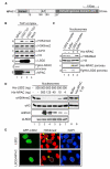
Figure 1
Figure 1. NPAC Is a Cofactor of LSD2 Positively Modulating Its H3K4 Demethylase Activity
(A) Schematic of NPAC domain structure. AT, AT-hook motif; PWWP, Pro-Trp-Trp-Pro domain. (B) LSD2 in the NPAC complex can efficiently demethylate nucleosomal H3K4me2. Tandem-affinity-purified NPAC and LSD2 complexes were incubated with nucleosomes purified from HeLa cells and analyzed by immunoblot using the indicated antibodies. (C) The addition of recombinant NPAC can improve H3K4 demethylase activity of the LSD2 complex. (D) Recombinant NPAC stimulates LSD2 nucleosomal H3K4 demethylation in a dose-dependent manner. The amount of LSD2-enzyme and NPAC-cofactor proteins used in each reaction is indicated. Demethylation was assessed by immunoblot using the indicated antibodies. Significantly larger amounts of His-LSD2 are required for the efficient demethylation of nucleosomes (see Figure S1A). (E) NPAC stimulates H3K4 demethylation mediated by LSD2 in cells. Immunofluorescence staining of U2OS transiently transfected with LSD2 alone or in combination with NPAC is shown. Green, GFP-LSD2; red, H3K4me2; blue, DAPI counterstain of DNA. Representatives with similar levels of GFP-LSD2 are shown. See also Figure S1.
Figure 2
Figure 2. The Linker Region of NPAC Is Sufficient for Its Cofactor Activity and LSD2 Interaction
(A) Schematic representation of the wild-type and deletion mutants of NPAC. DH, dehydrogenase domain; black box, AT-hook motif. (B) Nucleosome demethylation assays examining cofactor activities of NPAC mutants. Equal amounts of LSD2 were used in nucleosome demethylation reactions 2–11, in combination with various GST-tagged NPAC truncation proteins indicated above. GST was included as a negative control. (C) The linker region of NPAC can stimulate LSD2 histone demethylase activity toward short H3K4me2 peptides. Molecular masses corresponding to mono- and dimethylated H3K4 peptides (residues 1–21) are denoted as me1 and me2, respectively. (D) The linker region of NPAC is sufficient for LSD2 binding. Purified GST and GST-tagged wild-type and mutant NPAC proteins were used for GST pull-down of purified His-LSD2. Pull-down complexes were separated by SDS-PAGE and visualized by Coomassie blue staining. Asterisk, GST fusion protein.
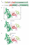
Figure 3
Figure 3. Binding of NPAC Does Not Induce Significant Conformational Changes in LSD2
(A) Comparison of the domain structures of human LSD2 and LSD1. Numbers indicate residue positions at the boundaries of each domain. The ZF domain is shown in lime, the Zf-CW domain in purple, the linker region in pink, the SWIRM domain in red, and the amine oxidase domain in green. The N-terminal flexible regions in both proteins and the tower domain of LSD1 are shown in gray. The same color scheme is used in all structural figures. (B–D) The crystal structures of LSD2 (B), NPAC-LSD2 heterodimer (C), and the ternary complex of LSD2-NPAC-H3 peptide (D). Disordered regions are shown in dashed lines, and FAD is shown in the stick representation (purple). Three zinc atoms are shown as gray balls. N and C termini of LSD2 are indicated. NPAC and H3K4M peptide are indicated and shown in the ribbon representation in blue and yellow, respectively. See also Figures S2 and S3 and Table S2.
Figure 4
Figure 4. LSD2 Exhibits Both Common and Distinctive Structural Features Compared to LSD1
(A) Structural overlay of LSD2 (colored as in Figure 3) and LSD1 (gray). (B) Structure of the N-terminal zinc finger of LSD2. Two zinc ions (gray balls) are coordinated with a Cys4His2Cys2 motif with the indicated residues shown in stick representation. This zinc-finger domain bears little resemblance to known zinc-finger structures. (C) Structure of the CW-type zinc finger of LSD2. Residues involved in zinc coordination are labeled and shown in stick representation. The disordered region is indicated by a dotted line. (D) Superimposed structures of the SWIRM domains of LSD2 and LSD1, colored as in (A). The extended loop and α9-helix in LSD2 SWIRM domain are denoted. The side chains of unconserved residues are presented in stick presentation. (E) Mutation of the extended loop in LSD2 SWIRM domain impairs its histone demethylase activity. MALDI-TOF MS analyses of the demethylation of H3K4me2 peptides incubated with the indicated proteins are shown. LSD2.WT, wild-type LSD2; LSD2.M, an LSD2 mutant replacing YQPNEC 273–278 with a flexible linker GSGSGS. See also Figure S4 and Table S2.
Figure 5
Figure 5. A Dodecapeptide of NPAC Interacts with LSD2
(A and B) Accommodation of NPAC residues in a hydrophobic pocket between the AO and SWIRM domains of LSD2. The surface rendering and ribbon representation of LSD2 are shown in (A) and (B), respectively. Critical residues involved in the interaction are shown in stick representation and colored in green (from the LSD2 AO domain), red (from the LSD2 SWIRM domain), and purple (from NPAC), respectively. (C) Structural superimposition of the NPAC binding site of LSD2 and the corresponding regions of LSD1. The LSD2 structures are nearly identical with or without NPAC binding. (D) Schematic of deletion mutants of the NPAC linker region (NP.d7–NP.d9). The red box marks the position of NPAC residues 214–225. (E) Examination of LSD2 binding of NPAC deletion mutants by GST pull-down. His-LSD2 in pull-down products were separated by SDS-PAGE and visualized by Coomassie blue staining. (F) Examination of the cofactor activity of NPAC mutants described in (D) using nucleosome demethylation assays. Immunoblots using the indicated antibodies are shown. (G) Partial sequences of NPAC mutations (NP.M1–NP.M3, residues 188–252) disrupting the potential NPAC binding site for LSD2 interaction. (H) GST-pull-down analyses of LSD2 binding by NPAC mutants in (G). Coomassie blue staining of His-LSD2 in input and GST-pull-down complexes is shown. (I) Cofactor activity analyses of NPAC mutants described in (G) using nucleosomal demethylation assays.
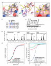
Figure 6
Figure 6. The NPAC Dodecapeptide Stimulates LSD2 Histone Demethylase Activity by Assisting Enzyme-Substrate Interaction
(A and B) A close-up view of the interplay of LSD2, NPAC, and H3K4M peptide in the cocrystal structure. The surface rendering and ribbon representation of LSD2 are shown in (A) and (B), respectively. H3K4M peptide (histone H3 residues 1–21, with Lys4 replaced with a Met) and NPAC (residues 214–225) are shown in the ribbon representation and colored in yellow and purple, respectively. Critical residues involved in H3 peptide interaction are shown in stick representation. Hydrogen bonds are indicated by dashed lines. (C) H3L20 makes a new contact with the NPAC-LSD2 complex, with its side chain inserted in a hydrophobic patch formed by NPAC F217 and LSD2 residues. (D) Sequences of wild-type (NP.WT) and mutant NPAC dodecapeptides (NP.M4–NP.M6) disrupting the potential interaction of NPAC with H3K4M peptide in the LSD2-NPAC-H3K4M peptide ternary complex. (E) NPAC F217 is essential for its cofactor activity. Immunoblots of nucleosome demethylation assays are shown. (F) The wild-type NPAC dodecapeptide, but not the F217A mutant, can stimulate LSD2-mediated demethylation of H3K4me2 peptides. (G) Mutations of D214, H216, and F217 of NPAC do not affect LSD2 binding. ITC enthalpy plots of wild-type and mutant NPAC dodecapeptide binding to LSD2 are shown. Indicated NPAC peptides were injected into LSD2 containing cuvettes. (H) ITC enthalpy plots of the binding of the H3K4M peptide to LSD2 and LSD2 in complex with either wild-type (WT) or F217A (M6) NPAC peptides (residues 214– 225). H3K4M peptide (residues 1–21) was injected into cuvettes containing the indicated combination of LSD2 and NPAC peptides. See also Figures S5 and S6 and Tables S3 and S4.

Similar articles

Cited by

References

    1. Adams PD, Grosse-Kunstleve RW, Hung LW, Ioerger TR, McCoy AJ, Moriarty NW, Read RJ, Sacchettini JC, Sauter NK, Terwilliger TC. PHENIX: building new software for automated crystallographic structure determination. Acta Crystallogr. D Biol. Crystallogr. 2002;58:1948–1954. - PubMed
    1. Allis CD, Berger SL, Cote J, Dent S, Jenuwien T, Kouzarides T, Pillus L, Reinberg D, Shi Y, Shiekhattar R, et al. New nomenclature for chromatin-modifying enzymes. Cell. 2007;131:633–636. - PubMed
    1. Bernstein BE, Meissner A, Lander ES. The mammalian epigenome. Cell. 2007;128:669–681. - PubMed
    1. Bhaumik SR, Smith E, Shilatifard A. Covalent modifications of histones during development and disease pathogenesis. Nat. Struct. Mol. Biol. 2007;14:1008–1016. - PubMed
    1. Binda C, Valente S, Romanenghi M, Pilotto S, Cirilli R, Karytinos A, Ciossani G, Botrugno OA, Forneris F, Tardugno M, et al. Biochemical, structural, and biological evaluation of tranylcypromine derivatives as inhibitors of histone demethylases LSD1 and LSD2. J. Am. Chem. Soc. 2010;132:6827–6833. - PubMed

Publication types