. 2012;8(5):e1002647.
doi: 10.1371/journal.pgen.1002647. Epub 2012 May 3.

The C. elegans H3K27 demethylase UTX-1 is essential for normal development, independent of its enzymatic activity

Affiliations

The C. elegans H3K27 demethylase UTX-1 is essential for normal development, independent of its enzymatic activity

Julien Vandamme et al. PLoS Genet. 2012.

Abstract

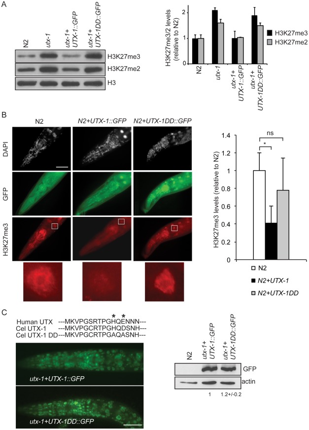
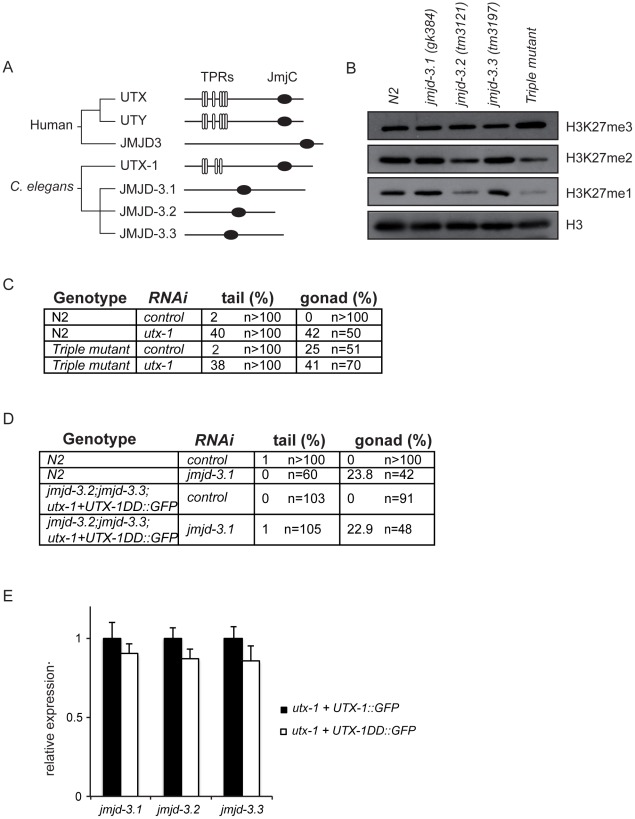
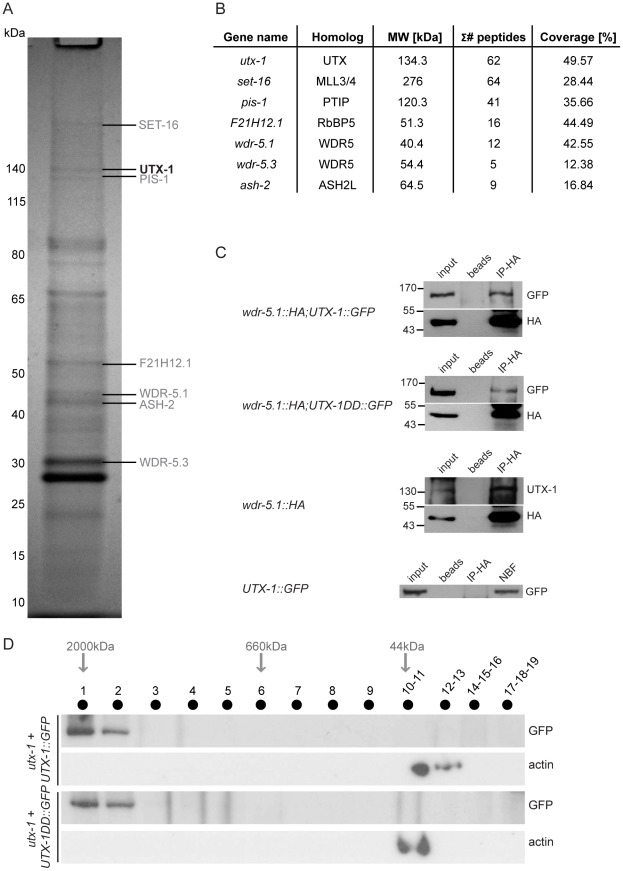
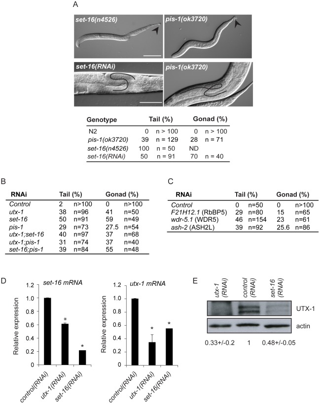
Epigenetic modifications influence gene expression and provide a unique mechanism for fine-tuning cellular differentiation and development in multicellular organisms. Here we report on the biological functions of UTX-1, the Caenorhabditis elegans homologue of mammalian UTX, a histone demethylase specific for H3K27me2/3. We demonstrate that utx-1 is an essential gene that is required for correct embryonic and postembryonic development. Consistent with its homology to UTX, UTX-1 regulates global levels of H3K27me2/3 in C. elegans. Surprisingly, we found that the catalytic activity is not required for the developmental function of this protein. Biochemical analysis identified UTX-1 as a component of a complex that includes SET-16(MLL), and genetic analysis indicates that the defects associated with loss of UTX-1 are likely mediated by compromised SET-16/UTX-1 complex activity. Taken together, these results demonstrate that UTX-1 is required for many aspects of nematode development; but, unexpectedly, this function is independent of its enzymatic activity.


Conflict of interest statement

The authors have declared that no competing interests exist.

Figures

Figure 1
Figure 1. UTX-1 expression and utx-1 embryonic phenotypes.
(A) Top: Human UTX and the C. elegans homologue UTX-1. TPRs, tetratricopeptide repeats; JmjC, Jumonji C domain. Bottom: Genomic organization of utx-1. Black H-shaped lines indicate the position of the tm3136 and tm3118 deletions. Black lines indicate the position of the different RNAi fragments (a, b, and c) used in this study. (B) UTX-1 expression during embryogenesis analyzed by immunostaining with an anti-UTX-1 antibody (top panel, right) and by epifluorescence (bottom panel, right). DAPI staining and Nomarski (DIC) images are also shown on the left. (C) UTX-1 expression by epifluorescence (right panels) during larval development. Nomarski (DIC) images are shown on the left panels. Asterisks indicate the distal tip cell, arrow head the forming vulva. Animals are oriented anterior to the left. (D) Brood size of wild type, utx-1 mutant worms and rescued utx-1 lines. Progeny is given as the average number of viable progeny per worm ± SD. The number of laid, not hatched, eggs counted in utx-1 (m+/z−) mutants is reported in the lower table. utx-1+UTX-1::GFP and utx-1+UTX-1DD::GFP, are utx-1 transgenic lines expressing wild-type or catalytically inactive mutant of UTX-1, respectively, as extrachromosomal arrays. (E) Representative Nomarski (DIC) images of a utx-1(tm 3136) mutant embryo and escaper L1 larva. Similar phenotypes are observed in utx-1(tm 3118) (not shown). Bars in B and E are 20 µm, in C 50 µm. Animals are oriented anterior to the left.
Figure 2
Figure 2. utx-1 postembryonic phenotypes.
(A) Representative DIC images of L1 larvae tails (upper panel) and L1 larvae (lower panel) of N2, utx-1(RNAi) and utx-1(tm3118) animals. Animals are oriented anterior to the left. Scale bar is 50 µm. (B) Representative DIC images of gonads in adults N2, utx-1(RNAi) and utx-1(tm3118) animals. Scale bar is 25 µm. In the upper panels, blacks lines indicate the migration of the gonad arm. In the lower panels, black lines indicate the contours of the oocytes. Animals are oriented anterior to the left. (C) Percentages of posterior (% tail defects) and gonad (% gonadal defects) defects in the indicated strains are shown. For RNAi, F1 larvae and adults from at least three independent experiments were analyzed. (D) utx-1 and jmjd-3.1 mRNA levels in embryos after control (black bars) or utx-1 (white bars) RNAi treatment as measured by qPCR, using rpl-26 mRNA as internal control. *P<0.01 (Student's t-test).
Figure 3
Figure 3. UTX-1 is an H3K27me2/3 demethylase.
(A) Protein lysates from embryos of the indicated strains were probed with antibodies against H3K27me3 and H3K27me2. H3 was used as loading control. Quantification of the western blot is shown in the graphic on the right. Error bars indicate the standard deviation calculated using at least 2 replicates. The signals were quantified using ImageJ software and normalized to H3. The values are relative to N2 levels. Similar results were obtained with at least two different transgenic lines and in the two utx-1 genetic backgrounds (tm3118 and tm3136). (B) Representative images of N2 expressing a translational construct for wild-type (N2+UTX-1::GFP) and catalytically inactive UTX-1 (N2+UTX-1DD::GFP) GFP fusion and GFP-negative siblings, fixed and stained with H3K27me3 antibody and DAPI. The white square encloses an intestinal cell, used for the H3K27me3 quantification. Enlargement of the white square is shown at the bottom of the panel. Quantification of H3K27me3 levels is shown in the graphic on the right. At least 25 cells for each genotype were quantified as described in Materials and Methods. Mean fluorescence + s.e.m. values of two independent experiments are displayed. *P<0.01. (Student's t-test). Animals are oriented anterior to the left. (C) Top: Alignment of a part of the Jumonji C domain of human UTX with UTX-1 and with the catalytically inactive UTX-1DD (DD = Demethylase Dead). Asterisks indicate two of the three conserved amino acids in the iron-binding domain (HXD/EXnH) of the JmjC-domain, modified in the UTX-1DD. Bottom: Epifluorescence of utx-1 mutant animals, carrying a translational GFP fusion of wild-type UTX-1 (utx-1+UTX-1::GFP) or catalytically inactive UTX-1 (utx-1+UTX-1DD::GFP). Anterior parts of the animals are shown, with anterior to the left. On the right, lysates from L1 carrying the two transgenes were probed with GFP antibody. Actin was used as loading control. The signal was quantified using ImageJ program and normalized to actin.
Figure 4
Figure 4. KDM6 family in C. elegans.
(A) Phylogenetic cluster of human UTX, UTY, JMJD3 and homologous proteins in C. elegans. TPRs, tetratricopeptide repeats; JmjC, Jumonji C domain. (B) Protein lysates from eggs of the indicated strains were analyzed by western blot using the indicated antibodies. H3 was used as loading control. Quantification of the western blot is shown in Figure S11. (C) Percentage of tail and gonad defects observed in N2 and the triple mutant (jmjd-3.1;jmjd-3.2;jmjd-3.3) after treatment with control or utx-1(RNAi). F1 animals from at least two independent experiments were scored. (D) Percentage of tail and gonad defects observed in N2 and the triple mutant (jmjd-3.2; jmjd-3.3;utx-1+UTX-1DD::GFP) after treatment with control or jmjd-3.1(RNAi). F1 animals from at least two independent experiments were scored. (E) Levels of expression of the JMJD3-like genes in eggs derived from the utx-1(tm3136) mutant strain rescued with the catalytically inactive utx-1 (utx-1+UTX-1DD::GFP) relative to utx-1(tm3136) rescued with utx-1 wild-type (utx-1+UTX-1::GFP). rpl-26 mRNA was used as internal control for normalization.
Figure 5
Figure 5. UTX-1 is part of a MLL-like complex.
(A) Immunoprecipitation of GFP tagged UTX-1 from a mixed population (eggs and adults) of utx-1(tm3118) rescued with UTX-1::GFP. Affinity purified proteins were resolved by SDS-PAGE and stained with colloidal Coomassie. Homologues of the mammalian UTX-MLL complex co-eluted with the bait and identified by LC-MS/MS are listed in grey. Position of the bait protein is shown in black. Molecular weight markers are indicated to the left of the gel. (B) Table summarizing the identified homologues of the components of the mammalian UTX-MLL complex. Gene names, molecular weight in kDa, number of unique peptides and sequence coverage in percentage are reported. (C) Co-immunoprecipitations of WDR-5.1::HA and UTX-1. Total protein extracts from eggs of the indicated strains were immunoprecipitated using anti-HA affinity gel beads. The precipitates were analyzed by SDS-PAGE followed by western blotting using antibodies against HA, GFP or endogenous UTX-1. Input = 30 µg of protein extract. NBF = non bound fraction. (D) UTX-1-associated protein complex assessed by size exclusion chromatography. Superose 6 gel filtration of total protein extracts derived from UTX-1 mutant rescued with wild-type (utx-1+UTX-1::GFP) and catalytically inactive UTX-1 (utx-1+UTX-1DD::GFP). Fractions were analyzed by western blotting using GFP and actin antibodies.
Figure 6
Figure 6. Functions of the SET-16/UTX-1 complex.
(A) (Upper panel) DIC images of set-16(n4526) and pis-1(ok3720) mutant larvae. Arrowhead indicates misshapen tails. Scale bar is 50 µm. (Lower panel) DIC images of set-16(RNAi) and pis-1(ok3720) adults. Gonadal migration defects are shown. The black line indicates the aberrant gonadal migration. Scale bar is 20 µm. Animals are oriented anterior to the left. The percentage of tail and gonadal defects associated to loss or reduction of set-16 and pis-1 are reported on the right. (B) Percentage of tail and gonad defects after RNAi of the indicated genes. F1 L1 larvae and adult animals from at least two independent experiments were scored. (C) Percentage of tail and gonad defects after RNAi of the indicated genes. F1 or F2 L1 larvae and adult animals from at least two independent experiments were scored. (D) utx-1 and set-16 mRNA levels in embryos of worms treated with control, utx-1 or set-16(RNAi). *P<0.01 (Student's t-test). (E) Protein lysates of embryos from worms treated with control, utx-1 or set-16(RNAi) were probed with an antibody against UTX-1. Actin was used as loading control. The signal was quantified using ImageJ program and normalized to actin. Indicated values are relative to control (RNAi) and derive from two independent experiments.

Similar articles

Cited by

References

    1. Strahl BD, Allis CD. The language of covalent histone modifications. Nature. 2000;403:41–45. - PubMed
    1. Kouzarides T. Chromatin modifications and their function. Cell. 2007;128:693–705. - PubMed
    1. Berger SL. The complex language of chromatin regulation during transcription. Nature. 2007;447:407–412. - PubMed
    1. Tsukada Y, Fang J, Erdjument-Bromage H, Warren ME, Borchers CH, et al. Histone demethylation by a family of JmjC domain-containing proteins. Nature. 2006;439:811–816. - PubMed
    1. Cloos PA, Christensen J, Agger K, Helin K. Erasing the methyl mark: histone demethylases at the center of cellular differentiation and disease. Genes Dev. 2008;22:1115–1140. - PMC - PubMed

Publication types

MeSH terms

Substances

LinkOut - more resources