. 2011 Sep 16;286(37):32355-65.
doi: 10.1074/jbc.M111.278408. Epub 2011 Jul 21.

COMMD1 (copper metabolism MURR1 domain-containing protein 1) regulates Cullin RING ligases by preventing CAND1 (Cullin-associated Nedd8-dissociated protein 1) binding

Affiliations

COMMD1 (copper metabolism MURR1 domain-containing protein 1) regulates Cullin RING ligases by preventing CAND1 (Cullin-associated Nedd8-dissociated protein 1) binding

Xicheng Mao et al. J Biol Chem. .

Abstract

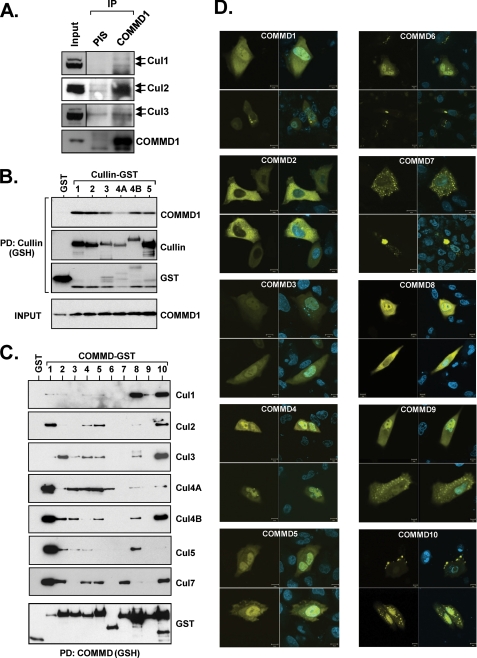
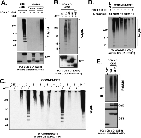
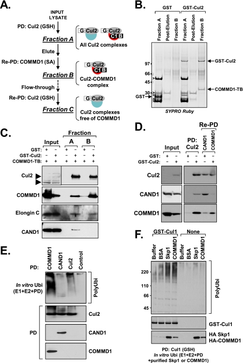
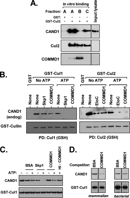
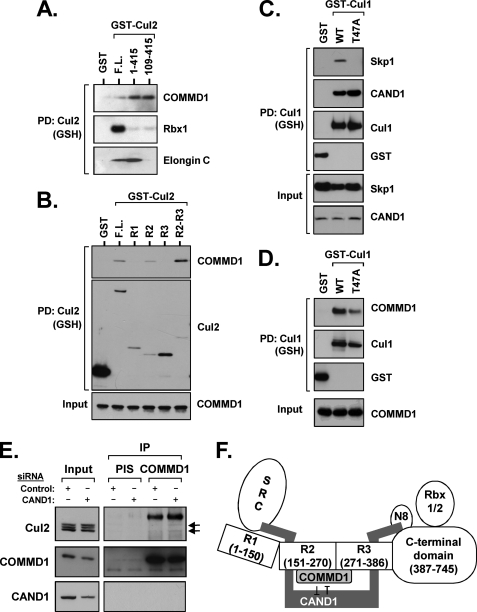
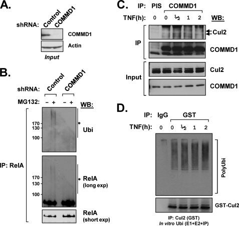
Cullin RING ligases (CRLs), the most prolific class of ubiquitin ligase enzymes, are multimeric complexes that regulate a wide range of cellular processes. CRL activity is regulated by CAND1 (Cullin-associated Nedd8-dissociated protein 1), an inhibitor that promotes the dissociation of substrate receptor components from the CRL. We demonstrate here that COMMD1 (copper metabolism MURR1 domain-containing 1), a factor previously found to promote ubiquitination of various substrates, regulates CRL activation by antagonizing CAND1 binding. We show that COMMD1 interacts with multiple Cullins, that the COMMD1-Cul2 complex cannot bind CAND1, and that, conversely, COMMD1 can actively displace CAND1 from CRLs. These findings highlight a novel mechanism of CRL activation and suggest that CRL regulation may underlie the pleiotropic activities of COMMD1.


Figures

FIGURE 1.
FIGURE 1.
COMMD1 and other COMMD proteins associate with CRL complexes. A, endogenous co-immunoprecipitation (IP) of COMMD1 and CRLs. COMMD1 was immunoprecipitated from HEK 293 cell lysates. The precipitated material was probed for the indicated Cullins. PIS, preimmune serum. B, COMMD1 binds to other Cullin family members. COMMD1 was expressed in HEK 293 cells together with Cullin proteins fused to GST, which were precipitated as above and immunoblotted for COMMD1. PD, pulldown. C, COMMD proteins interact with Cullins. In each experiment, one FLAG-tagged Cullin was expressed together with COMMD proteins fused to GST, which were subsequently precipitated. The presence of the Cullin in the precipitates was determined by immunoblotting with the FLAG antibody. D, cellular distribution of COMMD proteins. The indicated COMMD proteins fused to EYFP were expressed in HeLa cells. The cells were counterstained with Hoechst and imaged in a confocal microscope. The scale bar corresponds to 10 μm.
FIGURE 2.
FIGURE 2.
COMMD1 associates with active CRL complexes in a copper-independent manner. A, COMMD1 precipitates cellular factors with ubiquitin ligase activity. In vitro ubiquitination reactions were supplemented with precipitated proteins as indicated. Polyubiquitin chain formation was determined by Western blotting as an indication of ubiquitin ligase activity. COMMD1-GST or GST were expressed in HEK 293 cells (left panels) or prepared recombinantly in E. coli (right panels). Recombinant proteins either were offered directly (none) or were first mixed with a mammalian lysate and washed prior to the reaction (lysate). PD, pulldown. B, COMMD1 precipitates ubiquitin ligase activity through the COMM domain. COMMD1 full-length (FL), its amino terminus (NT), or the COMM domain (CD) fused to GST were precipitated from transfected HEK 293 cells and added to an in vitro ubiquitination reaction as in A. C, COMMD proteins precipitate ubiquitin ligase activity. COMMD proteins fused to GST were precipitated and added to an in vitro ubiquitination reaction as in A. D, depletion of Rbx1 reduces COMMD1-associated ligase activity. COMMD1-GST was utilized as a source of ubiquitin ligase activity as before. The protein was expressed in HEK 293 cells and either precipitated directly (−) or following immunodepletion of FLAG-Rbx1 (+). IP, immunoprecipitation. E, copper binding by COMMD1 is not involved in E3 and Cul2 binding. WT COMMD1 or an M110A/H134A mutant (MUT) unable to bind copper were expressed in HEK 293 cells. After precipitation, the protein complexes were examined for associated E3 activity as before (upper panel) and for the presence of co-precipitated endogenous Cul2 (middle panel). Ubi, ubiquitination.
FIGURE 3.
FIGURE 3.
The Cul2-COMMD1 complex excludes the CRL inhibitor CAND1. A, purification strategy used to isolate the Cul2-COMMD1 complex. Cul2 fused to GST (G) and COMMD1 fused to a biotinylation tag (B) were expressed in HEK 293 cells. Cul2 was purified through a GSH-Sepharose column (fraction A) and subsequently eluted. COMMD1 was precipitated from the eluted material using streptavidin (SA)-agarose beads (fraction B). Cul2 remaining in the flow-through after the streptavidin-agarose column was reprecipitated with a GSH column (Re-PD: Cul2 (GSH)) resulting in fraction C. PD, pulldown. B, Cul2-COMMD1 complex purification. Proteins from fractions A and B were separated by SDS-PAGE and stained with SYPRO Ruby; the identity of major bands was determined by paired Western blot analysis (nonspecific bands indicated by open arrowheads). C, the Cul2-COMMD1 complex excludes CAND1. Fractions A and B were immunoblotted for Cul2, COMMD1, Elongin C, and CAND1. Endogenous Cul2 and GST-Cul2 are noted by arrowheads to the left of the top panel. D, COMMD1 and CAND1 exist in separate Cul2 cellular pools. GST-Cul2, COMMD1 fused to a biotinylation tag, and FLAG-CAND1 were co-expressed in HEK 293 cells. Cul2 was purified through a GSH affinity column (PD: Cul2) and subsequently eluted. COMMD1 or CAND1 were then precipitated from the eluate using SA agarose beads or FLAG antibody, respectively. The resulting fractions were immunoblotted for Cul2, CAND1, or COMMD1. E, Cul2 complexes containing COMMD1 are active. GST-Cul2 was precipitated from transfected HEK 293 cells utilizing GSH beads (Cul2) or using protein G beads as a control (Control). FLAG-COMMD1 or FLAG-CAND1 co-expressed with GST-Cul2 were precipitated utilizing the FLAG antibody. After adjusting for equal Cul2 recovery, the resulting material was added to an in vitro ubiquitination reaction as in Fig. 2A and immunoblotted for ubiquitin. F, purified COMMD1 potentiates Cul1 E3 activity in vitro. GST-tagged Cul1 was expressed in HEK 293 cells and purified using GSH affinity matrix. Purified HA-Skp1 or HA-COMMD1 prepared from mammalian lysates was then added to the recovered Cul1 complex, and this preparation was used as a source for E3 activity as before.
FIGURE 4.
FIGURE 4.
COMMD1 promotes the dissociation of CAND1 from CRL complexes. A, COMMD1 prevents CAND1 binding to Cul2. Fractions A, B, and C were prepared as depicted in Fig. 3A, mixed for 2 h with a fresh HEK 293 cell lysate expressing FLAG-CAND1, washed, and immunoblotted for CAND1, Cul2, or COMMD1. B, COMMD1 can displace CAND1 from Cul1 or Cul2 complexes. GST-Cul1 or GST-Cul2 were expressed in HEK 293 cells and purified through a GSH affinity matrix. This material was then mixed with HA-Skp1 or HA-COMMD1 purified from mammalian cells. After incubation at 30 °C with ATP (15 mm), the GSH beads were washed, and the presence of endogenous CAND1 still bound to Cul1 or Cul2 was determined by immunoblotting. PD, pulldown. C, the activity of COMMD1 is heat-labile. GST-Cul1 complexes were purified as before from cells also co-expressing FLAG-CAND1. After a displacement reaction (performed as previously but using 7.5 mm ATP), the presence of remaining FLAG-CAND1 bound to Cul1 was determined by immunoblotting. HI-COMMD1, heat inactivated COMMD1 (95 °C for 10 min). D, recombinant COMMD1 made in E. coli is devoid of activity. Shown is the same reaction as in C, but utilizing mammalian HA-COMMD1 or bacterially made His6-COMMD1. Only the ATP containing reactions are shown; each subpanel was run in the same gel.
FIGURE 5.
FIGURE 5.
COMMD1 binds to CRLs in a manner distinct from substrate receptors. A, full-length and specific deletion mutants of Cul2 fused to GST were expressed in HEK 293 cells and precipitated from cell lysates with glutathione-Sepharose (GSH) beads. The recovered material was immunoblotted for endogenous COMMD1, Rbx1, and Elongin C. PD, pulldown. B, COMMD1 binds to Cullin repeat 2 in Cul2. COMMD1 was expressed in HEK 293 cells together with Cul2 deletion mutants fused to GST (R1, R2, or R3, Cullin repeats 1, 2, or 3, respectively), which were precipitated as in A and immunoblotted for COMMD1. C and D, Cul1 can bind to COMMD1 independently of Skp1. Cul1 WT or a T47A point mutant fused to GST were expressed in HEK 293 cells. Their interactions with HA-Skp1 or FLAG-CAND1 were tested by co-precipitation (C). The same Cul1 proteins were expressed together with HA-COMMD1 and Cul1-COMMD1 interactions were examined by co-precipitation (D). E, CAND1 deficiency promotes COMMD1-Cul2 binding. HEK 293 cells were transfected with siRNA duplexes targeting CAND1. Endogenous COMMD1 was immunoprecipitated (IP), and its interaction with endogenous Cul2 was evaluated as in Fig. 1A. F, model depicting the various domains in canonical Cullin proteins, their known interacting partners, and the competitive binding by COMMD1 and CAND1 for R2. The amino acid boundaries for R1, R2, and R3 in Cul2 are also depicted. N8, NEDD8.
FIGURE 6.
FIGURE 6.
COMMD1 is required for the ubiquitination of specific targets in vivo. A, Western blot demonstrating stable shRNA-mediated repression of COMMD1 in U2OS cells. B, RelA ubiquitination is greatly impaired in COMMD1-deficient cells. U2OS cells were treated with the proteasome inhibitor MG-132 as indicated, and cell nuclei were isolated and then lysed in a denaturing buffer. RelA was immunoprecipitated from nuclear extracts and immunoblotted for ubiquitin (top panel) or for RelA itself (middle and bottom panels). The asterisk indicates the high molecular weight smear consistent with ubiquitinated RelA. C, TNF stimulates COMMD1-Cul2 binding. HEK 293 cells were treated with TNF (1,000 units/ml) for the indicated time intervals, and endogenous COMMD1 was subsequently precipitated from cell lysates as in Fig. 1A. The recovery of endogenous Cul2 was determined by immunoblotting. D, cells stably expressing GST-Cul2 (see supplemental Fig. S3 for details) were treated with TNF (1,000 units/ml) for the indicated time points. Thereafter, GST-Cul2 was immunoprecipitated with a GST antibody. The recovered material was utilized as a source for E3 activity as previously. WB, Western blot; IP, immunoprecipitation; PIS, preimmune serum; Ubi, ubiquitination.

Similar articles

Cited by

References

    1. Weissman A. M. (2001) Nat. Rev. Mol. Cell Biol. 2, 169–178 - PubMed
    1. Wilkinson C. R. (2002) Trends Cell Biol. 12, 545–546 - PubMed
    1. Joazeiro C. A., Weissman A. M. (2000) Cell 102, 549–552 - PubMed
    1. Patterson C. (2002) Sci. STKE 2002, pe4. - PubMed
    1. Coscoy L., Ganem D. (2003) Trends Cell Biol. 13, 7–12 - PubMed

Publication types

MeSH terms