

Phox homology band 4.1/ezrin/radixin/moesin-like proteins function as molecular scaffolds that interact with cargo receptors and Ras GTPases
- PMID: 21512128
- PMCID: PMC3093529
- DOI: 10.1073/pnas.1017110108
Phox homology band 4.1/ezrin/radixin/moesin-like proteins function as molecular scaffolds that interact with cargo receptors and Ras GTPases
Abstract
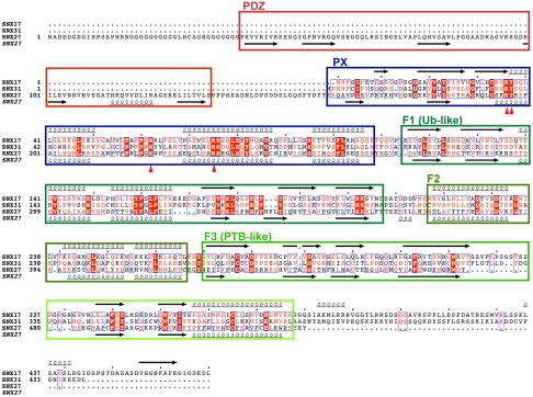
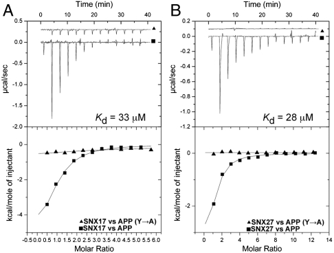
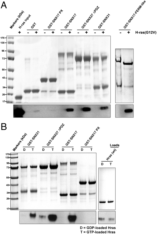
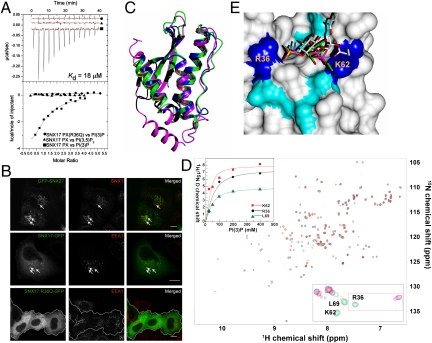
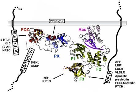
Following endocytosis, the fates of receptors, channels, and other transmembrane proteins are decided via specific endosomal sorting pathways, including recycling to the cell surface for continued activity. Two distinct phox-homology (PX)-domain-containing proteins, sorting nexin (SNX) 17 and SNX27, are critical regulators of recycling from endosomes to the cell surface. In this study we demonstrate that SNX17, SNX27, and SNX31 all possess a novel 4.1/ezrin/radixin/moesin (FERM)-like domain. SNX17 has been shown to bind to Asn-Pro-Xaa-Tyr (NPxY) sequences in the cytoplasmic tails of cargo such as LDL receptors and the amyloid precursor protein, and we find that both SNX17 and SNX27 display similar affinities for NPxY sorting motifs, suggesting conserved functions in endosomal recycling. Furthermore, we show for the first time that all three proteins are able to bind the Ras GTPase through their FERM-like domains. These interactions place the PX-FERM-like proteins at a hub of endosomal sorting and signaling processes. Studies of the SNX17 PX domain coupled with cellular localization experiments reveal the mechanistic basis for endosomal localization of the PX-FERM-like proteins, and structures of SNX17 and SNX27 determined by small angle X-ray scattering show that they adopt non-self-assembling, modular structures in solution. In summary, this work defines a novel family of proteins that participate in a network of interactions that will impact on both endosomal protein trafficking and compartment specific Ras signaling cascades.
Conflict of interest statement
The authors declare no conflict of interest.
Figures
 ion in the crystal structure (yellow) and PI(3)P from the SNX9∶PI(3)P complex (white).
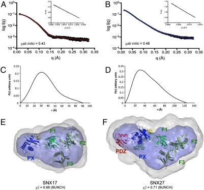
Similar articles
-
Structural basis for endosomal trafficking of diverse transmembrane cargos by PX-FERM proteins.Proc Natl Acad Sci U S A. 2013 Feb 19;110(8):E643-52. doi: 10.1073/pnas.1216229110. Epub 2013 Feb 4. Proc Natl Acad Sci U S A. 2013. PMID: 23382219 Free PMC article.
-
Measuring interactions of FERM domain-containing sorting Nexin proteins with endosomal lipids and cargo molecules.Methods Enzymol. 2014;534:331-49. doi: 10.1016/B978-0-12-397926-1.00019-6. Methods Enzymol. 2014. PMID: 24359963
-
Sorting nexin 31 binds multiple β integrin cytoplasmic domains and regulates β1 integrin surface levels and stability.J Mol Biol. 2014 Sep 9;426(18):3180-3194. doi: 10.1016/j.jmb.2014.07.003. Epub 2014 Jul 11. J Mol Biol. 2014. PMID: 25020227
-
Emerging Role of Sorting Nexin 17 in Human Health and Disease.Curr Protein Pept Sci. 2024;25(10):814-825. doi: 10.2174/0113892037284582240522155112. Curr Protein Pept Sci. 2024. PMID: 38874037 Review.
-
Two Sides of the Coin: Ezrin/Radixin/Moesin and Merlin Control Membrane Structure and Contact Inhibition.Int J Mol Sci. 2019 Apr 23;20(8):1996. doi: 10.3390/ijms20081996. Int J Mol Sci. 2019. PMID: 31018575 Free PMC article. Review.
Cited by
-
Sorting nexin 17 regulates ApoER2 recycling and reelin signaling.PLoS One. 2014 Apr 4;9(4):e93672. doi: 10.1371/journal.pone.0093672. eCollection 2014. PLoS One. 2014. PMID: 24705369 Free PMC article.
-
A unique PDZ domain and arrestin-like fold interaction reveals mechanistic details of endocytic recycling by SNX27-retromer.Proc Natl Acad Sci U S A. 2014 Sep 2;111(35):E3604-13. doi: 10.1073/pnas.1410552111. Epub 2014 Aug 18. Proc Natl Acad Sci U S A. 2014. PMID: 25136126 Free PMC article.
-
SNX17 regulates Notch pathway and pancreas development through the retromer-dependent recycling of Jag1.Cell Regen. 2012 Jun 28;1(1):4. doi: 10.1186/2045-9769-1-4. eCollection 2012. Cell Regen. 2012. PMID: 25408867 Free PMC article.
-
SNX27-Retromer directly binds ESCPE-1 to transfer cargo proteins during endosomal recycling.PLoS Biol. 2022 Apr 13;20(4):e3001601. doi: 10.1371/journal.pbio.3001601. eCollection 2022 Apr. PLoS Biol. 2022. PMID: 35417450 Free PMC article.
-
SNX17 protects integrins from degradation by sorting between lysosomal and recycling pathways.J Cell Biol. 2012 Apr 16;197(2):219-30. doi: 10.1083/jcb.201111121. Epub 2012 Apr 9. J Cell Biol. 2012. PMID: 22492727 Free PMC article.
References
-
- Cullen PJ. Endosomal sorting and signalling: An emerging role for sorting nexins. Nat Rev Mol Cell Biol. 2008;9:574–582. - PubMed
-
- Seet LF, Hong W. The Phox (PX) domain proteins and membrane traffic. Biochim Biophys Acta. 2006;1761:878–896. - PubMed
-
- Burden JJ, Sun XM, Garcia AB, Soutar AK. Sorting motifs in the intracellular domain of the low density lipoprotein receptor interact with a novel domain of sorting nexin-17. J Biol Chem. 2004;279:16237–16245. - PubMed
Publication types
MeSH terms
Substances
Associated data
- Actions
LinkOut - more resources
Full Text Sources
Other Literature Sources
Molecular Biology Databases
Research Materials
Miscellaneous
