. 2011 Mar 3;2(3):e125.
doi: 10.1038/cddis.2011.8.

Individual caspase-10 isoforms play distinct and opposing roles in the initiation of death receptor-mediated tumour cell apoptosis

Affiliations

Individual caspase-10 isoforms play distinct and opposing roles in the initiation of death receptor-mediated tumour cell apoptosis

A Mühlethaler-Mottet et al. Cell Death Dis. .

Abstract

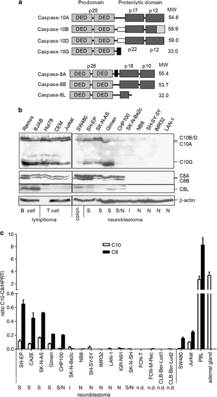
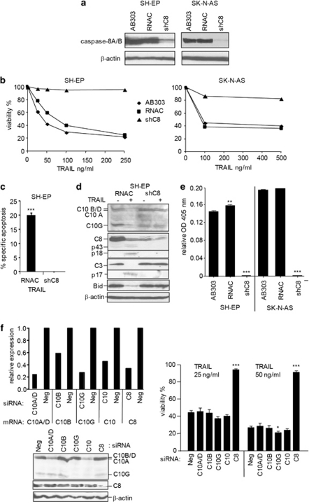
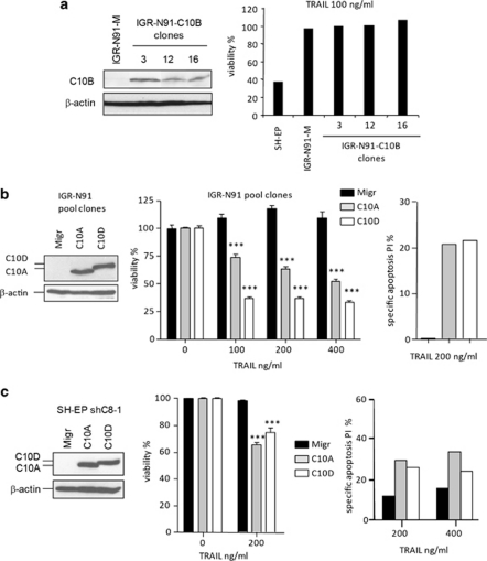
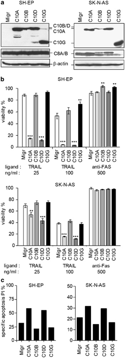
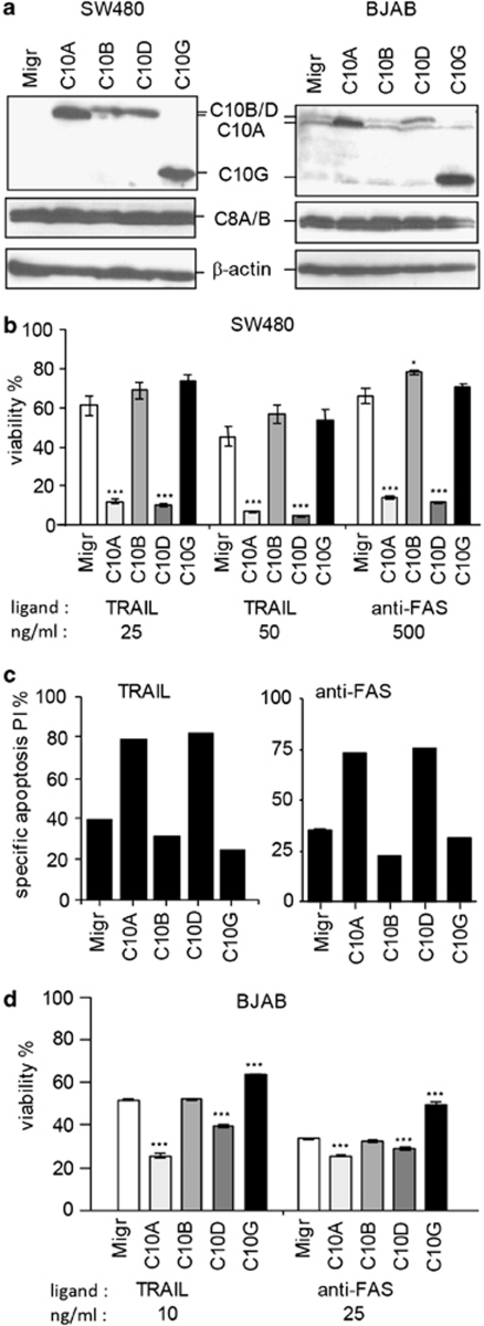
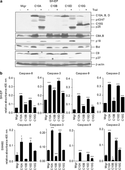
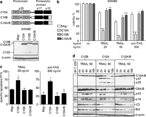
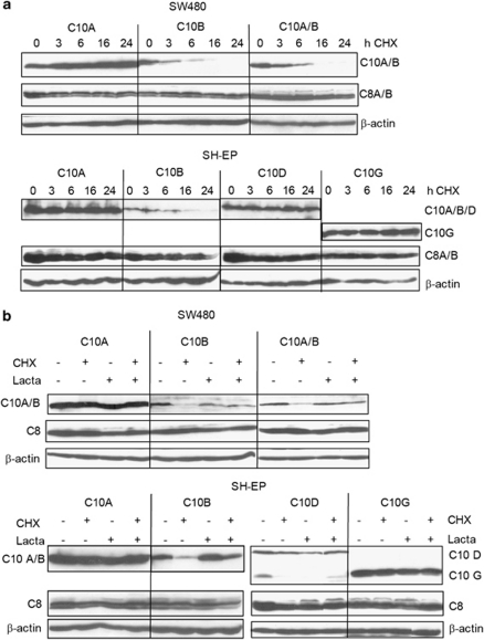
The cysteine protease caspase-8 is an essential executioner of the death receptor (DR) apoptotic pathway. The physiological function of its homologue caspase-10 remains poorly understood, and the ability of caspase-10 to substitute for caspase-8 in the DR apoptotic pathway is still controversial. Here, we analysed the particular contribution of caspase-10 isoforms to DR-mediated apoptosis in neuroblastoma (NB) cells characterised by their resistance to DR signalling. Silencing of caspase-8 in tumour necrosis factor-related apoptosis-inducing ligand (TRAIL)-sensitive NB cells resulted in complete resistance to TRAIL, which could be reverted by overexpression of caspase-10A or -10D. Overexpression experiments in various caspase-8-expressing tumour cells also demonstrated that caspase-10A and -10D isoforms strongly increased TRAIL and FasL sensitivity, whereas caspase-10B or -10G had no effect or were weakly anti-apoptotic. Further investigations revealed that the unique C-terminal end of caspase-10B was responsible for its degradation by the ubiquitin-proteasome pathway and for its lack of pro-apoptotic activity compared with caspase-10A and -10D. These data highlight in several tumour cell types, a differential pro- or anti-apoptotic role for the distinct caspase-10 isoforms in DR signalling, which may be relevant for fine tuning of apoptosis initiation.


Figures

Figure 1
Figure 1
Analysis of the expression levels of caspase-8 and -10 isoforms. (a) Schematic representation of caspase-10 and -8 protein isoforms. Regions encoded by alternative exons are indicated. DED, death effector domain. (b) Whole cell extracts from various cell lines were loaded on a 15% SDS-polyacrylamide gel as described, and analysed by immunoblotting for the expression of caspase-8 and -10 isoforms. The three types of NB cells (S, substrate adherent; I, intermediate; N, neuroblastic) are represented. n.d., not determined. The immunoblot for endogenous caspase-10 detection were revealed using the ECL Advanced western blotting substrate for stronger signal detection. (c) Caspase-8 and -10 mRNA expression levels in tumour cells were compared by semi-quantitative real-time PCR. Plots represent means of three separate experiments
Figure 2
Figure 2
Endogenous caspase-10 levels are not sufficient to initiate TRAIL-mediated apoptosis in NB cells. (a) SH-EP or SK-N-AS cells infected with the empty vector (AB303), a nonspecific shRNA (RNAC), or caspase-8 shRNA (shC8) were analysed by immunoblotting for the presence of caspase-8. β-Actin was used as loading control. (b) Cell viability was measured in SH-EP- and SK-N-AS-AB303 (diamond), -RNAC (square) and -shC8 (triangle) cells treated with increasing amount of TRAIL for 48 h. A representative experiment out of three is shown. (c) Percentage of sub-G1 apoptotic cells detected by the PI staining method after stimulation with TRAIL (100 ng/ml) are indicated, mean of three experiments are shown (***P<0.0001). (d) SH-EP cells were untreated or treated with 100 ng of TRAIL for 16 h. Immunoblotting analysis of caspase-8, -10, -3 and Bid processing. β-Actin was used as loading control, as in further immunoblotting analyses. (e) Hydrolysis of DEVD-pNA was measured in cell lysates from SH-EP and SK-N-AS-infected cells treated with 50 or 250 ng/ml of TRAIL for 16 h, respectively. Relative OD values at 405 nm of stimulated compared with unstimulated cells of one experiment performed in duplicates are represented. (***P<0.0001, **P<0.01). (f) SH-EP cells were transiently transfected with the indicated siRNAs (siNeg, negative control siRNA). Relative expression levels of the indicated mRNA compared with HPRT1 mRNA as measured by real-time PCR are plotted in the graph (upper left panel). Immunoblotting analysis of caspase-10 and -8 expression in SH-EP transfected for 48 h with the indicated siRNAs (bottom left panel). Cell viability of cells treated for 48 h with indicated doses of TRAIL (right panel) is represented (***P<0.0001)
Figure 3
Figure 3
Caspase-10A or -10D isoforms can substitute for caspase-8 in the initiation of the apoptotic cascade. (a) Immunoblotting analysis of caspase-10 expression in IGR-N-91 clones transduced with the empty vector (M) or with vector encoding for caspase-10B, or SH-EP cells (positive control). Cell viability was measured in cells treated with TRAIL (100 ng/ml) for 48 h. (b) Immunoblotting analysis of caspase-8, caspase-10, and β-actin expression in IGR-N-91 cells transduced with the empty vector (Migr) or with vector encoding for caspase-10A or -10D (pool of five clones each). Cell viability was measured in the pool of clones of IGR-N-91-Migr, -C10A and -C10D cells treated with the indicated doses of TRAIL for 48 h. Mean of two experiments performed in quadruplicates are shown (***P<0.0001). Percentage of sub-G1 apoptotic cells after stimulation with TRAIL are indicated, one representative experiment is shown. (c) SH-EP-shC8 cells transduced with the empty vector (Migr) or with vector encoding for caspase-10A or -10D were analysed as in (b). Mean of three experiments performed in quadruplicates are shown for the viability assay (***P<0.0001). One representative experiment is shown for apoptosis analysis
Figure 4
Figure 4
Caspase-10A and -10D have a pro-apoptotic role in NB cells. (a) Immunoblotting analysis of caspase-10 and -8 expressions in SH-EP and SK-N-AS cells transduced with the empty vector (Migr) or with vector encoding for C10A, C10B, C10D or C10G. (b) Cell viability of SH-EP and SK-N-AS transduced cells treated with the indicated doses of TRAIL and anti-Fas antibody for 48 h. Mean of three experiments performed in quadruplicates are shown (***P<0.0001, **P<0.005). (c) Percentage of sub-G1 apoptotic cells after stimulation with 25 ng/ml of TRAIL for SH-EP and 100 ng/ml for SK-N-AS are indicated. A representative experiment is shown
Figure 5
Figure 5
Caspase-10A and -10D have a pro-apoptotic role in SW480 and BJAB cell lines. (a) Immunoblotting analysis of caspase-10 and -8 expressions in SW480, or BJAB cells transduced with the empty Migr vector or with vector encoding for caspase-10A, -B, -D or -G. (b and d) Cell viability of SW480 (b), and BJAB (d) transduced cells treated with the indicated doses of TRAIL and anti-Fas antibody for 48 h. Mean of three experiments performed in quadruplicates are shown (***P<0.0001, *P<0.05). (c) Percentage of sub-G1 apoptotic cells after stimulation with 25 ng/ml of TRAIL and 500 ng/ml of anti-Fas antibody are indicated. A representative experiment is shown
Figure 6
Figure 6
Caspase-10A and -10D overexpression enhances the caspase cascade activation. (a) Immunoblotting analysis of caspases-10, -8, -9, and Bid cleavage in cells untreated or treated with 50 ng/ml of TRAIL for 16 h. (b) Caspase activity was measured in cell lysates from SH-EP (upper panels) and SW480 (lower panels) transduced cells treated with 50 ng/ml TRAIL for 8 h or 25 ng/ml of TRAIL for 16 h, respectively. Relative OD at 405 nm of stimulated compared with unstimulated cells of two experiments performed in duplicates are represented (***P<0.0001, **P<0.001, *P<0.05)
Figure 7
Figure 7
The C-terminal end of caspase-10B is responsible for its lack of activity in TRAIL and Fas sensitisation. (a) Up: schematic representation of the C10A/B fusion protein. Down: whole cell extracts from SW480-Migr, -C10A, -C10B or -C10A/B cells were analysed by immunoblotting for the presence of caspase-10 and β-actin. (b) Cell viability measured in SW480 cells treated with the indicated doses of TRAIL and anti-Fas antibody for 48 h. Mean of two experiments performed in quadruplicates are shown (***P<0.0005, **P<0.005, *P<0.05). (c) Percentage of sub-G1 apoptotic cells after stimulation with 50 ng/ml of TRAIL and 500 ng/ml of anti-Fas antibody are indicated. Mean of two experiments are shown (*P<0.05). (d) Whole cell extracts from SW480-C10A, -C10B, -C10A/B cells, untreated or treated with 50 ng/ml of TRAIL for various time length, or with 200 ng/ml of TRAIL for 8 h, were analysed by immunoblotting for the cleavage of caspases-10, -8, -3, and Bid
Figure 8
Figure 8
Caspase-10B is highly unstable, in contrast to other caspase-10 isoforms. (a) Whole cell extracts from SW480 and SH-EP cells overexpressing the indicated caspase-10 isoforms, untreated or treated with 20 ug/ml of CHX for various times, were analysed by immunoblotting for the presence of caspases-10, -8 and β-actin. (b) Whole cell extracts from SW480 and SH-EP-infected cells untreated or treated with 20 ug/ml of CHX, with or without lactacystin (10 μM for SH-EP or 50 μM for SW480) for 16 h, were analysed as in (a)

Similar articles

Cited by

References

    1. Hanahan D, Weinberg RA. The hallmarks of cancer. Cell. 2000;100:57–70. - PubMed
    1. Hengartner MO. The biochemistry of apoptosis. Nature. 2000;407:770–776. - PubMed
    1. Fulda S. Caspase-8 in cancer biology and therapy. Cancer Lett. 2009;281:128–133. - PubMed
    1. Wang J, Chun HJ, Wong W, Spencer DM, Lenardo MJ. Caspase-10 is an initiator caspase in death receptor signaling. Proc Natl Acad Sci USA. 2001;98:13884–13888. - PMC - PubMed
    1. Milhas D, Cuvillier O, Therville N, Clave P, Thomsen M, Levade T, et al. Caspase-10 triggers Bid cleavage and caspase cascade activation in FasL-induced apoptosis. J Biol Chem. 2005;280:19836–19842. - PubMed

Publication types

MeSH terms

LinkOut - more resources