NK-cell enteropathy: a benign NK-cell lymphoproliferative disease mimicking intestinal lymphoma: clinicopathologic features and follow-up in a unique case series
- PMID: 20966166
- PMCID: PMC3056587
- DOI: 10.1182/blood-2010-08-302737
NK-cell enteropathy: a benign NK-cell lymphoproliferative disease mimicking intestinal lymphoma: clinicopathologic features and follow-up in a unique case series
Abstract
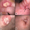
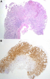
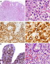
Intestinal T-cell and natural killer (NK)-cell lymphomas are clinically aggressive and can be challenging to diagnose in small endoscopic biopsies. We describe 8 patients in whom atypical NK-cell lymphoproliferative lesions mimicked NK- or T-cell lymphoma. The patients (2 men; 6 women; ages 27-68 years) presented with vague gastrointestinal symptoms with lesions involving stomach, duodenum, small intestine, and colon. At endoscopy, the lesions exhibited superficial ulceration, edema, and hemorrhage. Biopsies revealed a mucosal infiltrate of atypical cells with an NK-cell phenotype (CD56(+)/TIA-1(+)/Granzyme B(+)/cCD3(+)), which displaced but did not invade the glandular epithelium. Epstein-Barr virus-encoded RNA in situ hybridization was negative, and T-cell receptor-γ gene rearrangement showed no evidence of a clonal process. Based on an original diagnosis of lymphoma, 3 patients received aggressive chemotherapy followed by autologous bone marrow transplantation in 2. Five patients were followed without treatment. However, no patient developed progressive disease or died of lymphoma (median follow-up, 30 months). Repeat endoscopies in 6 of 8 patients showed persistence or recurrence of superficial gastrointestinal lesions. This unique entity mimics intestinal and NK-/T-cell lymphomas on endoscopic biopsies and can result in erroneous diagnosis, leading to aggressive chemotherapy. We propose the term "NK-cell enteropathy" for this syndrome of as yet unknown etiology.
Figures


Comment in
-
A sheep in wolf's clothing.Blood. 2011 Feb 3;117(5):1438-9. doi: 10.1182/blood-2010-11-319913. Blood. 2011. PMID: 21292781
Similar articles
-
CD8+, CD56+ (natural killer-like) T-cell lymphoma involving the small intestine with no evidence of enteropathy: clinicopathology and molecular study of five Japanese patients.Pathol Int. 2008 Oct;58(10):626-34. doi: 10.1111/j.1440-1827.2008.02281.x. Pathol Int. 2008. PMID: 18801082
-
Primary CD56 positive lymphomas of the gastrointestinal tract.Cancer. 2001 Feb 1;91(3):525-33. doi: 10.1002/1097-0142(20010201)91:3<525::aid-cncr1030>3.0.co;2-u. Cancer. 2001. PMID: 11169934 Review.
-
[Ocular natural killer/T cell lymphoma: a clinicopathologic analysis].Zhonghua Yan Ke Za Zhi. 2019 May 11;55(5):374-380. doi: 10.3760/cma.j.issn.0412-4081.2019.05.012. Zhonghua Yan Ke Za Zhi. 2019. PMID: 31137150 Chinese.
-
T-cell and T/natural killer-cell lymphomas involving ocular and ocular adnexal tissues: a clinicopathologic, immunohistochemical, and molecular study of seven cases.Ophthalmology. 1999 Nov;106(11):2109-20. doi: 10.1016/S0161-6420(99)90492-X. Ophthalmology. 1999. PMID: 10571346
-
Nonnasal lymphoma expressing the natural killer cell marker CD56: a clinicopathologic study of 49 cases of an uncommon aggressive neoplasm.Blood. 1997 Jun 15;89(12):4501-13. Blood. 1997. PMID: 9192774 Review.
Cited by
-
Clinical and genetic profile of Chinese patients with indolent natural killer-cell lymphoproliferative disorder of the gastrointestinal tract.Neoplasia. 2024 Nov;57:101048. doi: 10.1016/j.neo.2024.101048. Epub 2024 Sep 13. Neoplasia. 2024. PMID: 39276532 Free PMC article.
-
Mature B, T and NK-cell, plasma cell and histiocytic/dendritic cell neoplasms: classification according to the World Health Organization and International Consensus Classification.J Hematol Oncol. 2024 Jul 8;17(1):51. doi: 10.1186/s13045-024-01570-5. J Hematol Oncol. 2024. PMID: 38978094 Free PMC article. Review.
-
Intestinal T-cell lymphomas NOS presenting as a polypoidal lesion: A case report.Medicine (Baltimore). 2024 Jun 7;103(23):e38465. doi: 10.1097/MD.0000000000038465. Medicine (Baltimore). 2024. PMID: 38847694 Free PMC article.
-
What is new in the 5th edition of the World Health Organization classification of mature B and T/NK cell tumors and stromal neoplasms?J Hematop. 2024 Jun;17(2):71-89. doi: 10.1007/s12308-024-00585-8. Epub 2024 Apr 29. J Hematop. 2024. PMID: 38683440 Review.
-
Biology and genetics of extranodal mature T-cell and NKcell lymphomas and lymphoproliferative disorders.Haematologica. 2023 Dec 1;108(12):3261-3277. doi: 10.3324/haematol.2023.282718. Haematologica. 2023. PMID: 38037802 Free PMC article. Review.
References
-
- Chan JKC, Quintanilla-Martinez L, Ferry JA, Peh SC. Extranodal NK-/T-cell lymphoma, nasal type. In: Swerdlow S, Campo E, Harris NL, et al., editors. WHO Classification of Tumours of Haematopoietic and Lymphoid Tissues. 4th ed. Lyon, France: International Agency for Research on Cancer Press; 2008. pp. 285–288.
-
- Meijer JW, Mulder CJ, Goerres MG, Boot H, Schweizer JJ. Coeliac disease and (extra)intestinal T-cell lymphomas: definition, diagnosis and treatment. Scand J Gastroenterol Suppl. 2004;241:78–84. - PubMed
-
- van de Water JM, Cillessen SA, Visser OJ, Verbeek WH, Meijer CJ, Mulder CJ. Enteropathy associated T-cell lymphoma and its precursor lesions. Best Pract Res Clin Gastroenterol. 2010;24(1):43–56. - PubMed
-
- Kwong YL, Chan AC, Liang R, et al. CD56+ NK lymphomas: clinicopathological features and prognosis. Br J Haematol. 1997;97(4):821–829. - PubMed
-
- Au WY, Weisenburger DD, Intragumtornchai T, et al. Clinical differences between nasal and extranasal natural killer/T-cell lymphoma: a study of 136 cases from the International Peripheral T-Cell Lymphoma Project. Blood. 2009;113(17):3931–3937. - PubMed
Publication types
MeSH terms
Substances
Grants and funding
LinkOut - more resources
Full Text Sources
Research Materials
Miscellaneous

