Network organization of the human autophagy system
- PMID: 20562859
- PMCID: PMC2901998
- DOI: 10.1038/nature09204
Network organization of the human autophagy system
Abstract
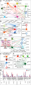
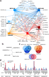
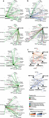
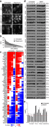
Autophagy, the process by which proteins and organelles are sequestered in autophagosomal vesicles and delivered to the lysosome/vacuole for degradation, provides a primary route for turnover of stable and defective cellular proteins. Defects in this system are linked with numerous human diseases. Although conserved protein kinase, lipid kinase and ubiquitin-like protein conjugation subnetworks controlling autophagosome formation and cargo recruitment have been defined, our understanding of the global organization of this system is limited. Here we report a proteomic analysis of the autophagy interaction network in human cells under conditions of ongoing (basal) autophagy, revealing a network of 751 interactions among 409 candidate interacting proteins with extensive connectivity among subnetworks. Many new autophagy interaction network components have roles in vesicle trafficking, protein or lipid phosphorylation and protein ubiquitination, and affect autophagosome number or flux when depleted by RNA interference. The six ATG8 orthologues in humans (MAP1LC3/GABARAP proteins) interact with a cohort of 67 proteins, with extensive binding partner overlap between family members, and frequent involvement of a conserved surface on ATG8 proteins known to interact with LC3-interacting regions in partner proteins. These studies provide a global view of the mammalian autophagy interaction landscape and a resource for mechanistic analysis of this critical protein homeostasis pathway.
Figures


Comment in
-
Autophagy: Snapshot of the network.Nature. 2010 Jul 1;466(7302):38-40. doi: 10.1038/466038a. Nature. 2010. PMID: 20596005 Free PMC article.
Similar articles
-
The LC3 interactome at a glance.J Cell Sci. 2014 Jan 1;127(Pt 1):3-9. doi: 10.1242/jcs.140426. Epub 2013 Dec 17. J Cell Sci. 2014. PMID: 24345374 Review.
-
Quantitative proteomics identifies NCOA4 as the cargo receptor mediating ferritinophagy.Nature. 2014 May 1;509(7498):105-9. doi: 10.1038/nature13148. Epub 2014 Mar 30. Nature. 2014. PMID: 24695223 Free PMC article.
-
Rab GTPase-activating proteins in autophagy: regulation of endocytic and autophagy pathways by direct binding to human ATG8 modifiers.Mol Cell Biol. 2012 May;32(9):1733-44. doi: 10.1128/MCB.06717-11. Epub 2012 Feb 21. Mol Cell Biol. 2012. PMID: 22354992 Free PMC article.
-
Autophagy: Snapshot of the network.Nature. 2010 Jul 1;466(7302):38-40. doi: 10.1038/466038a. Nature. 2010. PMID: 20596005 Free PMC article.
-
LC3/GABARAP family proteins: autophagy-(un)related functions.FASEB J. 2016 Dec;30(12):3961-3978. doi: 10.1096/fj.201600698R. Epub 2016 Sep 6. FASEB J. 2016. PMID: 27601442 Review.
Cited by
-
Dual proteome-scale networks reveal cell-specific remodeling of the human interactome.Cell. 2021 May 27;184(11):3022-3040.e28. doi: 10.1016/j.cell.2021.04.011. Epub 2021 May 6. Cell. 2021. PMID: 33961781 Free PMC article.
-
Tepsin binds LC3B to promote ATG9A trafficking and delivery.Mol Biol Cell. 2024 Apr 1;35(4):ar56. doi: 10.1091/mbc.E23-09-0359-T. Epub 2024 Feb 21. Mol Biol Cell. 2024. PMID: 38381558 Free PMC article.
-
Drosophila protein interaction map (DPiM): a paradigm for metazoan protein complex interactions.Fly (Austin). 2012 Oct-Dec;6(4):246-53. doi: 10.4161/fly.22108. Fly (Austin). 2012. PMID: 23222005 Free PMC article.
-
Regulation of the transcription factor EB-PGC1α axis by beclin-1 controls mitochondrial quality and cardiomyocyte death under stress.Mol Cell Biol. 2015 Mar;35(6):956-76. doi: 10.1128/MCB.01091-14. Epub 2015 Jan 5. Mol Cell Biol. 2015. PMID: 25561470 Free PMC article.
-
Bulk and selective autophagy cooperate to remodel a fungal proteome in response to changing nutrient availability.bioRxiv [Preprint]. 2024 Sep 26:2024.09.24.614842. doi: 10.1101/2024.09.24.614842. bioRxiv. 2024. PMID: 39386609 Free PMC article. Preprint.
References
Publication types
MeSH terms
Substances
Grants and funding
LinkOut - more resources
Full Text Sources
Other Literature Sources
Molecular Biology Databases

