

Conversion of 5-methylcytosine to 5-hydroxymethylcytosine in mammalian DNA by MLL partner TET1
- PMID: 19372391
- PMCID: PMC2715015
- DOI: 10.1126/science.1170116
Conversion of 5-methylcytosine to 5-hydroxymethylcytosine in mammalian DNA by MLL partner TET1
Abstract
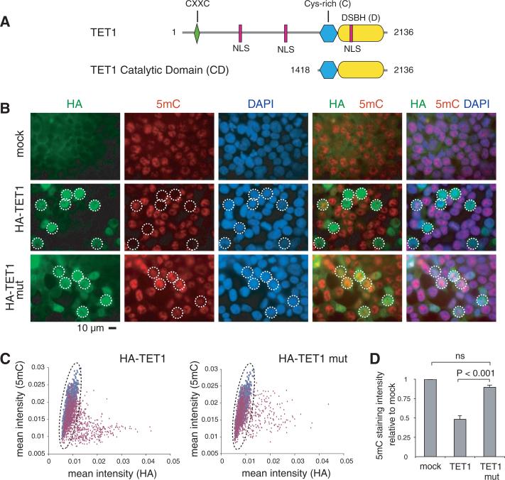
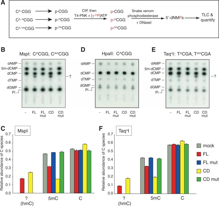
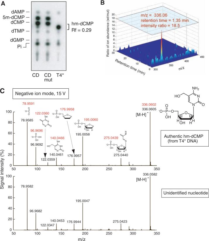
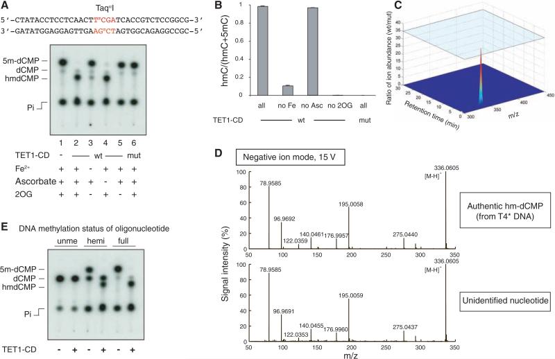
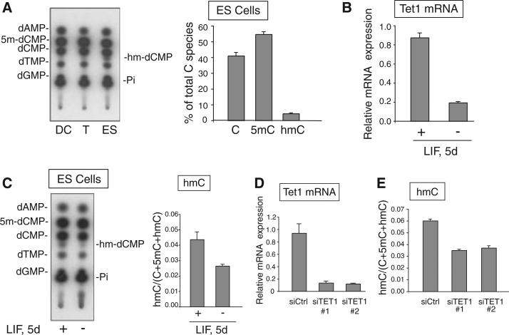
DNA cytosine methylation is crucial for retrotransposon silencing and mammalian development. In a computational search for enzymes that could modify 5-methylcytosine (5mC), we identified TET proteins as mammalian homologs of the trypanosome proteins JBP1 and JBP2, which have been proposed to oxidize the 5-methyl group of thymine. We show here that TET1, a fusion partner of the MLL gene in acute myeloid leukemia, is a 2-oxoglutarate (2OG)- and Fe(II)-dependent enzyme that catalyzes conversion of 5mC to 5-hydroxymethylcytosine (hmC) in cultured cells and in vitro. hmC is present in the genome of mouse embryonic stem cells, and hmC levels decrease upon RNA interference-mediated depletion of TET1. Thus, TET proteins have potential roles in epigenetic regulation through modification of 5mC to hmC.
Figures
Comment in
-
5-hydroxymethylcytosine, a modified mammalian DNA base with a potential regulatory role.Epigenomics. 2009 Oct;1(1):21-2. doi: 10.2217/epi.09.18. Epigenomics. 2009. PMID: 22122633 No abstract available.
Similar articles
-
Structure of a Naegleria Tet-like dioxygenase in complex with 5-methylcytosine DNA.Nature. 2014 Feb 20;506(7488):391-5. doi: 10.1038/nature12905. Epub 2013 Dec 25. Nature. 2014. PMID: 24390346 Free PMC article.
-
Genome-wide regulation of 5hmC, 5mC, and gene expression by Tet1 hydroxylase in mouse embryonic stem cells.Mol Cell. 2011 May 20;42(4):451-64. doi: 10.1016/j.molcel.2011.04.005. Epub 2011 Apr 21. Mol Cell. 2011. PMID: 21514197 Free PMC article.
-
Tet-mediated formation of 5-carboxylcytosine and its excision by TDG in mammalian DNA.Science. 2011 Sep 2;333(6047):1303-7. doi: 10.1126/science.1210944. Epub 2011 Aug 4. Science. 2011. PMID: 21817016 Free PMC article.
-
Structure and Function of TET Enzymes.Adv Exp Med Biol. 2016;945:275-302. doi: 10.1007/978-3-319-43624-1_12. Adv Exp Med Biol. 2016. PMID: 27826843 Review.
-
Epigenetic Function of TET Family, 5-Methylcytosine, and 5-Hydroxymethylcytosine in Hematologic Malignancies.Oncol Res Treat. 2019;42(6):309-318. doi: 10.1159/000498947. Epub 2019 May 3. Oncol Res Treat. 2019. PMID: 31055566 Review.
Cited by
-
Insights into the epigenetic mechanisms controlling pancreatic carcinogenesis.Cancer Lett. 2013 Jan 28;328(2):212-21. doi: 10.1016/j.canlet.2012.10.005. Epub 2012 Oct 13. Cancer Lett. 2013. PMID: 23073473 Free PMC article. Review.
-
Tissue-specific effects of genetic and epigenetic variation on gene regulation and splicing.PLoS Genet. 2015 Jan 29;11(1):e1004958. doi: 10.1371/journal.pgen.1004958. eCollection 2015 Jan. PLoS Genet. 2015. PMID: 25634236 Free PMC article.
-
TET2 mediated demethylation is involved in the protective effect of triptolide on podocytes.Am J Transl Res. 2021 Mar 15;13(3):1233-1244. eCollection 2021. Am J Transl Res. 2021. PMID: 33841652 Free PMC article.
-
Epigenetic regulation of monoallelic rearrangement (allelic exclusion) of antigen receptor genes.Front Immunol. 2014 Dec 5;5:625. doi: 10.3389/fimmu.2014.00625. eCollection 2014. Front Immunol. 2014. PMID: 25538709 Free PMC article. Review.
-
Methods for cancer epigenome analysis.Adv Exp Med Biol. 2013;754:313-38. doi: 10.1007/978-1-4419-9967-2_15. Adv Exp Med Biol. 2013. PMID: 22956508 Free PMC article. Review.
References
Publication types
MeSH terms
Substances
Grants and funding
LinkOut - more resources
Full Text Sources
Other Literature Sources
Molecular Biology Databases
Research Materials
