

Heme oxygenase-1, a critical arbitrator of cell death pathways in lung injury and disease
- PMID: 19362144
- PMCID: PMC3078523
- DOI: 10.1016/j.freeradbiomed.2009.04.007
Heme oxygenase-1, a critical arbitrator of cell death pathways in lung injury and disease
Abstract
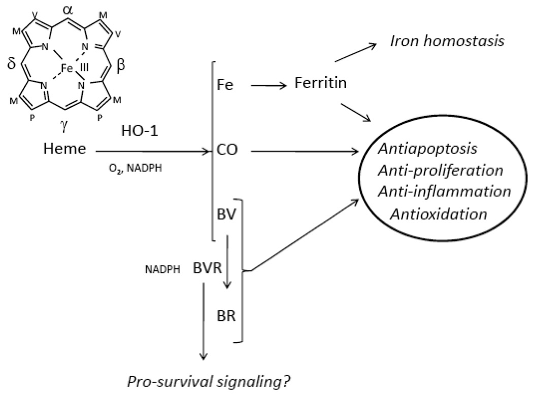
Increases in cell death by programmed (i.e., apoptosis, autophagy) or nonprogrammed mechanisms (i.e., necrosis) occur during tissue injury and may contribute to the etiology of several pulmonary or vascular disease states. The low-molecular-weight stress protein heme oxygenase-1 (HO-1) confers cytoprotection against cell death in various models of lung and vascular injury by inhibiting apoptosis, inflammation, and cell proliferation. HO-1 serves a vital metabolic function as the rate-limiting step in the heme degradation pathway and in the maintenance of iron homeostasis. The transcriptional induction of HO-1 occurs in response to multiple forms of chemical and physical cellular stress. The cytoprotective functions of HO-1 may be attributed to heme turnover, as well as to beneficial properties of its enzymatic reaction products: biliverdin-IXalpha, iron, and carbon monoxide (CO). Recent studies have demonstrated that HO-1 or CO inhibits stress-induced extrinsic and intrinsic apoptotic pathways in vitro. A variety of signaling molecules have been implicated in the cytoprotection conferred by HO-1/CO, including autophagic proteins, p38 mitogen-activated protein kinase, signal transducer and activator of transcription proteins, nuclear factor-kappaB, phosphatidylinositol 3-kinase/Akt, and others. Enhanced HO-1 expression or the pharmacological application of HO end-products affords protection in preclinical models of tissue injury, including experimental and transplant-associated ischemia/reperfusion injury, promising potential future therapeutic applications.
Figures

Similar articles
-
Heme oxygenase-1 and gut ischemia/reperfusion injury: A short review.World J Gastroenterol. 2013 Jun 21;19(23):3555-61. doi: 10.3748/wjg.v19.i23.3555. World J Gastroenterol. 2013. PMID: 23801856 Free PMC article. Review.
-
Heme oxygenase-1/carbon monoxide: from metabolism to molecular therapy.Am J Respir Cell Mol Biol. 2009 Sep;41(3):251-60. doi: 10.1165/rcmb.2009-0170TR. Epub 2009 Jul 17. Am J Respir Cell Mol Biol. 2009. PMID: 19617398 Free PMC article. Review.
-
Heme oxygenase system in ischemia and reperfusion injury.Ann Transplant. 2004;9(1):84-7. Ann Transplant. 2004. PMID: 15478901 Review.
-
Products of heme oxygenase and their potential therapeutic applications.Am J Physiol Renal Physiol. 2006 Mar;290(3):F563-71. doi: 10.1152/ajprenal.00220.2005. Am J Physiol Renal Physiol. 2006. PMID: 16461755 Review.
-
Therapeutic Potential of Heme Oxygenase-1/carbon Monoxide System Against Ischemia-Reperfusion Injury.Curr Pharm Des. 2017;23(26):3884-3898. doi: 10.2174/1381612823666170413122439. Curr Pharm Des. 2017. PMID: 28412905 Review.
Cited by
-
Si-Based Hydrogen-Producing Nanoagent Protects Fetuses From Miscarriage Caused by Mother-to-Child Transmission.Front Med Technol. 2021 May 13;3:665506. doi: 10.3389/fmedt.2021.665506. eCollection 2021. Front Med Technol. 2021. PMID: 35047922 Free PMC article.
-
Protective role of heme oxygenase-1 against inflammation in atherosclerosis.Front Biosci (Landmark Ed). 2011 Jun 1;16(6):2372-88. doi: 10.2741/3860. Front Biosci (Landmark Ed). 2011. PMID: 21622183 Free PMC article. Review.
-
BMP-6 inhibits MMP-9 expression by regulating heme oxygenase-1 in MCF-7 breast cancer cells.J Cancer Res Clin Oncol. 2011 Jun;137(6):985-95. doi: 10.1007/s00432-010-0963-z. Epub 2010 Dec 7. J Cancer Res Clin Oncol. 2011. PMID: 21136273
-
Ferritinophagy drives uropathogenic Escherichia coli persistence in bladder epithelial cells.Autophagy. 2016 May 3;12(5):850-63. doi: 10.1080/15548627.2016.1160176. Autophagy. 2016. PMID: 27002654 Free PMC article.
-
KRIT1 loss-of-function induces a chronic Nrf2-mediated adaptive homeostasis that sensitizes cells to oxidative stress: Implication for Cerebral Cavernous Malformation disease.Free Radic Biol Med. 2018 Feb 1;115:202-218. doi: 10.1016/j.freeradbiomed.2017.11.014. Epub 2017 Nov 21. Free Radic Biol Med. 2018. PMID: 29170092 Free PMC article.
References
-
- Ryter SW, Alam J, Choi AM. Heme oxygenase-1/carbon monoxide: from basic science to therapeutic applications. Physiol. Rev. 2006;86:583–650. - PubMed
-
- Morse D, Choi AM. Heme oxygenase-1: the “emerging molecule” has arrived. Am. J. Respir. Cell Mol. Biol. 2002;27:8–16. - PubMed
-
- Tenhunen R, Marver HS, Schmid R. Microsomal heme oxygenase. Characterization of the enzyme. J. Biol. Chem. 1969;244:6388–6394. - PubMed
Publication types
MeSH terms
Substances
Grants and funding
LinkOut - more resources
Full Text Sources
Other Literature Sources
