

Chemoresistant colorectal cancer cells, the cancer stem cell phenotype, and increased sensitivity to insulin-like growth factor-I receptor inhibition
- PMID: 19244128
- PMCID: PMC3198868
- DOI: 10.1158/0008-5472.CAN-08-2023
Chemoresistant colorectal cancer cells, the cancer stem cell phenotype, and increased sensitivity to insulin-like growth factor-I receptor inhibition
Abstract
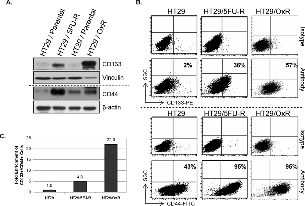
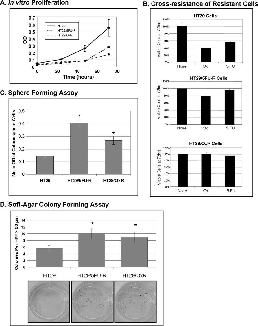
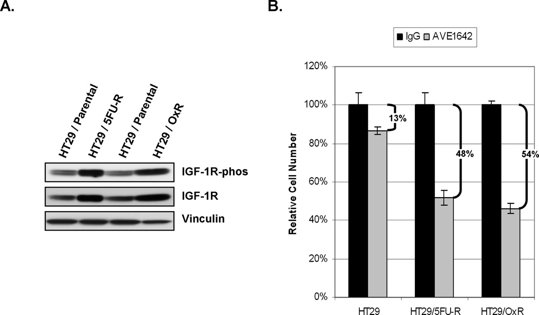
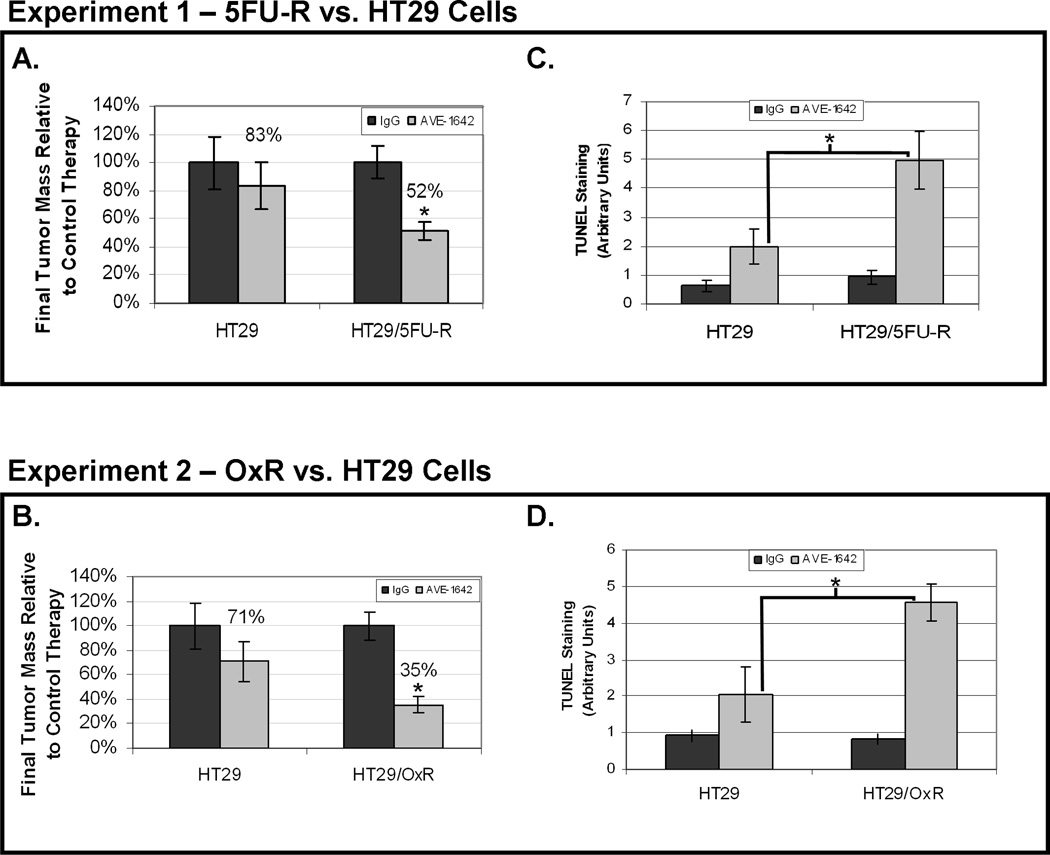
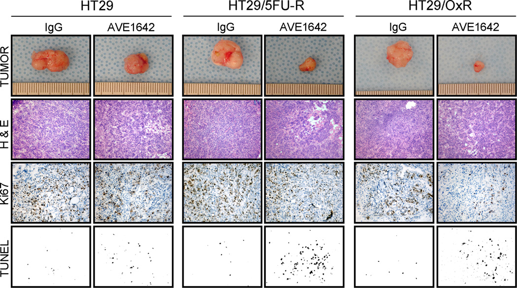
5-Fluorouracil (5FU) and oxaliplatin are standard therapy for metastatic colorectal cancer (CRC), but the development of chemoresistance is inevitable. Because cancer stem cells (CSC) are hypothesized to be chemoresistant, we investigated CSC properties in newly developed chemoresistant CRC cell lines and sought to identify targets for therapy. The human CRC cell line HT29 was exposed to increasing doses of 5FU (HT29/5FU-R) or oxaliplatin (HT29/OxR) to achieve resistance at clinically relevant doses. Western blotting and flow cytometry were done to determine molecular alterations. The insulin-like growth factor-I receptor (IGF-IR) monoclonal antibody (mAb) AVE-1642 was used to inhibit signaling in vitro and in vivo using murine xenograft models. HT29/5FU-R and HT29/OxR showed 16- to 30-fold enrichment of CD133(+) cells and 2-fold enrichment of CD44(+) cells (putative CRC CSC markers). Resistant cells were enriched 5- to 22-fold for double-positive (CD133(+)/CD44(+)) cells. Consistent with the CSC phenotype, resistant cells exhibited a decrease in cellular proliferation in vitro (47-59%; P < 0.05). Phosphorylated and total IGF-IR levels were increased in resistant cell lines. HT29/5FU-R and HT29/OxR cells were approximately 5-fold more responsive to IGF-IR inhibition relative to parental cells (P < 0.01) in vitro. Tumors derived from HT29/OxR cells showed significantly greater growth inhibition in response to an IGF-IR mAb than did parental cells (P < 0.05). Chemoresistant CRC cells are enriched for CSC markers and the CSC phenotype. Chemotherapy-induced IGF-IR activation provided for enhanced sensitivity to IGF-IR-targeted therapy. Identification of CSC targets presents a novel therapeutic approach in this disease.
Figures
Similar articles
-
Anti-EGFR antibody sensitizes colorectal cancer stem-like cells to Fluorouracil-induced apoptosis by affecting autophagy.Oncotarget. 2016 Dec 6;7(49):81402-81409. doi: 10.18632/oncotarget.13233. Oncotarget. 2016. PMID: 27833077 Free PMC article.
-
Colorectal cancer stem cell and chemoresistant colorectal cancer cell phenotypes and increased sensitivity to Notch pathway inhibitor.Mol Med Rep. 2015 Aug;12(2):2417-24. doi: 10.3892/mmr.2015.3694. Epub 2015 Apr 28. Mol Med Rep. 2015. PMID: 25936357 Free PMC article.
-
CD133+, CD166+CD44+, and CD24+CD44+ phenotypes fail to reliably identify cell populations with cancer stem cell functional features in established human colorectal cancer cell lines.Stem Cells Transl Med. 2012 Aug;1(8):592-603. doi: 10.5966/sctm.2012-0003. Epub 2012 Aug 6. Stem Cells Transl Med. 2012. PMID: 23197865 Free PMC article.
-
Targeting Colorectal Cancer Stem Cells as an Effective Treatment for Colorectal Cancer.Technol Cancer Res Treat. 2020 Jan-Dec;19:1533033819892261. doi: 10.1177/1533033819892261. Technol Cancer Res Treat. 2020. PMID: 32748700 Free PMC article. Review.
-
Lung cancer stem cell: fancy conceptual model of tumor biology or cornerstone of a forthcoming therapeutic breakthrough?J Thorac Oncol. 2014 Jan;9(1):7-17. doi: 10.1097/JTO.0000000000000028. J Thorac Oncol. 2014. PMID: 24346089 Review.
Cited by
-
Current understanding of epigenetics mechanism as a novel target in reducing cancer stem cells resistance.Clin Epigenetics. 2021 May 29;13(1):120. doi: 10.1186/s13148-021-01107-4. Clin Epigenetics. 2021. PMID: 34051847 Free PMC article. Review.
-
A Proteomic Investigation to Discover Candidate Proteins Involved in Novel Mechanisms of 5-Fluorouracil Resistance in Colorectal Cancer.Cells. 2024 Feb 14;13(4):342. doi: 10.3390/cells13040342. Cells. 2024. PMID: 38391955 Free PMC article.
-
Doxorubicin-enriched, ALDH(br) mouse breast cancer stem cells are treatable to oncolytic herpes simplex virus type 1.BMC Cancer. 2012 Nov 23;12:549. doi: 10.1186/1471-2407-12-549. BMC Cancer. 2012. PMID: 23176143 Free PMC article.
-
Drug resistance mechanisms of cancer stem-like cells and their therapeutic potential as drug targets.Cancer Drug Resist. 2019 Sep 19;2(3):457-470. doi: 10.20517/cdr.2019.36. eCollection 2019. Cancer Drug Resist. 2019. PMID: 35582573 Free PMC article. Review.
-
The emerging role of insulin and insulin-like growth factor signaling in cancer stem cells.Front Endocrinol (Lausanne). 2014 Feb 4;5:10. doi: 10.3389/fendo.2014.00010. eCollection 2014. Front Endocrinol (Lausanne). 2014. PMID: 24550888 Free PMC article. Review.
References
-
- Jemal A, Siegel R, Ward E, et al. Cancer statistics, 2008. CA Cancer J Clin. 2008;58(2):71–96. - PubMed
-
- Goldberg RM, Rothenberg ML, Van Cutsem E, et al. The continuum of care: a paradigm for the management of metastatic colorectal cancer. Oncologist. 2007;12(1):38–50. - PubMed
-
- Hurwitz H, Fehrenbacher L, Novotny W, et al. Bevacizumab plus irinotecan, fluorouracil, and leucovorin for metastatic colorectal cancer. N Engl J Med. 2004;350(23):2335–2342. - PubMed
-
- Walko CM, Lindley C. Capecitabine: a review. Clin Ther. 2005;27(1):23–44. - PubMed
-
- Kelland L. The resurgence of platinum-based cancer chemotherapy. Nat Rev Cancer. 2007;7(8):573–584. - PubMed
Publication types
MeSH terms
Substances
Grants and funding
LinkOut - more resources
Full Text Sources
Other Literature Sources
Medical
Research Materials
Miscellaneous
