Department of Diagnostic Radiology and Nuclear Medicine
Since German physicist Wilhelm Conrad Roentgen discovered the x-ray in 1895, radiology has become an integral part of our healthcare delivery system. With advances in technology, radiologic studies now establish or verify the diagnosis in three out of four cases of organic disease.
The development and integration of nuclear medicine, ultrasonography, computed tomography and magnetic resonance imaging (MRI) has provided diagnostic imaging with an even more central role in diagnosis and selected (interventional) therapeutic procedures. The radiology department at the University of Maryland has state-of-the-art facilities and cutting-edge technologies, making it one of the most sophisticated in the world.
Research Interests
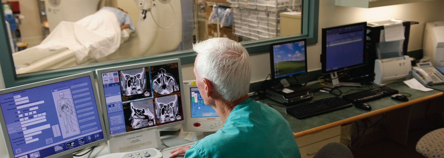
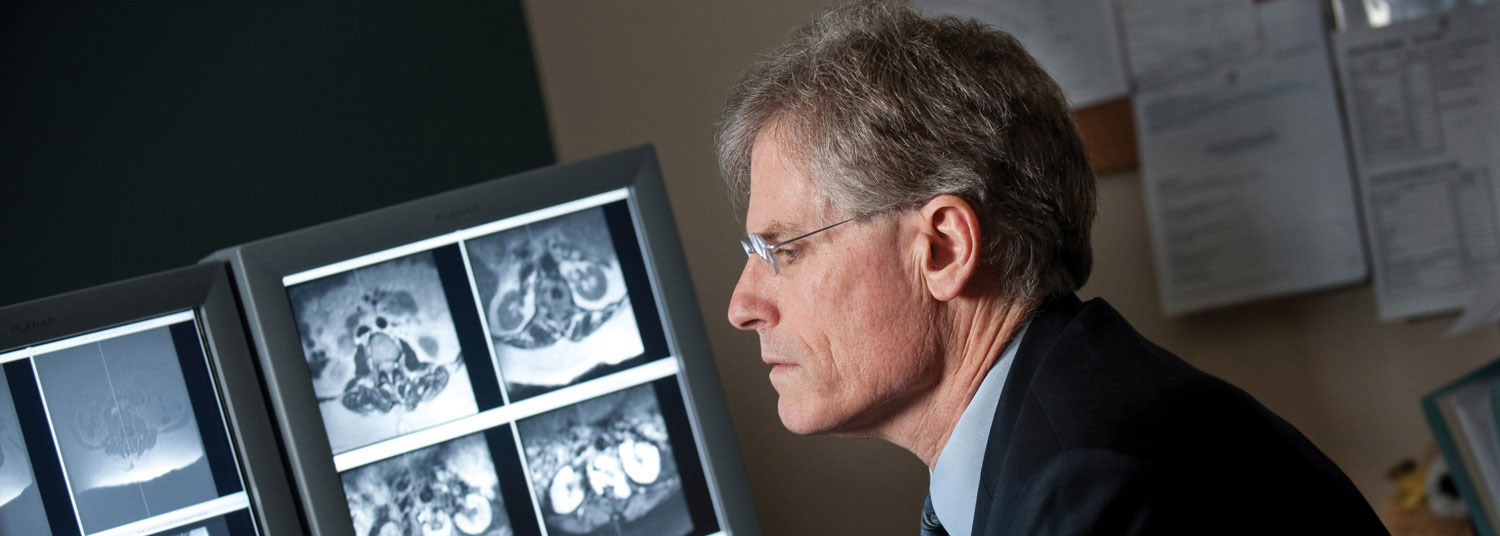
Clinical research is the main focus of departmental research activity. Multiple divisions within the department are pursuing a wide variety of research in state-of-the-art technologies such as spiral CT, MR imaging, SPECT imaging, teleradiology, and picture-archiving and communications system (PACS).
Recently, the department has become among the first in the nation to obtain CT fluoroscopy and portable CT. Specific projects include the evaluation of interventional and non-interventional applications of CT fluoroscopy, assessment of MR pulse sequences to improve diagnosis, use of spiral CT to decrease the intravenous contrast dose, and a comparison of the quality of conventional and PACS images. A complete PACS system is installed in University Hospital and the Baltimore VA Hospital.
The department is organized into the subspecialty sections of abdominal imaging, angiography/interventional radiology, chest radiology, musculoskeletal radiology, neuroradiology, nuclear medicine, pediatric radiology, trauma radiology and ultrasonography. The subspecialty organization and multiple interdepartmental conferences facilitate collaboration with diverse clinical specialties.
Current projects include cooperative studies with physicians in the University of Maryland Greenebaum Comprehensive Cancer Center, MR evaluation of renal-pancreas transplants and CT assessment of patients undergoing lung volume reduction surgery.
Other projects are underway in cooperation with MIEMSS physicians, evaluating the usefulness of CT and MRI in the diagnosis of multiple visceral and skeletal trauma, particularly involving the pelvis and acetabuli. Multiple cooperative cardiovascular nuclear medicine studies are progressing with the department of medicine's division of cardiology.
Center for Advanced Imaging Research (CAIR)
Housed in the new HSF-III facility at the University of Maryland School of Medicine, the CAIR was created in 2018 to consolidate major research resources in the Department of Diagnostic Radiology and Nuclear Medicine.
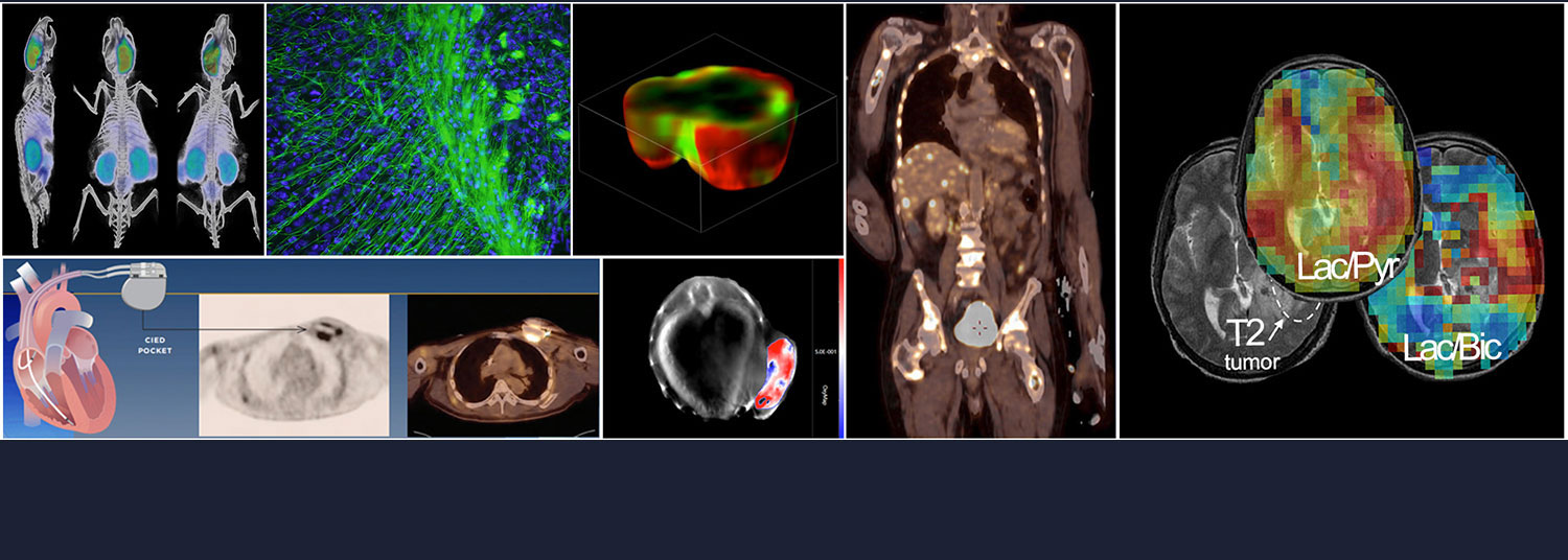
CAIR focuses on developing and using cutting edge imaging modalities such as advanced magnetic resonance imaging (MRI), hyperpolarized imaging, positron-emission tomography (PET), and focused ultrasound. These techniques are used in groundbreaking ways to help scientists understand disease processes and advance diagnostic imaging, with the ultimate goal of improving treatment to patients in the clinical setting.
Education
Undergraduate Medical Program
The department of radiology offers the medical student an opportunity to acquire a broad base of knowledge related to imaging in almost all aspects of medicine. Formal instruction begins during anatomy in the first year and pathology in the second year. During the third or fourth year, students may elect to take the basic radiology course (RADI 540). The curriculum is supplemented with small group case discussions with the faculty and contact through interdepartmental rounds and conferences involving radiology during clinical rotations.
Third or Fourth Years Basic Radiology Elective, RADI 540
Small groups of students are assigned for a period of four weeks to the department of radiology. Groups are subdivided to allow individual instruction as the student rotates through a series of observation periods in selected subspecialties within the department. Students also receive an introduction to the department of radiation oncology. Reading assignments, slide-tape exercises, a student teaching file and seminars form the core of the learning experience. Students attend departmental conferences and joint conferences with other departments. An objective final examination is included in the course.
Third and Fourth Years Subspecialty Radiology Elective
Students learn more about the appropriate use of diagnostic imaging and interpreting images. The curriculum is flexible, tailored to the needs of the student's career choice. Students are expected to investigate a small aspect of imaging within their area of interest and make a short presentation to the faculty and residents. This presentation and overall performance, as evaluated by the curriculum supervisor, serve as the evaluation criteria for this elective. Students are given the opportunity (in all sections) to perform clinical and/or lab research, correlate imaging evaluations, do statistical analysis, run literature reviews, etc.
Graduate Program
A four-year residency is offered in diagnostic radiology at the University of Maryland Medical System. Fellowships are offered in computed body tomography/ultrasonography/MRI, interventional and vascular radiology, neuroradiology, critical care trauma, musculoskeletal radiology, women's imaging, nuclear medicine and chest radiology.

