. 1999 Feb 1;13(3):295-306.
doi: 10.1101/gad.13.3.295.

Roles of ephrinB ligands and EphB receptors in cardiovascular development: demarcation of arterial/venous domains, vascular morphogenesis, and sprouting angiogenesis

Affiliations

Roles of ephrinB ligands and EphB receptors in cardiovascular development: demarcation of arterial/venous domains, vascular morphogenesis, and sprouting angiogenesis

R H Adams et al. Genes Dev. .

Abstract

Eph receptor tyrosine kinases and their cell-surface-bound ligands, the ephrins, regulate axon guidance and bundling in the developing brain, control cell migration and adhesion, and help patterning the embryo. Here we report that two ephrinB ligands and three EphB receptors are expressed in and regulate the formation of the vascular network. Mice lacking ephrinB2 and a proportion of double mutants deficient in EphB2 and EphB3 receptor signaling die in utero before embryonic day 11.5 (E11.5) because of defects in the remodeling of the embryonic vascular system. Our phenotypic analysis suggests complex interactions and multiple functions of Eph receptors and ephrins in the embryonic vasculature. Interaction between ephrinB2 on arteries and its EphB receptors on veins suggests a role in defining boundaries between arterial and venous domains. Expression of ephrinB1 by arterial and venous endothelial cells and EphB3 by veins and some arteries indicates that endothelial cell-to-cell interactions between ephrins and Eph receptors are not restricted to the border between arteries and veins. Furthermore, expression of ephrinB2 and EphB2 in mesenchyme adjacent to vessels and vascular defects in ephB2/ephB3 double mutants indicate a requirement for ephrin-Eph signaling between endothelial cells and surrounding mesenchymal cells. Finally, ephrinB ligands induce capillary sprouting in vitro with a similar efficiency as angiopoietin-1 (Ang1) and vascular endothelial growth factor (VEGF), demonstrating a stimulatory role of ephrins in the remodeling of the developing vascular system.


Figures
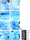
Figure 1
Figure 1
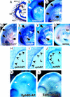
Expression of ephrinB ligands and EphB receptors in yolk sac blood vessels. Whole mount stainings of E9.5 yolk sacs with an anti-ephrinB2 antibody (A,B) or with alkaline phosphatase (AP) fusion proteins (C–H). Vitelline arteries were identified as the posterior vascular domain of the yolk sac (white arrowhead in A,C,E,G), vitelline veins as the anterior domain of the yolk sac (black arrowheads in B,D,F,H), respectively. Immunohistochemistry detects expression of ephrinB2 protein on yolk sac arteries (A), but not, or only at very low levels, on the venous domain (B). Arterial expression of ephrinB2 and possibly other ephrinB ligands was confirmed by binding of the EphB3–AP fusion protein (C). Weaker staining was observed on vitelline veins, revealing presence of ligands on both domains of the yolk sac vasculature (D). EphrinB2–AP detects receptors on vitelline veins (F), but gives weaker staining on arteries (E). Binding of ephrinB1–AP to all vessel types (G,H). (I) RT–PCR analysis. mRNAs for at least two ligands (ephrinB1 and ephrinB2) and three receptors (ephB2, ephB3, ephB4) were found in E9.5 yolk sac (y) and embryo (e). No PCR products were obtained in control reactions (c) without reverse transcription.
Figure 2
Figure 2
Expression of ephrinB ligands and EphB receptors in embryonic blood vessels. (A) Schematic drawing of the embryonic vasculature with veins in blue and arteries and aortic arches (black arrowheads) in red [modified from Streeter (1918)]. Whole-mount in situ hybridization on E9.5 wild-type embryos with antisense probes as indicated (B,C,E,F) and an ephB4 sense control (G), anterior is up, dorsal on the left. mRNA for ephrinB2 (B) is expressed in dorsal aorta (white arrow) and aortic arches (white arrowhead), whereas ephrinB1 transcripts (C) are found in all major vessels including dorsal aorta, aortic arches (white arrowhead), cardinal and umbilical veins (black arrows). Receptors binding ephrinB2–AP are localized in cardinal and umbilical veins, and in aortic arches (D). Expression of ephB3 (E) and ephB4 (F) in anterior and posterior cardinal and tail veins, sinus venosus. ephB3 is also expressed in aortic arches (white arrowhead, E). (H–J) Cross sections of whole-mount in situ hybridized embryos with the indicated antisense probes showing vessel walls of the anterior cardinal vein (approximately the same position for all three embryos). Dorsal is up. Note expression of ephrinB1, ephB3, and ephB4 in all or most endothelial cells (arrowheads). (K,L) AP stainings of E10.5 wild-type embryos. (K) Ligands of EphB3 in head vessels of different diameters including branches of anterior cardinal vein (arrowhead) and capillaries (white arrows). (L) Receptors of ephrinB2 in larger head vessels as well as capillaries. (ACV) anterior cardinal vein; (BA) branchial arches; (DA) dorsal aorta; (LB) limb bud; (SV) sinus venosus; (PCV) posterior cardinal vein; (UV) umbilical vein; (L) vessel lumen.
Figure 3
Figure 3
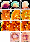
Cell–cell contacts between ligand- and receptor-expressing endothelial and mesenchymal cells. Whole-mount in situ hydrization on E9.5 embryos with antisense probes as indicated. The trunk region is shown, rostral is up, dorsal is left (A–D). Expression of flk-1 (A), ephB3 (C), and ephB4 (D) in intersomitic vessels. (B) ephrinB2 is expressed in the caudal half of somites. Somite boundaries are indicated with a bracket. (E,F) Whole-mount immunohistochemistry on E9.5 wild-type embryos with antibodies against PECAM-1 (brown), staining intersomitic vessels, and ephrinB2 (violet) staining dorsal somites. Tangential section, dorsal is up, rostral is left. A higher magnification of the area indicated by a white box (E) is shown (F). Note that endothelial cells are in direct contact with ephrinB2-expressing somitic cells. (G) Cross section through an E10.5 EphB2lacZ mutant heterozygote (Henkemeyer et al. 1996) at the level of the umbilical vein near the sinus venosus, stained for β-galactosidase activity, dorsal is up. Expression of EphB2 (β-gal) can be seen in mesenchymal cells adjacent to unstained endothelial cells of the vessel wall, as identified by PECAM-1 staining (H).
Figure 4
Figure 4
Vascular defects of ephrinB2−/− and ephB2/ephB3 double-mutant embryos. E9.5 yolk sacs of wild-type (A), ephrinB2−/− (B) and ephB2/ephB3 double-homozygous mutants (C). Lack of major vessel visible in freshly dissected homozygous mutant yolk sacs. (D–I) Whole-mount immunohistochemistry for PECAM-1. Vasculature in the trunk region of E10 embryos stained for PECAM-1, rostral is up, dorsal is left. Appearance of dorsal aorta (white arrow), aortic arches (white arrowheads), and anterior cardinal vein (black arrow) in a wild-type embryo (D) compared with ephrinB2−/− mutants (E,F) and an ephB2/B3 double mutant (G) showing irregular shapes or complete disruption of major vessels. (H) Trunk region of double heterozygous embryo (het/het) showing intact dorsal aorta and fourth aortic arch (white arrowheads). (I) Trunk region of ephB2/ephB3 double-mutant embryo with defective fourth aortic arch and dorsal aorta close to the arch (white arrowheads). (J,K) Cross section through aortic arches of embryos shown in H and I, rostral is up. Note that the normal vessel lumen (J) has not formed in the double mutant (K).
Figure 5
Figure 5
Defects in the vasculature of head, heart, and somitic vessels in ephrinB2−/− and ephB2/ephB3 receptor double mutants. Head region of PECAM-1-whole-mount stained E10 embryos (A–C). Branches of anterior cardinal vein of larger diameter are indicated by white arrowheads (A). Note the absence of large diameter vessels in the head of an ephB2/ephB3 double-mutant embryo (C). In more severe examples of receptor double mutants (data not shown) and in all ephrinB2−/− mutant embryos (B) head vasculature remains organized as a primitive capillary plexus. PECAM-1-stained hearts of wild-type (D), ephrinB2−/− (E), ephB2/ephB3 double mutants (F) at E10. Note smaller size of the heart and reduced trabeculation in ventricle (Ve) in ephrinB2−/− (E) and ephB2/ephB3 double mutants (F) compared with wild-type heart (D). (G,H) Sections of PECAM-1-stained hearts of wild-type (G) and ephB2/ephB3 double mutants (H). (I–M) Intersomitic vessels ofthe trunk at lumbar level stained for PECAM-1 (E10). (I) Wild-type embryo showing segmented pattern of intersomitic vessels (white arrows) and capillary network. Severe disorganization of intersomitic vessels and reduced capillary network in ephrinB2−/− mutants (J). ephB2/ephB3 double mutants show milder defects, e.g., abnormal dorsal sprouts from intersomitic vessels (arrowhead, K). (L,M) Sagittal sections of PECAM-1 stained embryos. (L) Wild-type embryo. (M) ephrinB2−/− embryo showing two somites with normal vessels at somitic borders (*) and several somites with abnormal sprouts penetrating into somites (black arrows).
Figure 6
Figure 6
EphrinB ligands induce sprouting angiogenesis in vitro. Adrenal-cortex-derived microvascular endothelial (ACE) cells (passage 11) on beads in three-dimensional fibrin gels incubated with control sample (A) or purified ephrinB1–Fc (B,C). (A,B) Phase-contrast photomicrographs. (C) Fluorescent nuclei of endothelial cells stained with Hoechst dye. (D,E) Quantitative analysis of sprout formation expressed as the number of sprouts with lengths exceeding the diameter of the bead per 50 MC beads. (D) EphrinB1–Fc (100 ng/ml) was used unclustered in either absence or presence of 20 μg/ml of the receptors EphB1–Fc, EphB2–Fc, or EphA5–Fc. EphB2–Fc alone had no sprouting activity in this assay. (E) EphrinB2–Fc (74 ng/ml) was used either unclustered or preclustered and compared with saturating amounts of Ang-1 (670 ng/ml) and VEGF (25 ng/ml). Values are mean ± s.e.m. (n = 4). (F) Ang1 receptor (Tie-2) phosphorylates the ephrinB1 cytoplasmic domain in vitro. Bacterially produced GST–Tie-2 or GST alone were purified and a fraction (200 ng) was combined with 200 ng of either GST–ephrinB1 extracellular domain (ext.) or GST–ephrinB1 cytoplasmic domain (cyto.) and subjected to an in vitro kinase reaction with the use of [γ32P]ATP. Reaction products were analyzed by 10% SDS–PAGE, stained with Coomassie brilliant blue (not shown), dried, and exposed to X-ray film. Note that the ephrinB1 cytoplasmic domain, but not ephrinB1 extracellular domain, is a direct in vitro substrate of Tie-2.
Figure 7
Figure 7
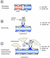
Presumed mechanisms and sites of action of ephrins and Eph receptors during remodeling of the vasculature. (A) Interaction of ephrinB2 ligand expressed on arteries and EphB3 and EphB4 receptors expressed on veins demarcates the boundary between arterial and venous domains. By analogy to the action in the nervous system, it was suggested that ephrin–Eph interactions may prevent intermixing of arterial and venous endothelial cells and, following sprouting, may result in the formation of a capillary network (Yancopoulos et al. 1998). However, this model was based on the exclusive and complementary expression of ephrinB2 and EphB4. (B) Coexpression of ligands and receptors on the same type of vessels (e.g., veins) provides a cell-to-cell signal for endothelial cells that may rather be stimulatory and help to promote morphogenesis and sprouting. (C) Mesenchymal cells adjacent to blood vessels also express ephrins or Eph receptors and may help patterning the vasculature. In the somites, this signal may be inhibitory and prevent sprouting, whereas in other regions, stimulatory signals are conceivable. Mesenchymal cells are also the source of angiogenic factors such as Ang1 and VEGF, which may modulate ephrinB–EphB receptor signaling.

Similar articles

Cited by

References

    1. Bergemann AD, Cheng H-J, Brambilla R, Klein R, Flanagan JG. Elf-2, a new member of the Eph ligand family, is segmentally expressed in mouse embryos in the region of the hindbrain and newly forming somites. Mol Cell Biol. 1995;15:4921–4929. - PMC - PubMed
    1. Bouillet P, Oulad-Abdelghani M, Vicaire S, Garnier J-M, Schuhbaur B, Dolle P, Chambon P. Efficient cloning of cDNAs of retinoic acid-responsive genes in P19 embryonal carcinoma cells and characterization of a novel mouse gene, Stra1 (mouse Lerk2/Eplg2) Dev Biol. 1995;170:420–433. - PubMed
    1. Brambilla R, Klein R. Telling axons where to grow: A role for Eph receptor tyrosine kinases in guidance. Mol Cell Neurosci. 1995;6:487–495. - PubMed
    1. Brambilla R, Schnapp A, Casagranda F, Labrador JP, Bergemann AD, Flanagan JG, Pasquale EB, Klein R. Membrane-bound LERK2 ligand can signal through three different Eph-related receptor tyrosine kinases. EMBO J. 1995;14:3116–3126. - PMC - PubMed
    1. Brennan C, Monschau B, Lindberg R, Guthrie B, Drescher U, Bonhoeffer F, Holder N. Two Eph receptor tyrosine kinase ligands control axon growth and may be involved in the creation of the retinotectal map in the zebrafish. Development. 1997;124:655–664. - PubMed

Publication types

MeSH terms