. 1999 Mar;73(3):1980-9.
doi: 10.1128/JVI.73.3.1980-1989.1999.

Mechanisms that regulate Epstein-Barr virus EBNA-1 gene transcription during restricted latency are conserved among lymphocryptoviruses of Old World primates

Affiliations

Mechanisms that regulate Epstein-Barr virus EBNA-1 gene transcription during restricted latency are conserved among lymphocryptoviruses of Old World primates

I K Ruf et al. J Virol. 1999 Mar.

Abstract

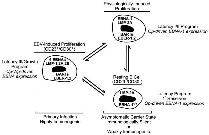
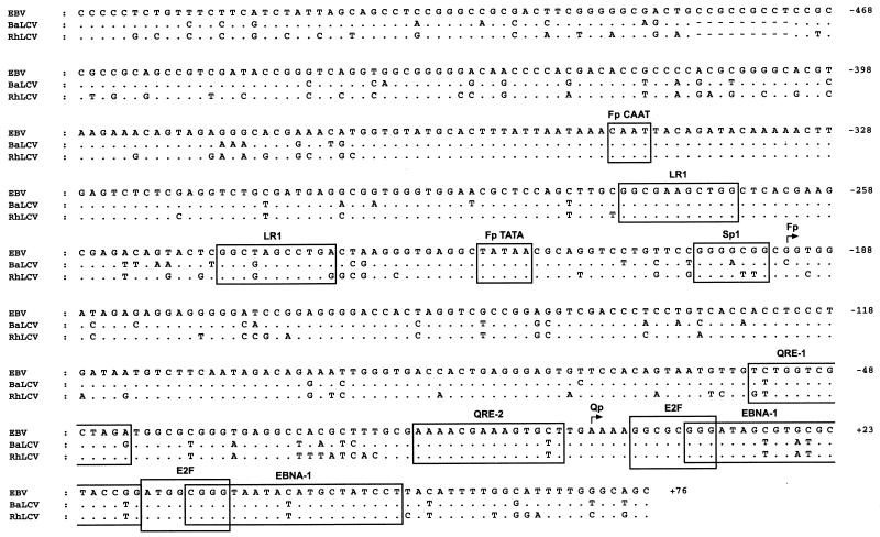
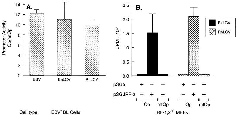
Epstein-Barr virus (EBV), the only known human lymphocryptovirus (LCV), displays a remarkable degree of genetic and biologic identity to LCVs that infect Old World primates. Within their natural hosts, infection by these viruses recapitulates many key aspects of EBV infection, including the establishment of long-term latency within B lymphocytes, and is therefore a potentially valuable animal model of EBV infection. However, it is unclear whether these LCVs have adopted or maintained the same mechanisms used by EBV to express essential viral proteins, such as EBNA-1, in the face of cell-mediated repression of EBV gene expression that occurs upon establishment of the asymptomatic carrier state. To address this issue, we determined whether the endogenous LCVs of baboon (Cercopithecine herpesvirus 12) and rhesus macaque (Cercopithecine herpesvirus 15) have the functional equivalent of the EBV promoter Qp, which mediates exclusive expression of EBNA-1 during the restricted programs of EBV latency associated with the carrier state. Our results indicate that (i) both the baboon and rhesus macaque LCVs have a genomic locus that is highly homologous to the EBV Qp region, (ii) key cis-regulatory elements of Qp are conserved in these LCV genomes and compose promoters that are functionally indistinguishable from EBV Qp, and (iii) EBNA-1 transcripts identical in structure to EBV Qp-specific EBNA-1 mRNAs are present in nonhuman LCV-infected cells, demonstrating that these Qp homologs are indeed utilized as alternative EBNA-1 promoters. These observations indicate that the molecular mechanisms which regulate EBV gene expression during restricted latency have been conserved among the LCVs. The contribution of these mechanisms to viral persistence in vivo can now be experimentally tested in nonhuman primate models of LCV infection.


Figures

FIG. 1
FIG. 1
Model for the maintenance of EBV latency in B lymphocytes. In primary infection, a rapid EBV-induced expansion of infected B cells that express the full complement of known latency-associated genes (see text) serves to establish a pool of infected cells. These are equivalent to EBV-immortalized LCLs which characteristically express each of the six EBNA proteins through the Cp or Wp promoter, and they are susceptible to the developing cellular immunity directed toward EBV proteins expressed exclusively during type III latency (50). In the asymptomatic carrier state, resolution of primary infection is concomitant with the establishment of resting B cells as the primary reservoir of EBV in the peripheral blood (37, 38). These cells display a restricted pattern of EBV gene expression more characteristic of EBV-associated tumors such as BL (type I latency) or nasopharyngeal carcinoma (type II latency), in which Cp and Wp are silent and EBNA gene transcription is limited to EBNA-1 driven by the promoter Qp (, , –11, 18, 22, 37, 45, 46, 53, 63, 67, 72, 75). Hypothetically, latently infected B cells are periodically subjected to EBV- or physiologically induced proliferation, e.g., in response to mitogenic signals such as CD40 ligand on activated T cells, that may serve to sustain a critical pool of infected B cells. Note that the precise pattern of EBV gene expression in the resting B cell is currently undefined, as expression of only LMP-2A and EBNA-1 has been evaluated for this population (37); however, studies of EBV gene expression in unfractionated B-lymphocyte populations (9, 10, 46, 72) have indicated that a broader pattern of expression including the BARTs and EBERs occurs in healthy carriers, and thus EBV gene expression in the resting B cell may be indistinguishable from that in the hypothetical infected B cell shown here that is proliferating in response to physiological signals.
FIG. 2
FIG. 2
DNA sequence conservation in the Qp promoter region. The Qp promoter region of the B95-8 EBV genome sequence (coordinates 61,886 to 62,498) (4) is aligned with the DNA sequences of the homologous regions of the BaLCV and RhLCV genomes and represents nucleotides −537 to +76 relative to the major transcription start site (+1) within the EBV Qp (45, 63). Known and predicted promoter-regulatory elements (see text) are boxed and include those for the lytic cycle-specific EBNA-1 promoter Fp (CAAT, LR-1, TATA, and Sp1) (8, 56) as well as for Qp (QRE-1, QRE-2, E2F, and EBNA-1) (–44, 57, 60, 61, 68). Sites of transcription initiation are denoted by bent arrows. Differences in the BaLCV and RhLCV sequences relative to that of EBV are indicated; dots and dashes represent identical and deleted nucleotides, respectively.
FIG. 3
FIG. 3
Activation of the BaLCV and RhLCV Qp promoters is IRF dependent. (A) The dependence of the BaLCV and RhLCV Qp promoters on the IRF binding site QRE-2, which is essential for EBV Qp function, was evaluated by an hGH reporter gene assay with EBV-negative Louckes BL cells. Promoter activity is presented as the ratio of hGH expression achieved from a wild-type promoter sequence (Qp) to that achieved from the respective LCV Qp in which the IRF binding site had been mutated (mtQp). (B) The BaLCV and RhLCV Qp promoters require IRF expression for activation. MEFs nullizygous for IRF-1 and IRF-2 were cotransfected with a Qp or mtQp hGH reporter plasmid and either an empty expression vector (pSG5) or the IRF-2 expression vector pSG.IRF-2. Promoter activity was measured by radioimmunoassay of hGH expression. The data shown are from a representative experiment in which all transfections were done in triplicate and hGH values were corrected for transfection efficiency. The 5′ and 3′ coordinates of the promoter DNA in each reporter plasmid were −143 to +75 relative to the EBV Qp transcription start site. Error bars indicate standard deviations.
FIG. 4
FIG. 4
The BaLCV and RhLCV Qp promoters are negatively autoregulated. (Left panel) EBV-negative Louckes BL cells were cotransfected with a BaLCV or RhLCV Qp reporter plasmid and either an empty expression vector (pSG5) or an EBNA-1 expression vector (pSG.E1) encoding BaLCV or RhLCV EBNA-1, respectively. (Right panel) BaLCV and RhLCV Qp reporter plasmids lacking a functional EBNA-1 binding domain (QpΔ34) are unresponsive to the respective LCV EBNA-1. Data are from a representative experiment in which all transfections were done in triplicate and hGH values were corrected for transfection efficiency. The reporter plasmids used in these experiments had the same 5′ and 3′ coordinates as indicated for Fig. 3. Error bars indicate standard deviations.
FIG. 5
FIG. 5
Northern blot analysis of EBNA-1 expression in BaLCV- and RhLCV-infected B lymphocytes. Each blot contained 10 μg of poly(A)+ RNA isolated from the BaLCV- and RhLCV-infected B-cell lines S594 and Mm278LCL, respectively. The positions to which the RNA size markers migrated in the gel (kilobases) are indicated to the left of the blots. Based on the sizes of the transcripts detected, those consistent with transcription initiating from either Fp or Qp (Fp/Qp) or from either Cp or Wp (Cp/Wp) are bracketed. Blots were reprobed for GAPDH (glyceraldehyde-3-phosphate dehydrogenase) mRNA to monitor RNA loading. The specific activities of the DNA probes were as follows: BaLCV, 2.9 × 10⁸ cpm/μg; RhLCV, 2.6 × 10⁸ cpm/μg; and GAPDH, 2.5 × 10⁸ cpm/μg. Autoradiographic exposure times were 7 days (EBNA-1) and 5 h (GAPDH). Data are representative of those from three experiments.
FIG. 6
FIG. 6
The BaLCV and RhLCV Qp promoters are functional within latently infected B lymphocytes. RT-PCR analysis was employed to detect, and distinguish between, Qp- and Fp-specific EBNA-1 gene transcription in S594 (BaLCV-infected) and Mm278LCL (RhLCV-infected) cells. The exon structure of an EBNA-1 mRNA (not to scale) derived from either Fp (active during lytic infection) or Qp is illustrated at the bottom; Fp and Qp transcription start sites (bent arrows) and the relative annealing sites of the oligonucleotide primers used for RT and subsequent amplification of cDNAs by PCR are depicted with respect to the mRNA structure. The upper two panels demonstrate detection of Qp-specific EBNA-1 transcription in latently infected BL cells (Akata), in which Fp is silent, and detection of Fp activity in lytically infected BL cells (P3HR-1 BL cells treated with 12-O-tetradecanoylphorbol-13-acetate and sodium butyrate), respectively. By comparison, the predominate origin of EBNA-1 transcription detected in the BaLCV- and RhLCV-infected cells was attributable to Qp. PCR products were detected by Southern blot hybridization with an EBV, BaLCV, or RhLCV cDNA probe that had been generated by the respective SP3-E1 primer pair and sequenced to confirm that they were EBNA-1 cDNAs. +/−, presence and absence, respectively, of reverse transcriptase in the cDNA synthesis reaction.
FIG. 7
FIG. 7
The EBV and BaLCV Qp promoters are similarly regulated in baboon and human LCLs. S595 and IB4 cells were transfected in triplicate with EBV and BaLCV Qp-hGH reporter plasmids (Fig. 4) that did (Qp) or did not (QpΔ34) contain a functional EBNA-1 autoregulatory domain. Data have been corrected for differences in transfection efficiency. Error bars indicate standard deviations.

Similar articles

Cited by

References

    1. Ablashi D V, Gerber P, Easton J. Oncogenic herpesviruses of nonhuman primates. Comp Immunol Microbiol Infect Dis. 1979;2:229–241. - PubMed
    1. Arrand J R, Rymo L. Characterization of the major Epstein-Barr virus-specific RNA in Burkitt lymphoma-derived cells. J Virol. 1982;41:376–389. - PMC - PubMed
    1. Ausubel F M, Brent R, Kingston R E, Moore D D, Seidman J G, Smith J A, Struhl K. Current protocols in molecular biology. New York, N.Y: John Wiley and Sons, Inc.; 1989.
    1. Baer R, Bankier A T, Biggin M D, Deininger P L, Farrell P J, Gibson T G, Hatfull G, Hudson G S, Satchwell S C, Sequin C, Tufnell P S, Barrell B G. DNA sequence and expression of the B95-8 Epstein-Barr virus genome. Nature. 1984;310:207–211. - PubMed
    1. Bodescot M, Perricaudet M, Farrell P. A promoter for the highly spliced EBNA family of RNAs of Epstein-Barr virus. J Virol. 1987;61:3424–3430. - PMC - PubMed

Publication types

Substances

LinkOut - more resources