. 1999 Feb 15;19(4):1179-88.
doi: 10.1523/JNEUROSCI.19-04-01179.1999.

Estrogen-induced activation of mitogen-activated protein kinase in cerebral cortical explants: convergence of estrogen and neurotrophin signaling pathways

Affiliations

Estrogen-induced activation of mitogen-activated protein kinase in cerebral cortical explants: convergence of estrogen and neurotrophin signaling pathways

M Singh et al. J Neurosci. .

Abstract

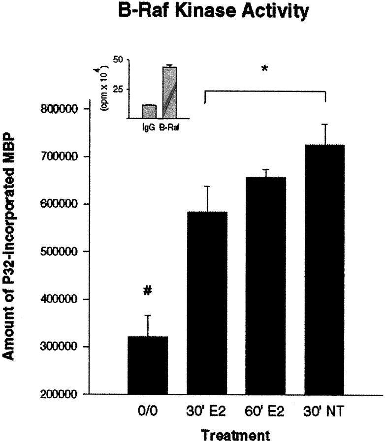
We have shown that estrogen elicits a selective enhancement of the growth and differentiation of axons and dendrites (neurites) in the developing CNS. We subsequently demonstrated widespread colocalization of estrogen and neurotrophin receptors (trk) within developing forebrain neurons and reciprocal transcriptional regulation of these receptors by their ligands. Using organotypic explants of the cerebral cortex, we tested the hypothesis that estrogen/neurotrophin receptor coexpression also may result in convergence or cross-coupling of their signaling pathways. Estradiol elicited rapid (within 5-15 min) tyrosine phosphorylation/activation of the mitogen-activated protein (MAP) kinases, ERK1 and ERK2, that persisted for at least 2 hr. This extracellular signal-regulated protein kinase (ERK) activation was inhibited successfully by the MEK1 inhibitor PD98059, but not by the estrogen receptor (ER) antagonist ICI 182,780, and did not appear to result from estradiol-induced activation of trk. Furthermore, we also found that estradiol elicited an increase in B-Raf kinase activity. The latter and subsequent downstream events leading to ERK activation may be a consequence of our documentation of a multimeric complex consisting of, at least, the ER, hsp90, and B-Raf. These novel findings provide an alternative mechanism for some of the estrogen actions in the developing CNS and could explain not only some of the very rapid effects of estrogen but also the ability of estrogen and neurotrophins to regulate the same broad array of cytoskeletal and growth-associated genes involved in neurite growth and differentiation.


Figures

Fig. 1.
Fig. 1.
Estradiol-induced ERK phosphorylation. Lysates derived from cerebral cortical explants were probed with a phosphospecific ERK1/ERK2 antibody. Shown is a time course for estradiol-induced ERK phosphorylation (top panel) and the reprobed blot for ERK1 and ERK2 protein (middle panel). Explants also were treated with a 100 ng/ml neurotrophin cocktail (NGF, BDNF, NT-3, and NT-4/5) for a single 30 min time point that served as the experimental positive control. Note the similarity in the intensity of the response to estrogen and the neurotrophins in the cortical explants. Untreated PC12 and NGF-treated PC12 cells served as negative and positive methodological controls, respectively. Densitometric representations of the relative intensities of the phosphorylated ERK1 and ERK2 bands are provided also (bottom panel).
Fig. 2.
Fig. 2.
Effect of estradiol on ERK phosphorylation in cerebellar cultures. Cerebellar explants were treated with 10 nm estradiol for 30 min or for 1 hr. Only marginal phosphorylation of ERK was observed, whereas the neurotrophin cocktail treatment (positive control) resulted in robust phosphorylation of ERK. The bottom panel represents the same blot, which was stripped and reprobed for ERK1 and ERK2 protein to verify equal loading of protein across lanes.
Fig. 3.
Fig. 3.
Temporal pattern of ERK activation after estradiol exposure. Shown is an in-gel kinase assay, using MBP as a substrate, for the temporal pattern of ERK1 and ERK2 activation in response to 10 nm estradiol. The effects of 100 ng/ml of NGF, BDNF, or NT-3 on ERK activation were used as controls to document the action of all of these neurotrophins on ERK activation in cerebral cortical explants. Untreated PC12 cells and NGF-treated PC12 cells served as methodological controls. A densitometric representation of the relative intensities of ERK2 activation is provided also (bottom panel).
Fig. 4.
Fig. 4.
Effect of ICI 182,780 on estradiol-induced ERK phosphorylation in rat cerebral cortical explants. Cultures were pretreated with either vehicle (DMSO, 0.1%) or 1 μm ICI 182,780 for 24 hr before being pulsed with either estradiol in combination with ICI 182,780 or estradiol with vehicle.A, Shown is the phospho-ERK blot documenting the inability of ICI 182,780 to block either estradiol- or neurotrophin-induced ERK phosphorylation. B, Documented is the positive control for the ICI 182,780 compound performed on the mammary tumor cell line, MCF-7, treated with either 10 nmestradiol or estradiol in combination with 10 μm of ICI 182,780. Thebottom panels in both A andB represent the same blot, which was stripped and reprobed for ERK1 and ERK2 protein to verify equal loading of protein across lanes.
Fig. 5.
Fig. 5.
Effect of the MEK inhibitor PD98059 on estrogen- and neurotrophin-induced ERK phosphorylation. After a 5 hr preincubation with 100 μm PD98059 the effects of both estradiol and the neurotrophins on ERK phosphorylation were abolished in the continued presence of the MEK inhibitor. The bottom panel represents the same blot, which was stripped and reprobed for ERK1 and ERK2 protein to verify equal loading of protein across lanes.
Fig. 6.
Fig. 6.
Effect of estradiol on B-Raf activity. The amount of 32P incorporated into MBP was evaluated from lysates of cerebral cortical explants immunoprecipitated with the B-Raf antibody (clone C-19). Both 30 and 60 min estradiol treatment significantly increased the kinase activity of B-Raf relative to untreated control explants. Treatment of the explants with the neurotrophin cocktail for 30 min served as the experimental positive control and also revealed a significant increase in B-Raf kinase activity. Values represent the average of four samples and were calculated by subtracting the activity level obtained from an IgG-immunoprecipitated control (background) from the values obtained in the experimental samples. The difference (in cpm) obtained after a control (untreated) sample was immunoprecipitated with preimmune IgG versus being immunoprecipitated with the B-Raf antibody is presented as the inset. Statistical analysis was performed by using a one-way ANOVA, followed by Scheffé’s analysis for group differences (#, different from all treated groups, p < 0.05; *difference between 30′ E2 group and 30′ NT group, with p < 0.05).
Fig. 7.
Fig. 7.
Effect of estradiol on trkphosphorylation. Lysates of cerebral cortical explants were immunoprecipitated with the pan-trk (203) antibody before Western analysis and probed with an anti-phosphotyrosine (4G10) antibody. Shown are the effects of a 30 min pulse of 10 nmestradiol and treatment with a 100 ng/ml neurotrophin cocktail for 30 min. Estradiol did not elicit tyrosine phosphorylation oftrk receptors, whereas the neurotrophins did. Untreated PC12 cells and the trkA-deficient NNR-5 cell line served as methodological negative controls, whereas NGF-treated PC12 cells served as the prototypical positive control.
Fig. 8.
Fig. 8.
Coprecipitation of the ER with B-Raf in cerebral cortical explants. A, Evaluation of B-Raf levels in the cerebral cortical explants by Western analysis revealed significant levels of a 95 kDa B-Raf band that migrated to a similar position as that observed in PC12 cells. An additional band of ∼68 kDa also was observed in the cerebral cortical lysates that was absent in PC12 cell lysates. B, After immunoprecipitation with an anti-ER antibody (ER715), probing with an anti-B-Raf antibody revealed an association between the ER and the 95 kDa isoform of B-Raf that was conspicuously absent in the IgG-immunoprecipitated lane.C, Confirmation of the identity of this specific B-Raf band as kinase-active Raf was performed by evaluating the amount of Raf activity present in cerebral cortical explants immunoprecipitated with the ER and comparing it with samples immunoprecipitated with preimmune IgG. Statistical analysis that used a two-tailed Student’st test revealed that the level of Raf activity in the ER-immunoprecipitated samples was significantly higher than that observed in the IgG-immunoprecipitated control (*p < 0.05).
Fig. 9.
Fig. 9.
Coprecipitation of the ER and B-Raf in P10 cerebral cortical tissue. A, Immunoprecipitation of cerebral cortical lysates derived from P10 rat pups with the ER antibody, followed by Western analysis for B-Raf, revealed a similar association of B-Raf with the ER (as seen in explants of similar age), which was not found in the IgG-immunoprecipitated control.B, Subsequent reprobing of the blot for the ER revealed a specific ER (67 kDa) band that was not present in the IgG-immunoprecipitated control lane.
Fig. 10.
Fig. 10.
Coimmunoprecipitation of the ER with hsp90, but not with MEK1. A, Immunoprecipitation with the ER and probing for hsp90 revealed an association in the ER with hsp90. The HeLa lysate served as a positive control for the detection of hsp90. Immunoprecipitation with either anti-MEK1 (B) or the ER (C) failed to reveal an association between the ER and MEK1. Recombinant estrogen receptor protein (C) and A-431 lysate (B) served as positive controls for the detection of the ER and MEK1, respectively.

Similar articles

Cited by

References

    1. Aronica SM, Kraus WL, Katzenellenbogen BS. Estrogen action via the cAMP signaling pathway: stimulation of adenylate cyclase and cAMP-regulated gene transcription. Proc Natl Acad Sci USA. 1994;91:8517–8521. - PMC - PubMed
    1. Barnier JV, Papin C, Eychene A, Lecoq O, Calothy G. The mouse B-raf gene encodes multiple protein isoforms with tissue-specific expression. J Biol Chem. 1995;270:23381–23389. - PubMed
    1. Black M, Aletta J, Greene L. Regulation of microtubule composition and stability during nerve growth factor-promoted neurite outgrowth. J Cell Biol. 1986;103:545–557. - PMC - PubMed
    1. Bunone G, Briand PA, Miksicek RJ, Picard D. Activation of the unliganded estrogen receptor by EGF involves the MAP kinase pathway and direct phosphorylation. EMBO J. 1996;15:2174–2183. - PMC - PubMed
    1. Chiaia N, Foy M, Teyler TJ. The hamster hippocampal slice. II. Neuroendocrine modulation. Behav Neurosci. 1983;97:839–843. - PubMed

Publication types

MeSH terms

Substances