. 1999 Feb 2;96(3):1140-5.
doi: 10.1073/pnas.96.3.1140.

ARG1 (altered response to gravity) encodes a DnaJ-like protein that potentially interacts with the cytoskeleton

Collaborators, Affiliations

ARG1 (altered response to gravity) encodes a DnaJ-like protein that potentially interacts with the cytoskeleton

J C Sedbrook et al. Proc Natl Acad Sci U S A. .

Abstract

Gravitropism allows plant organs to direct their growth at a specific angle from the gravity vector, promoting upward growth for shoots and downward growth for roots. Little is known about the mechanisms underlying gravitropic signal transduction. We found that mutations in the ARG1 locus of Arabidopsis thaliana alter root and hypocotyl gravitropism without affecting phototropism, root growth responses to phytohormones or inhibitors of auxin transport, or starch accumulation. The positional cloning of ARG1 revealed a DnaJ-like protein containing a coiled-coil region homologous to coiled coils found in cytoskeleton-interacting proteins. These data suggest that ARG1 participates in a gravity-signaling process involving the cytoskeleton. A combination of Northern blot studies and analysis of ARG1-GUS fusion-reporter expression in transgenic plants demonstrated that ARG1 is expressed in all organs. Ubiquitous ARG1 expression in Arabidopsis and the identification of an ortholog in Caenorhabditis elegans suggest that ARG1 is involved in other essential processes.


Figures
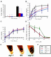
Figure 1
Figure 1
Phenotypic characterization of arg1 seedlings (28). (A) Average hypocotyl tip angles from the horizontal of dark-grown 4-day-old wild-type (WS) and arg1–1 and arg1–2 mutant seedlings, 0 and 72 hours after rotating the plates by 90°. The two series of measurements were done on separate plates and involved 96 and 112 plants, respectively. SDs are shown by thin lines on the top of each bar. A legend to AD is provided in F. (B) The kinetics of root bending on gravistimulation. Seedlings (4 days old) were reoriented 90° at time zero. Root-tip angles from the horizontal were measured over time, and average angles along with SD (vertical bars) were calculated (n = 57–78). (C) The kinetics of hypocotyl bending on exposure to a horizontal light source. Seedlings (4 days old) were exposed to a horizontal light source at time zero. Hypocotyl tip angles from the vertical were measured over time, and average angles along with SDs (vertical bars) were calculated (n = 23 and 30). (D) The effect of 2,4-D on average root growth of wild-type WS and of mutant arg1–1 and rgr1 (auxin-resistant control; ref. 17) seedlings. Seedlings (4 days old) were placed on media containing the indicated concentrations of 2,4-D (x axis) at time zero. Average root growth rates (mm per 12 hours) along with SDs (vertical bars) were calculated and plotted (n = 12–19). (E) arg1–1 and arg1–2 seedlings accumulate starch in their statocytes like wild type. Wild-type WS (Center) and arg1–1 (Left) and arg1–2 (Right) mutant seedlings (7 days old) were stained with IKI and analyzed under a light microscope equipped with Nomarski optics (×20).
Figure 2
Figure 2
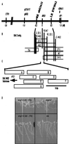
Genetic and physical map of the genomic region spanning ARG1. (A) Genetic map of a region of chromosome 1 between the genes ETR1 and APETELA 1 (AP1). Genetic and molecular markers are listed above the solid line, whereas their distances (in cM) from ARG1 are listed below. The YAC contig described in ref. stretches from the RFLP marker g4552 to AP1. ARG1 was mapped between the YAC-end markers RE abi13A11 and LE EW19A2 by RFLP analysis. (B) Depiction of the BAC contig (drawn to a cM scale) assembled to cover the chromosomal region between YAC-end markers RE abi13A11 and LE EW19A2. Vertical bars represent BAC- and YAC-end probes that hybridized to DNA isolated from the BAC clones (open rectangles). ARG1 was mapped on BACs 4N22 and 2E12 by RFLP analysis. (C) The relative positions of BAC 4N22 subclones (open rectangles labeled A–G). The ARG1 gene is represented by an arrow indicating the direction of its transcription. (D) Fragment A rescues the wavy-root growth phenotype of arg1–2. Wild-type WS seedlings (Lower Right), arg1–2 seedlings (Upper Right), T2 progeny of a fragment A-transformed arg1–2 plant (Lower Left), and T2 progeny of a fragment E-transformed arg1–2 plant (Upper Left) were subjected to the wavy-root growth assay described in Materials and Methods. The asterisk indicates a segregating T2 progeny that develops a mutant root-waving phenotype; others develop a wild-type root-wave phenotype that cosegregates with the transgene (not shown).
Figure 3
Figure 3
Structure of the ARG1 protein. (A) A scale-drawn representation of the putative domains (shaded rectangles) within the ARG1 protein (open rectangle). The locations of the arg1–1 and arg1–2 mutations are depicted by arrows. TM, transmembrane domain. (BC) Amino acid sequence alignments of the ARG1 J domain (B) and coiled-coil region (C) with the corresponding domains from other proteins (–, –63). The name of the protein and the contributing organism is to the right of the sequences, whereas the beginning positions of the sequences within the protein are listed to the left. Amino acid residues within the black boxes are identical to the corresponding ARG1 residue, whereas shaded residues are conserved, as determined by the blosom 62 substitution matrix (64). Alignments were made by using the DNAstar program dna*. H. Ch., Heavy Chain.
Figure 4
Figure 4
Representation of amino acid sequence alignments between ARG1 and its putative orthologs in A. thaliana [ARL1, GenBank accession no. AC002396 (F3I6.4)] and C. elegans [R74.4, GenBank accession no. Z36238 (R74.4)]. The ARG1 sequence is represented by an open box at the middle of the figure, whereas the A. thaliana and C. elegans orthologs are represented by open boxes at the top and bottom of the figure, respectively. The conserved J, potential transmembrane and coiled coil domains are represented by shaded boxes in all cases. The percentage of amino acids found identical within each domain between each ortholog and ARG1 is indicated by a number inserted in that domain of the ortholog. Sequence alignment gaps including more than five residues are represented by grey lines joining the two orthologs. The number of amino acids involved in each gap is represented by a number inserted between the corresponding grey lines.
Figure 5
Figure 5
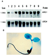
The ARG1 gene is expressed ubiquitously in A. thaliana plants. (A) Northern blot analysis of total RNAs extracted from roots (lane 1) or hypocotyls and cotyledons (lane 2) of 8-day-old light-grown seedlings, from hypocotyls and cotyledons of etiolated 5-day-old seedlings (lane 3), or from roots (lane 4), rosette leaves (lane 5), cauline leaves (lane 6), inflorescence stems (lane 7), flowers (lane 8), or siliques (lane 9) of 24-day-old soil-grown plants. The Northern membrane was first hybridized with a 32P-labeled full-length ARG1 cDNA probe (Upper). After exposure, the blot was stripped and rehybridized with a UBQ4 probe (65) to control for loading differences (Lower). (B) Cytochemical detection of GUS activity in a 3-day-old transgenic A. thaliana seedling carrying the GUS gene fused in-frame within the fourth exon of ARG1.

Similar articles

Cited by

References

    1. Masson P H. BioEssays. 1995;17:119–127. - PubMed
    1. Knight T A. Phil Trans R Soc. 1806;99:108–120.
    1. Darwin C. The Power of Movement in Plants. London: John Murray; 1880.
    1. Sack F D. Int Rev Cytol. 1991;127:193–252. - PubMed
    1. Kuznetsov O A, Hasenstein K H. Planta. 1996;198:87–94. - PubMed

Publication types

Associated data