. 1999 Jan 25;144(2):373-84.
doi: 10.1083/jcb.144.2.373.

Membrane-type 1 matrix metalloprotease (MT1-MMP) enables invasive migration of glioma cells in central nervous system white matter

Affiliations

Membrane-type 1 matrix metalloprotease (MT1-MMP) enables invasive migration of glioma cells in central nervous system white matter

A T Beliën et al. J Cell Biol. .

Abstract

Invasive glioma cells migrate preferentially along central nervous system (CNS) white matter fiber tracts irrespective of the fact that CNS myelin contains proteins that inhibit cell migration and neurite outgrowth. Previous work has demonstrated that to migrate on a myelin substrate and to overcome its inhibitory effect, rat C6 and human glioblastoma cells require a membrane-bound metalloproteolytic activity (C6-MP) which shares several biochemical and pharmacological characteristics with MT1-MMP. We show now that MT1-MMP is expressed on the surface of rat C6 glioblastoma cells and is coenriched with C6-MP activity. Immunodepletion of C6-MP activity is achieved with an anti-MT1-MMP antibody. These data suggest that MT1-MMP and the C6-MP are closely related or identical. When mouse 3T3 fibroblasts were transfected with MT1-MMP they acquired the ability to spread and migrate on the nonpermissive myelin substrate and to infiltrate into adult rat optic nerve explants. MT1-MMP-transfected fibroblasts and C6 glioma cells were able to digest bNI-220, one of the most potent CNS myelin inhibitory proteins. Plasma membranes of both MT1-MMP-transfected fibroblasts and C6 glioma cells inactivated inhibitory myelin extracts, and this activity was sensitive to the same protease inhibitors. Interestingly, pretreatment of CNS myelin with gelatinase A/MMP-2 could not inactivate its inhibitory property. These data imply an important role of MT1-MMP in spreading and migration of glioma cells on white matter constituents in vitro and point to a function of MT1-MMP in the invasive behavior of malignant gliomas in the CNS in vivo.


Figures
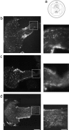
Figure 9
Figure 9
(a) Optic nerve explant infiltration assay. Adult rat optic nerves were placed underneath a teflon ring sealed to a culture dish with silicon grease. Hoechst dye-prelabeled C6 cells, MT1-MMP–transfected fibroblasts, or mock-transfected fibroblasts (100,000 cells) were placed in the middle chamber, in contact with the tips of the nerves (a). After 7 d Hoechst dye-labeled cells are observed infiltrating the optic nerve explants in longitudinal sections. MT1-MMP–transfected fibroblasts (b) but not mock-transfected fibroblasts (c) infiltrated the optic nerves. Enlargements show a high density of MT1-MMP–transfected fibroblasts at a distance of 1.6 mm. Very few mock-transfected fibroblasts were found at this depth. C6 cells infiltrated the optic nerve to the same extent as the MT1-MMP– transfected fibroblasts (d). Bar, 0.3 mm.
Figure 1
Figure 1
MT1-MMP immunoreactivity in C6 glioblastoma cells and MT1-MMP–transfected fibroblasts 1 h after plating the cells on CNS substrate. (a) Well-spread C6 cell shows diffuse MT1-MMP staining over the plasma membrane, especially along the cell periphery. (b) When the plane of focus is at the level of the substrate, MT1-MMP immunoreactivity is detected at the substrate contact sites. (c) MT1-MMP–transfected fibroblasts show immunoreactivity enriched on lamellipodia (arrows). (d) 24 h after plating MT1-MMP–transfected fibroblasts cells formed long processes on which foci of MT1-MMP are observed (arrowhead). Bar, 14 μm.
Figure 1
Figure 1
MT1-MMP immunoreactivity in C6 glioblastoma cells and MT1-MMP–transfected fibroblasts 1 h after plating the cells on CNS substrate. (a) Well-spread C6 cell shows diffuse MT1-MMP staining over the plasma membrane, especially along the cell periphery. (b) When the plane of focus is at the level of the substrate, MT1-MMP immunoreactivity is detected at the substrate contact sites. (c) MT1-MMP–transfected fibroblasts show immunoreactivity enriched on lamellipodia (arrows). (d) 24 h after plating MT1-MMP–transfected fibroblasts cells formed long processes on which foci of MT1-MMP are observed (arrowhead). Bar, 14 μm.
Figure 2
Figure 2
MT1-MMP Western blot and remodeling of the CNS inhibitory substrate. (a) Western blots of cell lysates and salt-washed plasma membranes using a monoclonal antibody against MT1-MMP. The protein fractions were electrophoresed on a 10% SDS polyacrylamide gel, blotted onto a nitrocellulose membrane, and probed with MT1-MMP antibody. Bound antibody was visualized by peroxidase-conjugated second antibody and chemiluminescence. Lane 1, C6 cell homogenate (CH C6); lane 2, C6 cell plasma membranes (PM C6); lane 3, plasma membranes of MT1-MMP–transfected fibroblasts (PM MT); lane 4, plasma membranes of mock-transfected fibroblasts (PM Mo). Molecular mass markers are indicated at the left side, proMT1-MMP and processed MT1-MMP (arrows) on the right. Two nonspecific bands are seen between the pro- and mature MT1-MMP forms since they are visualized also in the absence of primary antibody. (b) Culture dishes coated with inhibitory CNS membrane proteins were pretreated in different ways and their inhibitory activity assayed for 3T3 fibroblast spreading. Treatment with C6 homogenate (50 μg) or plasma membranes (30 μg) of C6 glioma cells or MT1-MMP–transfected fibroblasts reduced the inhibitory property significantly, whereas plasma membranes of mock-transfected fibroblasts did not. Pretreatment of the CNS substrate with gelatinase A/MMP-2 (0.1, 1, and 10 μM) did not influence its nonpermissive substrate property. Values are means ± SEM of three experiments performed in triplicate. Significance with the two-tailed Student's t test: P < 0.01 (**).
Figure 2
Figure 2
MT1-MMP Western blot and remodeling of the CNS inhibitory substrate. (a) Western blots of cell lysates and salt-washed plasma membranes using a monoclonal antibody against MT1-MMP. The protein fractions were electrophoresed on a 10% SDS polyacrylamide gel, blotted onto a nitrocellulose membrane, and probed with MT1-MMP antibody. Bound antibody was visualized by peroxidase-conjugated second antibody and chemiluminescence. Lane 1, C6 cell homogenate (CH C6); lane 2, C6 cell plasma membranes (PM C6); lane 3, plasma membranes of MT1-MMP–transfected fibroblasts (PM MT); lane 4, plasma membranes of mock-transfected fibroblasts (PM Mo). Molecular mass markers are indicated at the left side, proMT1-MMP and processed MT1-MMP (arrows) on the right. Two nonspecific bands are seen between the pro- and mature MT1-MMP forms since they are visualized also in the absence of primary antibody. (b) Culture dishes coated with inhibitory CNS membrane proteins were pretreated in different ways and their inhibitory activity assayed for 3T3 fibroblast spreading. Treatment with C6 homogenate (50 μg) or plasma membranes (30 μg) of C6 glioma cells or MT1-MMP–transfected fibroblasts reduced the inhibitory property significantly, whereas plasma membranes of mock-transfected fibroblasts did not. Pretreatment of the CNS substrate with gelatinase A/MMP-2 (0.1, 1, and 10 μM) did not influence its nonpermissive substrate property. Values are means ± SEM of three experiments performed in triplicate. Significance with the two-tailed Student's t test: P < 0.01 (**).
Figure 3
Figure 3
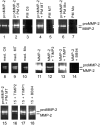
Gelatin zymography. ProMMP-2 (lane 1) is partially converted to mature MMP-2 by C6 plasma membranes (PM C6) (lane 2), or MT1-MMP–transfected fibroblast plasma membranes (PM MT) (lane 4), but not by mock-transfected fibroblast plasma membranes (PM Mo) (lane 6). Plasma membranes alone (PM C6, lane 3; PM MT, lane 5; PM Mo, lane 7) neither contain mature nor proMMP-2. Conditioned media of C6 cells (lane 8), MT1-MMP–transfected fibroblasts (lane 9) and mock-transfected fibroblasts (lane 10) contain active and proMMP-2. Effects of protease inhibitors: MMP-2 is strongly inhibited by 100 nM TIMP2 (lane 12 versus lane 11) and 1 μM BB94 (lane 14), but not by 100 nM TIMP1 (lane 13). Conversion of proMMP-2 to mature MMP-2 by MT1-MMP–transfected fibroblast plasma membranes is decreased by TIMP2 (100 nM, lane 16 versus lane 15), but not by TIMP1 (100 nM, lane 17) or BB94 (1 μM).
Figure 4
Figure 4
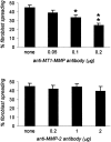
C6 plasma membrane extracts depleted of MT1-MMP by immunoprecipitation can no more inactivate the inhibitory activity of CNS myelin for fibroblast spreading, whereas immunodepletion of gelatinase A/MMP-2 did not affect the inactivation potential of the extract. 40 μg of C6 plasma membrane detergent extracts were immunodepleted with the indicated amounts anti–MT1-MMP antibody or anti–MMP-2 antibody. Inhibitory CNS substrate-coated wells were subsequently incubated for 30 min at 37°C with the immunodepleted extracts (supernatants), washed, and then used to evaluate the spreading ability of normal fibroblasts. Values are means ± SEM of two experiments performed in triplicate. Significance with the two-tailed Student's t test: P < 0.1 (*); P < 0.01 (**).
Figure 5
Figure 5
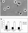
(a) Spreading of MT1-MMP–transfected fibroblasts on culture dishes coated with inhibitory CNS membrane protein extracts. 1 h after plating ∼35% of the MT1-MMP–transfected cells were well attached and showed a flat appearance (arrow). The nonspreading cells appear as round, phase-bright cells. (b) Quantification of the spreading of MT1-MMP–, mock-, and nontransfected fibroblast on CNS inhibitory substrate and control substrate (PLYS). Significantly more MT1-MMP–transfected fibroblasts spread on CNS myelin as compared with mock- or nontransfected fibroblasts. In all three conditions, cells spread equally well on the control substrate PLYS. Values are means ± SEM of three experiments, performed in triplicate. Significance with the two-tailed Student's t test: P < 0.01 (**).
Figure 6
Figure 6
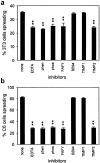
Effect of various metalloprotease inhibitors on the spreading ability of MT1-MMP–transfected fibroblasts and C6 glioma cells on CNS myelin. (a) Transiently MT1-MMP–transfected fibroblasts of which only ∼15% express high MT1-MMP levels. The proportion of spreading cells on CNS myelin substrate was reduced by 15% by 1 mM EDTA, 200 μM o-phenanthroline (phen), 300 μM phosphoramidon (phos), 10 μM Cbz-Phe-Ala-Phe-Tyr-amide (FAFY), and 100 nM TIMP2. No inhibition was observed in presence of 1 μM BB94 or 100 nM TIMP1. (b) About 80% of the C6 cells spread on the inhibitory CNS myelin substrate. This was reduced to ∼25% by the same inhibitors found to affect the MT1-MMP–transfected fibroblasts. Values are means ± SEM of three experiments, performed in triplicate. Significance with the two-tailed Student's t test: P < 0.01 (**).
Figure 7
Figure 7
MT1-MMP–transfected fibroblasts migrate on CNS myelin. (a) MT1-MMP– transfected fibroblasts were plated in a defined area (marked by circle). After 24 h the area covered with cells doubled in size. (b and c) Enlargements of the rim of the migration zone show the migration of single MT1-MMP– transfected cells. (d) These “pioneer” cells are expressing high levels of MT1-MMP with foci of MT1-MMP on their processes as shown by MT1-MMP immunostaining (same field as c). Bars: (a) 300 μm; (b) 150 μm; (c and d) 50 μm.
Figure 8
Figure 8
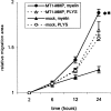
Time-course and quantification of 3T3 fibroblast migration. MT1-MMP–transfected fibroblasts migrated extensively on inhibitory CNS protein substrate within 24 h, in contrast to mock-transfected fibroblasts, whereas both migrated equally well on the control substrate (PLYS). Values are means ± SEM of four experiments, performed in quadruplicate. Significance with the two-tailed Student's t test: P < 0.01 (**).
Figure 10
Figure 10
MT1-MMP–transfected fibroblasts infiltrated the optic nerves over longer distances than mock-transfected fibroblasts. Values are means ± SEM of 12 nerves infiltrated with MT1-MMP–transfected fibroblasts, 12 nerves with mock-transfected fibroblasts, and eight nerves with C6 cells. Significance with the two-tailed Student's t test: P < 0.01 (**).
Figure 11
Figure 11
Western blot of bNI-220–enriched CNS protein extract using a polyclonal rabbit antibody against bNI-220 (Huber et al., 1998). CNS protein extract enriched for bNI-220 was preincubated for 1 h at 37°C in the presence of plasma membranes of C6 cells and transfected fibroblasts. The protein fractions then were electrophoresed on a 5% SDS polyacrylamide gel, blotted onto a nitrocellulose membrane, and then probed with purified anti–bNI-220 polyclonal rabbit antibody. Bound antibody was visualized with an peroxidase-conjugated secondary antibody followed by chemiluminescent detection. Plasma membranes of C6 glioma cells (PM C6, lane 1) and MT1-MMP–transfected fibroblasts (PM MT, lane 2) could degrade the bNI-220 completely (C6 glioma cells) or partially (MT1-MMP–transfected fibroblasts), whereas mock-transfected fibroblast plasma membranes could not (PM Mo, lane 3). Digestion of bNI-220 in the presence of PM MT was blocked by 100 nM TIMP2 (lane 4), but not by 100 nM TIMP1 (lane 5) or 1 μM BB94 (lane 6). Arrow indicates the bNI-220 level, arrowheads point to a nonspecific band, indicating that in all lanes equal amounts of protein were loaded.

Similar articles

Cited by

References

    1. Amberger VR, Paganetti PA, Seulberger H, Eldering JA, Schwab ME. Characterization of a membrane-bound metalloendoprotease of rat C6 glioblastoma cells. Cancer Res. 1994;54:4017–4025. - PubMed
    1. Amberger VR, Hensel T, Ogata N, Schwab ME. Spreading and migration of human glioma and rat C6 cells on central nervous system myelin in vitro is correlated with tumor malignancy and involves a metalloproteolytic activity. Cancer Res. 1998;58:149–158. - PubMed
    1. Atkinson SJ, Crabbe T, Cowell S, Ward RV, Butler MJ, Sato H, Seiki M, Reynolds JJ, Murphy G. Intermolecular autolytic cleavage can contribute to the activation of progelatinase A by cell membranes. J Biol Chem. 1995;270:30479–30485. - PubMed
    1. Benda ME, Lightbody J, Sato G, Levine L, Sweet W. Differential rat glia cell strain in tissue culture. Science. 1968;161:370–371. - PubMed
    1. Berens ME, Rief MD, Loo MA, Giese A. The role of extracellular matrix in human astrocytoma migration and proliferation studied in a microliter scale assay. Clin Exp Met. 1994;12:405–415. - PubMed

Publication types

MeSH terms