Comparative analysis of cytotoxic T lymphocytes in lymph nodes and peripheral blood of simian immunodeficiency virus-infected rhesus monkeys
- PMID: 9882363
- PMCID: PMC103982
- DOI: 10.1128/JVI.73.2.1573-1579.1999
Comparative analysis of cytotoxic T lymphocytes in lymph nodes and peripheral blood of simian immunodeficiency virus-infected rhesus monkeys
Abstract
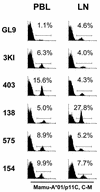
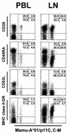
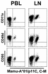
Most studies of human immunodeficiency virus type 1 (HIV-1)-specific cytotoxic T lymphocytes (CTL) have been confined to the evaluation of these effector cells in the peripheral blood. What has not been clear is the extent to which CTL activity in the blood actually reflects this effector cell function in the lymph nodes, the major sites of HIV-1 replication. To determine the concordance between CTL activity in lymph nodes and peripheral blood lymphocytes (PBL), CTL specific for simian immunodeficiency virus of macaques (SIVmac) have been characterized in lymph nodes of infected, genetically selected rhesus monkeys by using both Gag peptide-specific functional CTL assays and tetrameric peptide-major histocompatibility complex (MHC) class I molecule complex staining techniques. In studies of six chronically SIVmac-infected rhesus monkeys, Gag epitope-specific functional lytic activity and specific tetrameric peptide-MHC class I staining were readily demonstrated in lymph node T lymphocytes. Although the numbers of tetramer-binding cells in some animals differed from those documented in their PBL, the numbers of tetramer-binding cells from these two different compartments were not statistically different. Phenotypic characterization of the tetramer-binding CD8(+) lymph node T lymphocytes of the infected monkeys demonstrated a high level of expression of the activation-associated adhesion molecules CD11a and CD49d, the Fas molecule CD95, and MHC class II-DR. These studies documented a low expression of the naive T-cell marker CD45RA and the adhesion molecule CD62L. This phenotypic profile of the tetramer-binding lymph node CD8(+) T cells was similar to that of tetramer-binding CD8(+) T cells from PBL. These observations suggest that characterization of AIDS virus-specific CTL activity by sampling of cells in the peripheral blood should provide a reasonable estimation of CTL in an individual's secondary lymphoid tissue.
Figures


Similar articles
-
Simian immunodeficiency virus-specific cytotoxic T lymphocytes and cell-associated viral RNA levels in distinct lymphoid compartments of SIVmac-infected rhesus monkeys.Blood. 2000 Aug 15;96(4):1474-9. Blood. 2000. PMID: 10942394
-
Analysis of Gag-specific cytotoxic T lymphocytes in simian immunodeficiency virus-infected rhesus monkeys by cell staining with a tetrameric major histocompatibility complex class I-peptide complex.J Exp Med. 1998 May 4;187(9):1373-81. doi: 10.1084/jem.187.9.1373. J Exp Med. 1998. PMID: 9565630 Free PMC article.
-
Use of major histocompatibility complex class I/peptide/beta2M tetramers to quantitate CD8(+) cytotoxic T lymphocytes specific for dominant and nondominant viral epitopes in simian-human immunodeficiency virus-infected rhesus monkeys.J Virol. 1999 Jul;73(7):5466-72. doi: 10.1128/JVI.73.7.5466-5472.1999. J Virol. 1999. PMID: 10364294 Free PMC article.
-
Cytotoxic T lymphocytes specific for the simian immunodeficiency virus.Immunol Rev. 1999 Aug;170:127-34. doi: 10.1111/j.1600-065x.1999.tb01334.x. Immunol Rev. 1999. PMID: 10566147 Review.
-
Simian immunodeficiency virus-specific cytotoxic T lymphocytes in rhesus monkeys: characterization and vaccine induction.Semin Immunol. 1993 Jun;5(3):215-23. doi: 10.1006/smim.1993.1025. Semin Immunol. 1993. PMID: 8394161 Review.
Cited by
-
The first T cell response to transmitted/founder virus contributes to the control of acute viremia in HIV-1 infection.J Exp Med. 2009 Jun 8;206(6):1253-72. doi: 10.1084/jem.20090365. Epub 2009 Jun 1. J Exp Med. 2009. PMID: 19487423 Free PMC article.
-
Migration of antigen-specific T cells away from CXCR4-binding human immunodeficiency virus type 1 gp120.J Virol. 2004 May;78(10):5184-93. doi: 10.1128/jvi.78.10.5184-5193.2004. J Virol. 2004. PMID: 15113900 Free PMC article.
-
Immune distribution and localization of phosphoantigen-specific Vgamma2Vdelta2 T cells in lymphoid and nonlymphoid tissues in Mycobacterium tuberculosis infection.Infect Immun. 2008 Jan;76(1):426-36. doi: 10.1128/IAI.01008-07. Epub 2007 Oct 8. Infect Immun. 2008. PMID: 17923514 Free PMC article.
-
Detection of simian immunodeficiency virus Gag-specific CD8(+) T lymphocytes in semen of chronically infected rhesus monkeys by cell staining with a tetrameric major histocompatibility complex class I-peptide complex.J Virol. 1999 May;73(5):4508-12. doi: 10.1128/JVI.73.5.4508-4512.1999. J Virol. 1999. PMID: 10196357 Free PMC article.
-
Weak anti-HIV CD8(+) T-cell effector activity in HIV primary infection.J Clin Invest. 1999 Nov;104(10):1431-9. doi: 10.1172/JCI7162. J Clin Invest. 1999. PMID: 10562305 Free PMC article.
References
-
- Allen T M, Sidney J, del Guercio M F, Glickman R L, Lensmeyer G L, Wiebe D A, DeMars R, Pauza C D, Johnson R P, Sette A, Watkins D I. Characterization of the peptide binding motif of a rhesus MHC class I molecule (Mamu-A*01) that binds an immunodominant CTL epitope from simian immunodeficiency virus. J Immunol. 1998;160:6062–6071. - PubMed
-
- Altman J D, Moss P A H, Goulder P J R, Barouch D H, McHeyzer-Williams M G, Bell J I, McMichael A J, Davis M M. Phenotypic analysis of antigen-specific T lymphocytes. Science. 1996;274:94–96. - PubMed
-
- Azuma M, Phillips J H, Lanier L L. CD28− T lymphocytes. Antigenic and functional properties. J Immunol. 1993;150:1147–1159. - PubMed
-
- Baume D M, Caligiuri M A, Manley T J, Daley J F, Ritz J. Differential expression of CD8 alpha and CD8 beta associated with MHC-restricted and non-MHC-restricted cytolytic effector cells. Cell Immunol. 1990;131:352–365. - PubMed
-
- Borthwick N J, Bofill M, Gombert W M, Akbar A N, Medina E, Sagawa K, Lipman M C, Johnson M A, Janossy G. Lymphocyte activation in HIV-1 infection. II. Functional defects of CD28− T cells. J Acquired Immune Defic Syndr. 1994;8:431–441. - PubMed
Publication types
MeSH terms
Substances
Grants and funding
LinkOut - more resources
Full Text Sources
Research Materials
Miscellaneous

