Mature follicular dendritic cell networks depend on expression of lymphotoxin beta receptor by radioresistant stromal cells and of lymphotoxin beta and tumor necrosis factor by B cells
- PMID: 9874572
- PMCID: PMC1887694
- DOI: 10.1084/jem.189.1.159
Mature follicular dendritic cell networks depend on expression of lymphotoxin beta receptor by radioresistant stromal cells and of lymphotoxin beta and tumor necrosis factor by B cells
Abstract
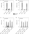
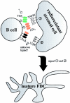
The formation of germinal centers (GCs) represents a crucial step in the humoral immune response. Recent studies using gene-targeted mice have revealed that the cytokines tumor necrosis factor (TNF), lymphotoxin (LT) alpha, and LTbeta, as well as their receptors TNF receptor p55 (TNFRp55) and LTbetaR play essential roles in the development of GCs. To establish in which cell types expression of LTbetaR, LTbeta, and TNF is required for GC formation, LTbetaR-/-, LTbeta-/-, TNF-/-, B cell-deficient (BCR-/-), and wild-type mice were used to generate reciprocal or mixed bone marrow (BM) chimeric mice. GCs, herein defined as peanut agglutinin-binding (PNA+) clusters of centroblasts/centrocytes in association with follicular dendritic cell (FDC) networks, were not detectable in LTbetaR-/- hosts after transfer of wild-type BM. In contrast, the GC reaction was restored in LTbeta-/- hosts reconstituted with either wild-type or LTbetaR-/- BM. In BCR-/- recipients reconstituted with compound LTbeta-/-/BCR-/- or TNF-/-/BCR-/- BM grafts, PNA+ cell clusters formed in splenic follicles, but associated FDC networks were strongly reduced or absent. Thus, development of splenic FDC networks depends on expression of LTbeta and TNF by B lymphocytes and LTbetaR by radioresistant stromal cells.
Figures


Similar articles
-
Distinct contributions of TNF and LT cytokines to the development of dendritic cells in vitro and their recruitment in vivo.Blood. 2003 Feb 15;101(4):1477-83. doi: 10.1182/blood.V101.4.1477. Blood. 2003. PMID: 12560241
-
Distinct roles of lymphotoxin alpha and the type I tumor necrosis factor (TNF) receptor in the establishment of follicular dendritic cells from non-bone marrow-derived cells.J Exp Med. 1997 Dec 15;186(12):1997-2004. doi: 10.1084/jem.186.12.1997. J Exp Med. 1997. PMID: 9396768 Free PMC article.
-
TNF and lymphotoxin beta cooperate in the maintenance of secondary lymphoid tissue microarchitecture but not in the development of lymph nodes.J Immunol. 1999 Dec 15;163(12):6575-80. J Immunol. 1999. PMID: 10586051
-
The lymphotoxin pathway: beyond lymph node development.Immunol Res. 2006;35(1-2):41-54. doi: 10.1385/IR:35:1:41. Immunol Res. 2006. PMID: 17003508 Review.
-
Lymphotoxin-alpha-deficient and TNF receptor-I-deficient mice define developmental and functional characteristics of germinal centers.Immunol Rev. 1997 Apr;156:137-44. doi: 10.1111/j.1600-065x.1997.tb00965.x. Immunol Rev. 1997. PMID: 9176705 Review.
Cited by
-
Distinct TLR-mediated cytokine production and immunoglobulin secretion in human newborn naïve B cells.Innate Immun. 2016 Aug;22(6):433-43. doi: 10.1177/1753425916651985. Epub 2016 Jun 1. Innate Immun. 2016. PMID: 27252169 Free PMC article.
-
Follicular dendritic cell dedifferentiation reduces scrapie susceptibility following inoculation via the skin.Immunology. 2005 Feb;114(2):225-34. doi: 10.1111/j.1365-2567.2004.02074.x. Immunology. 2005. PMID: 15667567 Free PMC article.
-
B cell- and T cell-intrinsic regulation of germinal centers by thymic stromal lymphopoietin signaling.Sci Immunol. 2023 Jan 6;8(79):eadd9413. doi: 10.1126/sciimmunol.add9413. Epub 2023 Jan 6. Sci Immunol. 2023. PMID: 36608149 Free PMC article.
-
B cells and autoantibodies in the pathogenesis of multiple sclerosis and related inflammatory demyelinating diseases.Adv Immunol. 2008;98:121-49. doi: 10.1016/S0065-2776(08)00404-5. Adv Immunol. 2008. PMID: 18772005 Free PMC article. Review.
-
Attrition of T Cell Zone Fibroblastic Reticular Cell Number and Function in Aged Spleens.Immunohorizons. 2018 May;2(5):155-163. doi: 10.4049/immunohorizons.1700062. Epub 2018 Jul 23. Immunohorizons. 2018. PMID: 30706058 Free PMC article.
References
-
- Kosco-Vilbois MH, Bonnefoy JY, Chvatchko Y. The physiology of murine germinal center reactions. Immunol Rev. 1997;156:127–136. - PubMed
-
- Kelsoe G, Zheng B. Sites of B-cell activation in vivo. Curr Opin Immunol. 1993;5:418–422. - PubMed
-
- MacLennan IC. Germinal centers. Annu Rev Immunol. 1994;12:117–139. - PubMed
-
- Tew JG, Wu J, Qin D, Helm S, Burton GF, Szakal AK. Follicular dendritic cells and presentation of antigen and costimulatory signals to B cells. Immunol Rev. 1997;156:39–52. - PubMed
-
- Liu YJ, Arpin C. Germinal center development. Immunol Rev. 1997;156:111–126. - PubMed
Publication types
MeSH terms
Substances
LinkOut - more resources
Full Text Sources
Other Literature Sources
Molecular Biology Databases
Miscellaneous

