. 1998 Dec 28;143(7):1871-81.
doi: 10.1083/jcb.143.7.1871.

Redundant and distinct functions for dynamin-1 and dynamin-2 isoforms

Affiliations

Redundant and distinct functions for dynamin-1 and dynamin-2 isoforms

Y Altschuler et al. J Cell Biol. .

Abstract

A role for dynamin in clathrin-mediated endocytosis is now well established. However, mammals express three closely related, tissue-specific dynamin isoforms, each with multiple splice variants. Thus, an important question is whether these isoforms and splice variants function in vesicle formation from distinct intracellular organelles. There are conflicting data as to a role for dynamin-2 in vesicle budding from the TGN. To resolve this issue, we compared the effects of overexpression of dominant-negative mutants of dynamin-1 (the neuronal isoform) and dynamin-2 (the ubiquitously expressed isoform) on endocytic and biosynthetic membrane trafficking in HeLa cells and polarized MDCK cells. Both dyn1(K44A) and dyn2(K44A) were potent inhibitors of receptor-mediated endocytosis; however neither mutant directly affected other membrane trafficking events, including transport mediated by four distinct classes of vesicles budding from the TGN. Dyn2(K44A) more potently inhibited receptor-mediated endocytosis than dyn1(K44A) in HeLa cells and at the basolateral surface of MDCK cells. In contrast, dyn1(K44A) more potently inhibited endocytosis at the apical surface of MDCK cells. The two dynamin isoforms have redundant functions in endocytic vesicle formation, but can be targeted to and function differentially at subdomains of the plasma membrane.


Figures

Figure 1
Figure 1
Tetracycline-regulatable expression of dynamin mutants in adenovirally infected tTA-HeLa cells. tTA-HeLa cells were infected with recombinant adenovirus encoding mutant dyn1 and dyn2 under control of a tetracycline (tet)-regulatable promoter. After infection, cells were incubated for 16–18 h in the presence of the indicated concentrations of tet. Tfn endocytosis was measured as indicated in Materials and Methods and is shown by the solid bars relative to uninfected control cells. Inserts show Western blots of dynamin expression that was quantitated by ECL detection and scanning densitometry and shown by the hatched bars.
Figure 2
Figure 2
Tfn endocytosis is potently inhibited by dyn1 (K44A) and dyn2(K44A). tTA-HeLa cells were infected with recombinant adenovirus encoding dyn1(wt) (□), dyn2(wt) (Δ), dyn1 (K44A) (▪), or dyn2(K44A) (▴). Cells were incubated in the presence of 5 ng/ml tet for 18 h to induce expression. Tfn endocytosis was measured by incubating cells with 4 μg/ml B-Tfn for the indicated times as 37°C before measuring intracellular B-Tfn based on its inaccessibility to avidin as previously described (55). Results shown are expressed as the percentage internalized at 37°C vs. total bound at 4°C and are >100% owing to internalization of recycled Tfn-receptors. They are representative of >5 experiments.
Figure 3
Figure 3
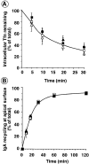
Recycling from endosomes is unaffected by dyn1(K44A) or dyn2(K44A). Adenovirally infected tTA-HeLa cells (A) or MDCK T23 cells (B) overexpressing dyn1(wt) (□); dyn1(K44A) (▪); dyn2(wt) (Δ), or dyn2 (K44A) (▴) were cultured for 18 h after infection in the presence (A) or absence (B) of 5 ng/ml tet. (A) HeLa cells were then loaded with B-Tfn at 37°C to steady state. Surface bound B-Tfn was masked with avidin at 4°C and the recycling of intracellular B-Tfn was determined after incubation at 37°C for the indicated times as described in Materials and Methods. (B) MDCK T23 cells were incubated with 125I-IgA at 4°C for 60 min and unbound IgA was eliminated by three quick washes with cold media. Surface bound ligand was internalized by warming the cells to 37°C for 5 min. Cells were then cooled down, cell surface IgA was removed by trypsin treatment, and the recycling of intracellular 125I-IgA was determined, as described in Materials and Methods, after incubation at 37°C for the indicated times. Results shown are average of three experiments.
Figure 4
Figure 4
Initial rates of fluid phase endocytosis are unaffected by overexpression of dyn2(K44A). Endocytosis of HRP into noninfected cells (○) or adenovirally infected tTA-HeLa cells expressing either wt (Δ) or K44A (▴) dyn2 was assessed as described in Materials and Methods. Results shown are representative of >4 experiments.
Figure 5
Figure 5
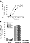
Delivery of three classes of biosynthetic transport vesicles from the TGN to the plasma membrane in cells overexpressing dyn1 and dyn2 mutants. (A) Adenovirally infected tTA HeLa cells overexpressing dyn2(wt) (Δ), dyn2(K44A) (▴), or dyn1 (K44A) (▪) were pulse-labeled with 35S-translabel for 30 min at 37°C and then incubated in methionine-containing chase media for the indicated times. The appearance of newly synthesized TfnR on the cell surface was determined by its susceptibility to trypsin digestion as described in Materials and Methods. The results shown are representative of three experiments. (B) Adenovirally infected MDCK T23 cells or uninfected control cells were pulse-labeled for 10 min with 35S-methionine followed by incubation at 37°C in chase media. The distribution of biosynthetically labeled pIgR was determined following immunoprecipitation of intracellular or apical or basolateral membrane associated pools as previously described (2).
Figure 6
Figure 6
Cathepsin D processing is not significantly inhibited in cells overexpressing dyn1 or dyn2 mutants. Adenovirally infected tTA HeLa cells overexpressing dyn1(wt) (□), dyn1(K44A) (▪), or dyn2(K44A) (▴) were pulse-labeled with 35S-translabel for 30 min at 37°C and then incubated in methionine-containing chase media for the indicated times. The appearance of the mature form of cathepsin D in late endosomes/lysosomes was determined following immunoprecipitation and quantitation using PhosphorImager analysis as described in Materials and Methods. The results are expressed as percent of wt control and are the average of three experiments.
Figure 7
Figure 7
Immunofluorescence localization of endogenous dyn2 in HeLa cells. tTA-HeLa cells were processed for indirect immunofluorescence as described in Materials and Methods. Shown is the punctate localization of endogenous dyn2, as detected using the mAb Hudy-1 (a and b), its colocalization with puncta located at the cell periphery detected using anti-clathrin light chain rabbit sera (a′) and the lack of colocalization with the human TGN48, detected using monospecific rabbit sera (b′). Secondary antibodies were Alexa 488-conjugated goat anti–mouse and Texas red– conjugated goat anti–rabbit. Images were obtained using a Zeiss Axiovert 100TV, with the Bio-Rad MRC1024 confocal system and show a projection of either 4 optical sections taken through the middle of the cell (a and a′) or of 12 optical sections from the bottom to top of the cell (b and b′).
Figure 8
Figure 8
Immunofluores-cence localization of endogenous and overexpressed dyn2 in HeLa cells. Uninfected tTA-HeLa cells (a, d, and g) or adenovirally infected tTA-HeLa cells expressing dyn2(wt) (b, e, h, j, and l) or dyn2(K44A) (c, f, i, k, and m) were fixed with 4% PFA and processed for indirect immunofluorescence as described in Materials and Methods. For dynamin localization in adenovirally infected cells overexpressing recombinant dynamin, cells were permeabilized with streptolysin O and cytosol was removed before fixation (b, c, e, f, h, and i). Primary antibodies used are indicated on the figure and their sources are given in Materials and Methods. Dyn2 = mAb anti-dynamin antibody, hudy1; clathrin = rabbit polyclonal anti-clathrin light chain; AP1 = rabbit polyclonal anti-γ adaptin; TGN48 = rabbit polyclonal anti–human TGN48; MPR = rabbit polyclonal anti-MPR (j and k) or goat polyclonal anti-MPR (l and m). Images were obtained using a Zeiss Axiovert 100TV, with the Bio-Rad MRC1024 confocal system and show sections through the top (a–c) or middle (d–k) of the cell.
Figure 9
Figure 9
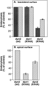
Differential effects of dyn1(K44A) and dyn2 (K44A) on endocytosis in polarized MDCK cells. MDCK T23 cells were infected with recombinant adenovirus encoding either dyn2(wt), dyn1(K44A) or dyn2(K44A) and further incubated for 18 h at 37°C. Endocytosis from the basolateral (A) or apical surface (B) was determined by incubating cells for 1 h at 4°C in the presence of 125I-Tfn (solid bars) or 125I-IgA (striped bars) as described in Materials and Methods. Cells were next washed to remove unbound ligand and then rapidly warmed to 37°C for 5 min. Surface associated ligands were stripped by extensive washing with 0.15 M glycine in PBS. The extent of internalization relative to noninfected control cells is shown.
Figure 10
Figure 10
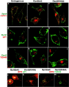
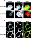
Dyn1 and dyn2 are differentially targeted to the apical and basolateral plasma membrane in polarized MDCK cells MDCK. T23 cells were infected with recombinant adenovirus encoding either HA-tagged dyn1(K44A) or dyn2(K44A), as indicated. The cells were further incubated for 18 h in the presence of 0.1 ng/ml doxycyclin (to reduce levels of overexpression) before fixation of the cells and processing for indirect immunofluorescence as described in Materials and Methods. A is a section through the apical surface, labeled with the apical plasma membrane protein, gp135, showing the targeting of dyn1(K44A) and dyn2(K44A) as detected using a rat anti-HA antibody. B is a section through a basolateral surface at the level of the nucleus showing targeting of dyn1(K44A) and dyn2(K44A) to the basolateral surface, as indicated by colocalization with E-cadherin. The excess label in the cytoplasm reflects overexpression of the recombinant dynamins.

Similar articles

Cited by

References

    1. Aroeti B, Kosen PA, Kuntz ID, Cohen FE, Mostov KE. Mutational and secondary structural analysis of the basolateral sorting signal of the polymeric immunoglobulin receptor. J Cell Biol. 1993;123:1149–1160. - PMC - PubMed
    1. Aroeti B, Mostov KE. Polarized sorting of the polymeric immunoglobulin receptor in the exocytotic and endocytotic pathways is controlled by the same amino acids. EMBO (Eur Mol Biol Organ) J. 1994;13:2297–2304. - PMC - PubMed
    1. Banting G, Maile R, Roquemore EP. The steady state distribution of humTGN46 is not significantly altered in cells defective in clathrin-mediated endocytosis. J Cell Sci. 1998;111:3451–3458. - PubMed
    1. Barth AIM, Pollack AL, Altschuler Y, Mostov KE, Nelson WJ. NH2-terminal detection of beta-catenin with adenomatous polyposis coli protein and altered MDCK cell adhesion. J Cell Biol. 1997;136:693–706. - PMC - PubMed
    1. Cao H, Garcia F, Krueger EW, McNiven MA. Differential distribution of dynamin isoforms in mammalian cells. Mol Biol Cell. 1997;8:424a. - PMC - PubMed

Publication types

MeSH terms