Nucleoporins nup98 and nup214 participate in nuclear export of human immunodeficiency virus type 1 Rev
- PMID: 9847314
- PMCID: PMC103815
- DOI: 10.1128/JVI.73.1.120-127.1999
Nucleoporins nup98 and nup214 participate in nuclear export of human immunodeficiency virus type 1 Rev
Abstract
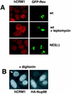
Human immunodeficiency virus type 1 (HIV-1) Rev contains a leucine-rich nuclear export signal that is essential for its nucleocytoplasmic export mediated by hCRM1. We examined the role of selected nucleoporins, which are located in peripheral structures of the nuclear pore complex and are thought to be involved in export, in Rev function in human cells. First, we found that upon actinomycin D treatment, Nup98, but not Nup214 or Nup153, is able to translocate to the cytoplasm of HeLa cells, demonstrating that Nup98 may act as a soluble factor. We further showed that Rev can recruit Nup98 and Nup214, but not Nup153, to the nucleolus. We also found that the isolated FG-containing repeat domains of Nup98 and Nup214, but not those of Nup153, competitively inhibit the Rev/RRE-mediated expression of HIV. Taken together, the recruitment of Nup98 and Nup214 by Rev and the competitive inhibition exhibited by their NP domains demonstrate direct participation of Nup98 and Nup214 in the Rev-hCRM1-mediated export.
Figures


Similar articles
-
Inhibition of human immunodeficiency virus Rev and human T-cell leukemia virus Rex function, but not Mason-Pfizer monkey virus constitutive transport element activity, by a mutant human nucleoporin targeted to Crm1.J Virol. 1998 Nov;72(11):8627-35. doi: 10.1128/JVI.72.11.8627-8635.1998. J Virol. 1998. PMID: 9765402 Free PMC article.
-
Epstein-Barr virus EB2 protein exports unspliced RNA via a Crm-1-independent pathway.J Virol. 2000 Jul;74(13):6068-76. doi: 10.1128/jvi.74.13.6068-6076.2000. J Virol. 2000. PMID: 10846090 Free PMC article.
-
Cofactor requirements for nuclear export of Rev response element (RRE)- and constitutive transport element (CTE)-containing retroviral RNAs. An unexpected role for actin.J Cell Biol. 2001 Mar 5;152(5):895-910. doi: 10.1083/jcb.152.5.895. J Cell Biol. 2001. PMID: 11238447 Free PMC article.
-
The HIV-1 Rev protein.Annu Rev Microbiol. 1998;52:491-532. doi: 10.1146/annurev.micro.52.1.491. Annu Rev Microbiol. 1998. PMID: 9891806 Review.
-
Retroviruses as model systems for the study of nuclear RNA export pathways.Virology. 1998 Sep 30;249(2):203-10. doi: 10.1006/viro.1998.9331. Virology. 1998. PMID: 9791012 Review. No abstract available.
Cited by
-
Oxidative stress inhibits nuclear protein export by multiple mechanisms that target FG nucleoporins and Crm1.Mol Biol Cell. 2009 Dec;20(24):5106-16. doi: 10.1091/mbc.e09-05-0397. Mol Biol Cell. 2009. PMID: 19828735 Free PMC article.
-
Patterns of HIV-1 protein interaction identify perturbed host-cellular subsystems.PLoS Comput Biol. 2010 Jul 29;6(7):e1000863. doi: 10.1371/journal.pcbi.1000863. PLoS Comput Biol. 2010. PMID: 20686668 Free PMC article.
-
Structural and functional analysis of the avian leukemia virus constitutive transport element.RNA. 1999 Dec;5(12):1645-55. doi: 10.1017/s1355838299991616. RNA. 1999. PMID: 10606274 Free PMC article.
-
Distinct functional domains within nucleoporins Nup153 and Nup98 mediate transcription-dependent mobility.Mol Biol Cell. 2004 Apr;15(4):1991-2002. doi: 10.1091/mbc.e03-10-0743. Epub 2004 Jan 12. Mol Biol Cell. 2004. PMID: 14718558 Free PMC article.
-
Ribozyme-mediated inhibition of HIV 1 suggests nucleolar trafficking of HIV-1 RNA.Proc Natl Acad Sci U S A. 2000 Aug 1;97(16):8955-60. doi: 10.1073/pnas.97.16.8955. Proc Natl Acad Sci U S A. 2000. PMID: 10922055 Free PMC article.
References
-
- Arai Y, Hosoda F, Kobayashi H, Arai K, Hayashi Y, Kamada N, Kaneko Y, Ohki M. The inv(11)(p15q22) chromosome translocation of de novo and therapy-related myeloid malignancies results in fusion of the nucleoporin gene, NUP98, with the putative RNA helicase gene, DDX10. Blood. 1997;89:3936–3944. - PubMed
-
- Boer J M, van Deursen J M, Croes H J, Fransen J A, Grosveld G C. The nucleoporin CAN/Nup214 binds to both the cytoplasmic and the nucleoplasmic sides of the nuclear pore complex in overexpressing cells. Exp Cell Res. 1997;232:182–185. - PubMed
-
- Bogerd H P, Fridell R A, Madore S, Cullen B R. A novel cellular co-factor for HIV-1 Rev. Cell. 1995;82:485–494. - PubMed
Publication types
MeSH terms
Substances
LinkOut - more resources
Full Text Sources
Miscellaneous

