. 1998 Nov 15;12(22):3512-27.
doi: 10.1101/gad.12.22.3512.

The interaction between HIV-1 Tat and human cyclin T1 requires zinc and a critical cysteine residue that is not conserved in the murine CycT1 protein

Affiliations

The interaction between HIV-1 Tat and human cyclin T1 requires zinc and a critical cysteine residue that is not conserved in the murine CycT1 protein

M E Garber et al. Genes Dev. .

Abstract

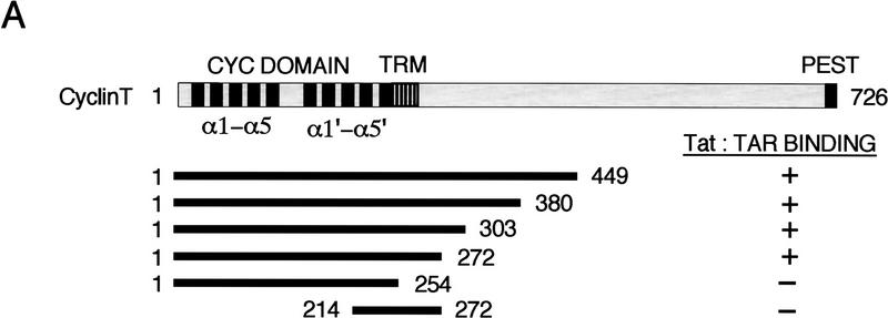
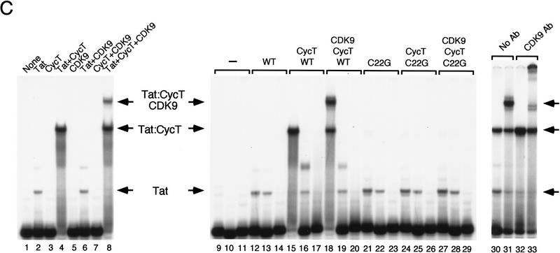
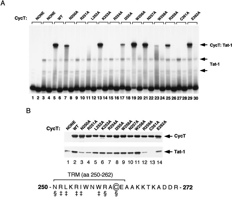
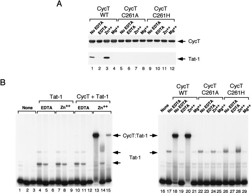
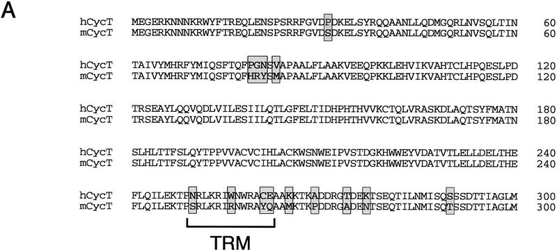
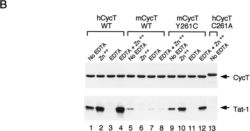
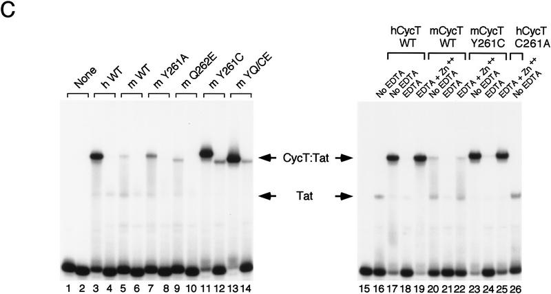
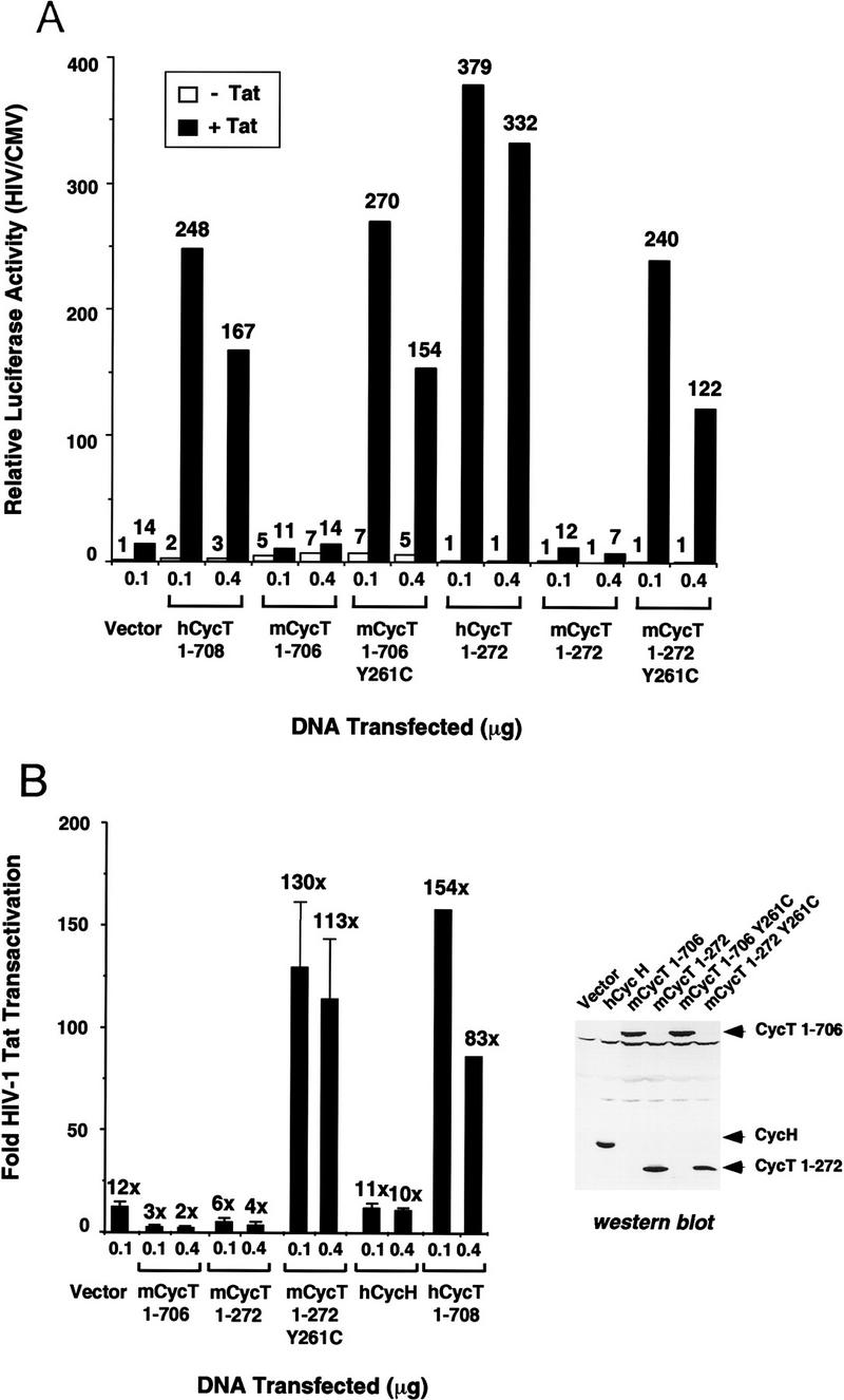
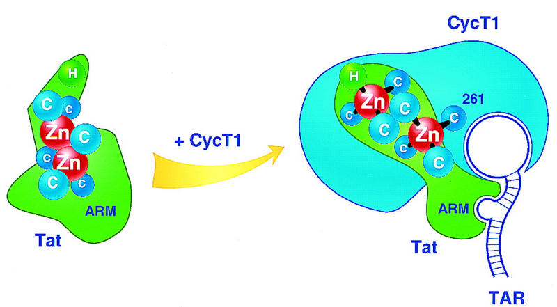
HIV-1 Tat activates transcription through binding to human cyclin T1, a regulatory subunit of the TAK/P-TEFb CTD kinase complex. Here we show that the cyclin domain of hCycT1 is necessary and sufficient to interact with Tat and promote cooperative binding to TAR RNA in vitro, as well as mediate Tat transactivation in vivo. A Tat:TAR recognition motif (TRM) was identified at the carboxy-terminal edge of the cyclin domain, and we show that hCycT1 can interact simultaneously with Tat and CDK9 on TAR RNA in vitro. Alanine-scanning mutagenesis of the hCycT1 TRM identified residues that are critical for the interaction with Tat and others that are required specifically for binding of the complex to TAR RNA. Interestingly, we find that the interaction between Tat and hCycT1 requires zinc as well as essential cysteine residues in both proteins. Cloning and characterization of the murine CycT1 protein revealed that it lacks a critical cysteine residue (C261) and forms a weak, zinc-independent complex with HIV-1 Tat that greatly reduces binding to TAR RNA. A point mutation in mCycT1 (Y261C) restores high-affinity, zinc-dependent binding to Tat and TAR in vitro, and rescues Tat transactivation in vivo. Although overexpression of hCycT1 in NIH3T3 cells strongly enhances transcription from an integrated proviral promoter, we find that this fails to overcome all blocks to productive HIV-1 infection in murine cells.


Figures

Figure 1
Figure 1
Identification of the TRM of hCycT1. (A) Summary of the ability of different truncated hCycT1 proteins to bind cooperatively with HIV-1 Tat to TAR RNA in gel-mobility shift experiments. The location of the TRM relative to the five helices (α1–5 and α1′–5′) of each fold of the cyclin domain are indicated. (B) Analysis of the ability of mutant hCycT1 proteins to interact directly with Tat. GST–hCycT1 (amino acids 1–303) proteins were coupled to beads and incubated with wild-type or mutant HIV-1 Tat (lanes 3–8) or HIV-2 Tat (lanes 11–16), as indicated above each lane. Aliquots corresponding to 10% of the input protein are shown for wild-type HIV-1 Tat (lane 1); HIV-1 Tat C22G (lane 2); wild-type HIV-2 Tat (lane 9); and HIV-2 Tat C59A (lane 10). Proteins were visualized by Western blot. (C) The cyclin domain of hCycT1 can interact simultaneously with HIV-1 Tat and CDK9. Binding of recombinant wild-type Tat, CDK9, and hCycT1 (amino acids 1–303) to HIV-1 TAR RNA was analyzed with gel-mobility shift experiments. Binding reactions contained 100 ng of (GST-cleaved) HIV-1 Tat (amino acids 1–86); 250 ng (GST-cleaved) hCycT1 (amino acids 1–303); and 200 ng of baculovirus-expressed CDK9, as indicated above each lane. Lanes 10, 13, 16, 19, 22, 25, and 28 contained a loop mutant TAR (+29/+34), and lanes 11, 14, 17, 20, 23, 26, and 29 contained a bulge mutant TAR RNA(U22A); all other lanes contained wild-type HIV-1 TAR RNA. Reactions in lanes 12–20 contained wild-type (WT) HIV-1 Tat, whereas lanes 21–29 contained an activation domain HIV-1 Tat mutant (C22G). (Right panel) CDK9 antibody alters the mobility of the Tat–hCycT1–CDK9 complex on HIV-1 TAR RNA. Reactions contained 100 ng (GST-cleaved) of HIV-1 Tat and 80 ng (GST-cleaved) of hCycT1 (amino acids 1–303) (lanes 30–33); 120 ng of CDK9 (lanes 31 and 33); and 100 ng of antisera specific to CDK9 (lanes 32 and 33). Arrows indicate the positions of the Tat:TAR, hCycT1–Tat:TAR, and CDK9–hCycT1–Tat:TAR complexes.
Figure 1
Figure 1
Identification of the TRM of hCycT1. (A) Summary of the ability of different truncated hCycT1 proteins to bind cooperatively with HIV-1 Tat to TAR RNA in gel-mobility shift experiments. The location of the TRM relative to the five helices (α1–5 and α1′–5′) of each fold of the cyclin domain are indicated. (B) Analysis of the ability of mutant hCycT1 proteins to interact directly with Tat. GST–hCycT1 (amino acids 1–303) proteins were coupled to beads and incubated with wild-type or mutant HIV-1 Tat (lanes 3–8) or HIV-2 Tat (lanes 11–16), as indicated above each lane. Aliquots corresponding to 10% of the input protein are shown for wild-type HIV-1 Tat (lane 1); HIV-1 Tat C22G (lane 2); wild-type HIV-2 Tat (lane 9); and HIV-2 Tat C59A (lane 10). Proteins were visualized by Western blot. (C) The cyclin domain of hCycT1 can interact simultaneously with HIV-1 Tat and CDK9. Binding of recombinant wild-type Tat, CDK9, and hCycT1 (amino acids 1–303) to HIV-1 TAR RNA was analyzed with gel-mobility shift experiments. Binding reactions contained 100 ng of (GST-cleaved) HIV-1 Tat (amino acids 1–86); 250 ng (GST-cleaved) hCycT1 (amino acids 1–303); and 200 ng of baculovirus-expressed CDK9, as indicated above each lane. Lanes 10, 13, 16, 19, 22, 25, and 28 contained a loop mutant TAR (+29/+34), and lanes 11, 14, 17, 20, 23, 26, and 29 contained a bulge mutant TAR RNA(U22A); all other lanes contained wild-type HIV-1 TAR RNA. Reactions in lanes 12–20 contained wild-type (WT) HIV-1 Tat, whereas lanes 21–29 contained an activation domain HIV-1 Tat mutant (C22G). (Right panel) CDK9 antibody alters the mobility of the Tat–hCycT1–CDK9 complex on HIV-1 TAR RNA. Reactions contained 100 ng (GST-cleaved) of HIV-1 Tat and 80 ng (GST-cleaved) of hCycT1 (amino acids 1–303) (lanes 30–33); 120 ng of CDK9 (lanes 31 and 33); and 100 ng of antisera specific to CDK9 (lanes 32 and 33). Arrows indicate the positions of the Tat:TAR, hCycT1–Tat:TAR, and CDK9–hCycT1–Tat:TAR complexes.
Figure 2
Figure 2
The TRM of hCycT1 is required for Tat transactivation in vivo but is not needed for hCycT1 to bind CDK9 or to enhance GST–CTD phosphorylation. (A) Analysis of the ability of hCycT1 truncation mutants to support HIV-1 Tat transactivation in murine (NIH-3T3) cells. NIH-3T3 cells were transfected with 100 ng of either pHIV-1/LUC (+TAR) or pHIV-1/LUC+30/+33 (−TAR), 50 ng of either pSV/Tat (+Tat) or pSV/TatZX (−Tat), 10 ng of pRL/CMV, 100 ng of either pCGN vector (vector) or the different pCMV/HA–hCycT1 truncation mutants, as indicated in the figure. The firefly luciferase activity produced from the HIV-1 promoter was normalized to Renilla luciferase activity from the CMV promoter to correct for transfection efficiency. The graph plots the fold HIV-1 Tat trans-activation as the ratio of corrected firefly luciferase activity observed in the presence and absence of Tat. Standard deviations were calculated from three independent transfections. The Western blot displays the relative level of expression of each cyclin T1 protein in transfected Chinese hamster ovary (CHO) cells. (B) Analysis of the ability of the hCycT1 truncation mutants to bind to purified CDK9 in vitro. Antisera specific to CDK9 was used to immunoprecipitate complexes formed between CDK9 and the truncated hCycT1 proteins. hCycT1 binding was visualized by Western blot using an anti-GST monoclonal antibody and CDK9 was visualized with an anti-FLAG monoclonal antibody. Reactions in lanes 3–5 each contained 200 ng of purified (FLAG-tagged) CDK9 protein and 500 ng of the different GST–hCycT1 proteins, as indicated above each lane. Control reactions contained 500 ng of GST–hCycT1 (amino acids 1–303) alone (lane 1), or 200 ng of CDK9 alone (lane 2). (C) Analysis of the ability of hCycT1 truncation mutants to enhance phosphorylation of GST–CTD by CDK9 in vitro. Reactions contained 250 ng of purified (FLAG-tagged) CDK9, 250 ng of the different hCycT1 truncation mutants (as indicated above each lane), and 200 ng of purified GST–CTD. Arrows indicate the position of autophosphorylated CDK9, and the hypophosphorylated (IIa) and hyperphosphorylated (IIo) forms of the CTD.
Figure 3
Figure 3
Alanine-scanning mutagenesis of the TRM of hCycT1. (A) Analysis of the ability of different alanine-substituted hCycT1 (amino acids 1–303) proteins to enhance the binding of HIV-1 Tat to TAR RNA in a gel-mobility shift experiment. Reactions contained either wild-type TAR RNA (odd-numbered lanes) or TAR loop mutant (+29/+34) RNAs (even-numbered lanes). Reactions included 175 ng (GST-cleaved) of Tat-1 (lanes 3–30) and 40 ng (GST-cleaved) of hCycT1 (amino acids 1–303). The position of each mutation is indicated above each lane. (WT) Wild-type hCycT1 (amino acids 1–303). (B) Analysis of the ability of the different hCycT1 mutants to interact directly with HIV-1 Tat in vitro. Reactions included 60 ng (GST-cleaved) of HIV-1 Tat and 250 ng of GST–hCycT1 (amino acids 1–303), either wild-type (WT) or mutant, as indicated above each lane. The hCycT1 protein was visualized with a monoclonal antibody to GST, and the (GST-cleaved, HA-tagged) Tat was visualized with an anti-HA monoclonal antibody. Lane 1 represents 10% of the input protein (200 ng) of the wild-type Tat protein. The TRM within hCycT1 is shown at the bottom. The residues designated with the ‡ symbol are required for TAR RNA recognition, whereas the residues indicated with the § symbol are needed to bind Tat.
Figure 4
Figure 4
The interaction between HIV-1 Tat and hCycT1 requires zinc. (A) Zinc is required for the binding of HIV-1 Tat to hCycT1 in vitro. The wild-type hCycT1 (WT amino acids 1–303; lanes 1–4), or mutant hCycT1 proteins that contain a substitution of cysteine 261 to either alanine (C261A; lanes 5–8), or to histidine (C261H; lanes 9–12) were coupled to beads and incubated with (GST-cleaved) HIV-1 Tat. The Tat and hCycT1 proteins were incubated together in buffer lacking EDTA (No EDTA; lanes 1, 5, and 9), or were treated with EDTA (EDTA; lanes 2, 6, and 10), or treated with EDTA and subsequently incubated with either zinc sulfate (Zn++; lanes 3, 7, and 11) or magnesium sulfate (Mg++; lanes 4, 8, and 12), as described in Materials and Methods. Proteins were visualized by Western blot. (B) Zinc-dependent binding of HIV-1 Tat and hCycT1 to TAR RNA. (Left panel) Zinc is required for the formation of hCycT1-Tat: TAR, but not Tat:TAR, complexes. Where indicated, reactions contained 150 ng of EDTA-treated HIV-1 Tat and 60 ng of EDTA-treated (GST-cleaved) hCycT1 (amino acids 1–303). Binding reactions contained either wild-type HIV-1 TAR RNA (lanes 1,4,7,10,13); loop-substituted HIV-1 TAR RNA (+29/+34; lanes 2,5,8,11,14); or bulge mutant RNA (U22A; lanes 3,6,9,12,15). Zinc sulfate was added to the EDTA-treated proteins (Zn++) in lanes 7–9 and lanes 13–15. (Right panel) Zinc-dependent binding of the hCycT1-Tat complex to wild-type HIV-1 TAR RNA. Reactions contained 150 ng of (GST-cleaved) HIV-1 Tat and 60 ng of either wild-type GST–hCycT1 amino acids 1–303 (WT; lanes 18–21); a cysteine to alanine mutant (C261A; lanes 22–25); or a cysteine to histidine mutant (C261H; lanes 26–29), as indicated above each lane. The HIV-1 Tat and GST–hCycT1 proteins were incubated in buffer lacking EDTA (no EDTA; lanes 18,22,26), or treated with EDTA (EDTA; lanes 19,23,27), or EDTA-treated and subsequently reconstituted with either zinc sulfate (Zn++; lanes 20,24,28) or magnesium sulfate (Mg++; lanes 21,25,29). The Tat preparations used in this experiment differed in the extent of dimer (Tat, top arrow) versus monomer (Tat, bottom arrow) formed, which had no effect on the results obtained.
Figure 5
Figure 5
Analysis of the ability of the murine CycT1 protein to interact with HIV-1 Tat and TAR RNA in vitro. (A) Sequence comparison of the cyclin domains of the murine and human CycT1 proteins. Amino acid differences between the two proteins are shown in shaded boxes and the TRM at the carboxy-terminal boundary of the cyclin domain is indicated with brackets. (B) The murine CycT1 protein forms a weak, zinc-independent complex with HIV-1 Tat in vitro. Wild-type or mutant versions of the human and mouse GST–CycT1 proteins (amino acids 1–272) were coupled to beads and incubated with purified (GST-cleaved) HA-tagged HIV-1 Tat. Reactions either lacked EDTA (no EDTA; lanes 1,5,9), or contained additional zinc sulfate (Zn++; lanes 2,6,10), or were treated with EDTA in the absence (EDTA; lanes 3,7,11), or presence of exogeneous zinc sulfate (EDTA+Zn++; lanes 4,8,12). Tat was incubated with resins containing either wild-type hCycT1 (lanes 1–4), wild-type mCycT1 (lanes 5–8), or the mutants mCycT1 Y261C (lanes 9–12), or hCycT1 C261A (amino acids 1–303; lane 13). The (GST-cleaved; HA-tagged) HIV-1 Tat and various GST–CycT1 proteins were visualized by Western blot using monoclonal antisera specific to the hemagglutinin (HA) tag and GST, respectively. (C) Analysis of the ability of murine CycT1 to enhance the binding of HIV-1 Tat to TAR RNA. (Left panel) Wild-type or mutant mCycT1 (amino acids 1–272) proteins were incubated in the presence or absence of HIV-1 Tat with wild-type (odd-numbered lanes) or loop mutant (even-numbered lanes) HIV-1 TAR RNA probes. Where indicated, the reactions contained 50 ng of (GST-cleaved) HIV-1 Tat and 50 ng of (GST-cleaved) mCycT1 (amino acids 1–272). The mCycT1 YQ/CE is a double mutant containing both Y261C and Q262E substitutions. hWT refers to the GST–hCycT1 (amino acids 1–272) control. (Right panel) Zinc-dependent binding of the mCycT1–Tat complex to wild-type TAR-1 RNA. Reactions included 175 ng of HIV-1 (GST-cleaved) Tat and 50 ng of the various (GST-cleaved) CycT1 proteins, as indicated above each lane. The EDTA treatment and metal reconstitution conditions are described in B.
Figure 5
Figure 5
Analysis of the ability of the murine CycT1 protein to interact with HIV-1 Tat and TAR RNA in vitro. (A) Sequence comparison of the cyclin domains of the murine and human CycT1 proteins. Amino acid differences between the two proteins are shown in shaded boxes and the TRM at the carboxy-terminal boundary of the cyclin domain is indicated with brackets. (B) The murine CycT1 protein forms a weak, zinc-independent complex with HIV-1 Tat in vitro. Wild-type or mutant versions of the human and mouse GST–CycT1 proteins (amino acids 1–272) were coupled to beads and incubated with purified (GST-cleaved) HA-tagged HIV-1 Tat. Reactions either lacked EDTA (no EDTA; lanes 1,5,9), or contained additional zinc sulfate (Zn++; lanes 2,6,10), or were treated with EDTA in the absence (EDTA; lanes 3,7,11), or presence of exogeneous zinc sulfate (EDTA+Zn++; lanes 4,8,12). Tat was incubated with resins containing either wild-type hCycT1 (lanes 1–4), wild-type mCycT1 (lanes 5–8), or the mutants mCycT1 Y261C (lanes 9–12), or hCycT1 C261A (amino acids 1–303; lane 13). The (GST-cleaved; HA-tagged) HIV-1 Tat and various GST–CycT1 proteins were visualized by Western blot using monoclonal antisera specific to the hemagglutinin (HA) tag and GST, respectively. (C) Analysis of the ability of murine CycT1 to enhance the binding of HIV-1 Tat to TAR RNA. (Left panel) Wild-type or mutant mCycT1 (amino acids 1–272) proteins were incubated in the presence or absence of HIV-1 Tat with wild-type (odd-numbered lanes) or loop mutant (even-numbered lanes) HIV-1 TAR RNA probes. Where indicated, the reactions contained 50 ng of (GST-cleaved) HIV-1 Tat and 50 ng of (GST-cleaved) mCycT1 (amino acids 1–272). The mCycT1 YQ/CE is a double mutant containing both Y261C and Q262E substitutions. hWT refers to the GST–hCycT1 (amino acids 1–272) control. (Right panel) Zinc-dependent binding of the mCycT1–Tat complex to wild-type TAR-1 RNA. Reactions included 175 ng of HIV-1 (GST-cleaved) Tat and 50 ng of the various (GST-cleaved) CycT1 proteins, as indicated above each lane. The EDTA treatment and metal reconstitution conditions are described in B.
Figure 6
Figure 6
Analysis of the ability of wild-type and mutant (Y261C) murine cyclin T1 (mCycT1) proteins to support HIV-1 Tat trans-activation in vivo. (A) Overexpression of the murine cyclin T1 protein in NIH3T3 cells enhances basal, but not HIV-1 Tat-activated, transcription from the HIV-1 promoter. NIH3T3 cells were transfected with 100 ng of pHIV-1/LUC, and 50 ng of either pSV/Tat (+Tat) or pSV/TatZX (−Tat), 10 ng of pRL/CMV, and either 100 ng or 400 ng of pCGN (vector) or the different human or murine CycT1 expression vectors, as indicated at the bottom of the graph. The relative luciferase activity was calculated following normalization for Renilla luciferase activity expressed from the CMV promoter from the pRL/CMV internal control plasmid. Both the human (amino acids 1–708) and murine (amino acids 1–706) CycT1 constructs express the full-length proteins without the 18-amino-acid PEST sequence at the carboxyl terminus of each protein. (B) Comparison of the fold-increase in HIV-1 Tat transactivation in NIH3T3 cells on transient expression of human and murine CycT1. Standard deviations were calculated from three independent transfections. The Western blot displays the relative level of expression of each cyclin T1 protein in transfected CHO cells.
Figure 7
Figure 7
Effect of overexpression of hCycT1 on HIV-1 production in murine cells. (A) Expression of hCycT1 increases the production of infectious HIV-1 in transient expression experiments carried out in murine cells. NIH3T3 cells were transiently transfected with DNAs depicted along the abscissa. HIV-1 NFNSX is a full-length HIV-1 proviral construct; HIV-1 Tat is encoded by pCMV/Tat; and hCycT1 is encoded by pBABE–CycT. Cells were additionally transfected with pSV40/LUC to assess relative transfection efficiencies. Virus supernatants were titered on GHOST X4/R5 cells. Virus infectivity is depicted along the ordinate and reflects virus titers normalized to transfection efficiency. (B) Development of murine lines that stably express functional hCycT1. NIH3T3 lines expressing different HIV-1 receptor molecules, human CD4, CXCR4, and/or CCR5, were additionally stably transduced with hCycT1, as depicted beneath the panel. These stable cell lines were examined for their respective abilities to support transcription from an HIV-1 LTR construct (pHIV-1/LUC) that directs expression of the firefly luciferase. pHIV-1/LUC was transiently transfected in these lines in the presence and absence of pCMV/Tat. The parental NIH3T3 cell line was also transfected with pBABE–CycT as a positive control. (C) Increased gene expression of an HIV-1 provirus in hCycT1+ stable murine lines. NIH3T3 cells stably expressing hCycT1 (plots on right) vs. progenitors that do not (plots on left) were infected with a replication defective HIV-1 vector encoding eGFP. Dots in the upper right quadrants of the FACS profiles represent cells that detectably express eGFP. (D) The presence of hCycT1 and human receptors is not sufficient to enable spreading replication of HIV-1 in murine cell cultures. Different cell lines are depicted in the legend below the panel, (HOS) Human osteosarcoma line. HIV-1 replication after initial challenge on day 0 was measured by accumulation of HIV-1 CA (p24) antigen in the culture supernatants by ELISA. p24 values plotted represent averages of duplicate sets. (T4) Human CD4; (X4) human CXCR4; (R5) human CCR5; (NFNSX) R5-tropic HIV-1 isolate; (LAI) X4-tropic HIV-1 isolate.
Figure 8
Figure 8
Hypothetical view of the metal binding site at the HIV-1 Tat–hCycT1 interaction surface. HIV-1 Tat contains six cysteine and one histidine residues that are essential for trans-activation and could have a role in metal-binding. Free Tat coordinates two atoms of zinc per monomer (Frankel et al. 1988a), potentially in an arrangement involving five cysteine residues (Huang and Wang 1996). Binding of hCycT1 to Tat is proposed to induce structural changes in Tat that allow high-affinity, loop-specific binding to TAR RNA. Part of the interaction surface between Tat and hCycT1 could involve the shared binding of a zinc atom that coordinates to residue C261 in the cyclin.

Similar articles

Cited by

References

    1. Adamczewski J, Rossignol M, Tassan J, Nigg E, Moncollin V, Egly J-M. MAT1, cdk7 and cyclin H form a kinase complex which is UV light-sensitive upon association with TFIIH. EMBO J. 1996;15:1877–1884. - PMC - PubMed
    1. Alonso A, Cujec T, Peterlin BM. Effects of human chromosome 12 on interactions between Tat and TAR of human immunodeficiency virus type 1. J Virol. 1994;68:6505–6513. - PMC - PubMed
    1. Andersen G, Busso D, Poterszman A, Hwang J, Wurtz J-M, Ripp R, Thierry J-C, Egly J-M, Moras D. The structure of cyclin H: Common mode of kinase activation and specific features. EMBO J. 1997;16:958–967. - PMC - PubMed
    1. Bagby S, Kim S, Maldonado E, Tong K, Reinberg D, Ikura M. Solution structure of the carboxy-terminal core domain of human TFIIB: Similarity to cyclin A and interaction with TATA-binding protein. Cell. 1995;82:857–867. - PubMed
    1. Bartz S, Vodicka M. Production of high-titer human immunodeficiency virus type 1 pseudotyped with vesicular stomatitis virus glycoprotein. Methods. 1997;12:337–342. - PubMed

Publication types

MeSH terms

Associated data