. 1998 Dec;18(12):7499-509.
doi: 10.1128/MCB.18.12.7499.

Identification and characterization of pancreatic eukaryotic initiation factor 2 alpha-subunit kinase, PEK, involved in translational control

Affiliations

Identification and characterization of pancreatic eukaryotic initiation factor 2 alpha-subunit kinase, PEK, involved in translational control

Y Shi et al. Mol Cell Biol. 1998 Dec.

Abstract

In response to various environmental stresses, eukaryotic cells down-regulate protein synthesis by phosphorylation of the alpha subunit of eukaryotic translation initiation factor 2 (eIF-2alpha). In mammals, the phosphorylation was shown to be carried out by eIF-2alpha kinases PKR and HRI. We report the identification and characterization of a cDNA from rat pancreatic islet cells that encodes a new related kinase, which we term pancreatic eIF-2alpha kinase, or PEK. In addition to a catalytic domain with sequence and structural features conserved among eIF-2alpha kinases, PEK contains a distinctive amino-terminal region 550 residues in length. Using recombinant PEK produced in Escherichia coli or Sf-9 insect cells, we demonstrate that PEK is autophosphorylated on both serine and threonine residues and that the recombinant enzyme can specifically phosphorylate eIF-2alpha on serine-51. Northern blot analyses indicate that PEK mRNA is expressed in all tissues examined, with highest levels in pancreas cells. Consistent with our mRNA assays, PEK activity was predominantly detected in pancreas and pancreatic islet cells. The regulatory role of PEK in protein synthesis was demonstrated both in vitro and in vivo. The addition of recombinant PEK to reticulocyte lysates caused a dose-dependent inhibition of translation. In the Saccharomyces model system, PEK functionally substituted for the endogenous yeast eIF-2alpha kinase, GCN2, by a process requiring the serine-51 phosphorylation site in eIF-2alpha. We also identified PEK homologs from both Caenorhabditis elegans and the puffer fish Fugu rubripes, suggesting that this eIF-2alpha kinase plays an important role in translational control from nematodes to mammals.


Figures

FIG. 1
FIG. 1
The predicted sequence of pancreatic eIF-2α kinase, PEK. PEK is 1,108 residues in length. The kinase catalytic sequences are underlined. A predicted signal sequence and a hydrophobic region are boxed, and possible amino-terminal myristoylation sites are in boldface.
FIG. 2
FIG. 2
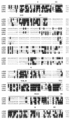
The sequences of PEK are related to eIF-2α kinases. Shown is a sequence alignment of the kinase catalytic domains of rat PEK, human PKR, rat HRI, and yeast GCN2 generated by the program Pileup. Identical amino acid residues of the eIF-2α kinases are designated by black boxes, and gaps in the alignment are indicated by dashes. Kinase subdomains are highlighted by bars above the multisequence alignment.
FIG. 3
FIG. 3
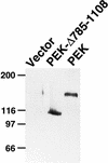
Expression of PEK in E. coli as measured by immunoblotting. PEK or PEK-Δ785-1108 fused to amino-terminal polyhistidine tags was expressed in E. coli by using the T7 promoter system, as described in Materials and Methods. Equal amounts of total cell lysates were analyzed by SDS-PAGE, and PEK was detected by immunoblotting with a polyclonal antibody that recognizes the polyhistidine sequences. Vector, lysates prepared from E. coli containing only the parent vector, pET28a. Molecular weight markers, in kilodaltons, are on the left.
FIG. 4
FIG. 4
PEK in an in vitro kinase assay is autophosphorylated on both serine and threonine residues. (A) Lysates prepared from E. coli expressing PEK or vector alone were incubated with [γ-32P]ATP and analyzed by SDS-PAGE, followed by autoradiography. The radiolabeled PEK is indicated by an arrow on the right. Sizes of protein standards in kilodaltons are on the left. (B) Phosphoamino acid analysis of 32P-labeled PEK from the in vitro assay. Radiolabeled PEK was hydrolyzed with HCl, applied to cellulose-coated sheets, and separated by one-dimensional thin-layer electrophoresis. In parallel, eIF-2α that was phosphorylated by PEK in an in vitro assay was similarly analyzed. The 32P-labeled amino acids were detected by autoradiography. The positions of serine, threonine, and tyrosine are indicated by one-letter codes.
FIG. 5
FIG. 5
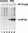
Immunoprecipitated PEK phosphorylates human eIF-2α. PEK or an unrelated bacterial protein was expressed in Sf-9 cells and the PEK was immunoprecipitated with polyclonal antisera prepared against a synthetic peptide derived from the carboxy terminus of the kinase. Immunocomplexes prepared from lysates expressing an unrelated bacterial protein (lanes 1 to 3) or PEK (lanes 4 to 6) were incubated with [γ-32P]ATP and increasing amounts of human eIF-2α as described in Materials and Methods. Radiolabeled proteins were separated by electrophoresis with an SDS-polyacrylamide gel and visualized by autoradiography. Kinase assay mixtures contained 1 (lane 1 and 4), 2 (lanes 2 and 5), or 4 (lanes 3 and 6) μg of purified human eIF-2α protein. The arrowhead indicates the position of the phosphorylated eIF-2α.
FIG. 6
FIG. 6
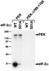
PEK phosphorylation of eIF-2α is dependent on residue serine-51. Full-length PEK and truncated PEK-Δ785-1108 were partially purified and added to kinase reaction mixtures containing [γ-32P]ATP and a modified form of yeast eIF-2 containing wild-type serine-51 (WT) or alanine substituted for serine at this phosphorylation site (S51A). Radiolabeled proteins were analyzed by SDS-PAGE, followed by autoradiography. The reason full-length PEK is a doublet appears to be in vitro proteolysis. In other preparations, as determined by autophosphorylation or immunoblotting, a single species of PEK was detected. Phosphorylated PEK and eIF-2α are indicated to the right. The sizes of protein standards in kilodaltons are on the left.
FIG. 7
FIG. 7
Northern blot analysis of tissue distributions of the PEK mRNA. A Northern blot containing 2 μg of poly(A)+ RNA purified from the different rat tissues indicated (A) and a separate blot containing ∼2 μg of mRNA from rat skeletal muscle, kidney, testis, and pancreas tissue (B) were hybridized with an α-32P-labeled cDNA probe encoding PEK. Following autoradiography to visualize the ∼5.2-kb PEK mRNA (top panel), the membranes were rehybridized with a radiolabeled rat β-actin probe, and the resulting autoradiogram is shown in the bottom panel. The PEK and β-actin mRNAs are indicated by arrowheads.
FIG. 8
FIG. 8
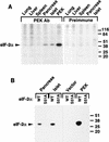
PEK immunoprecipitated from mammalian tissues phosphorylates eIF-2α at serine-51. (A) PEK was immunoprecipitated from the indicated tissues by using polyclonal PEK antibody and incubated with [γ-32P]ATP and human eIF-2α as described in Materials and Methods. As a control, a similar immunoprecipitation assay was carried out with preimmune serum. Radiolabeled proteins were separated by SDS-PAGE and were visualized by autoradiography. The arrowhead indicates the position of phosphorylated eIF-2α. All tissues used in this study are from rats, with the exception of islets, which were isolated from dogs. (B) PEK was immunoprecipitated from pancreas, islets, and Sf-9 cells. As a control, similar immunoprecipitations were carried out with lysates prepared from Sf-9 insect cells containing only the vector. Immunoprecipitates were added to kinase reaction mixtures containing [γ-32P]ATP and a modified form of yeast eIF-2 containing wild-type serine-51 (WT) or alanine substituted for serine at this phosphorylation site (S51A). Radiolabeled proteins were analyzed by SDS-PAGE, followed by autoradiography. Sizes of protein standards in kilodaltons on the right sides of both panels.
FIG. 9
FIG. 9
PEK functionally substitutes for the eIF-2α kinase GCN2 in a yeast model system. (A) Strain H1894 (Δgcn2) was transformed with plasmid pC102-2, encoding GCN2 kinase (GCN2), p504, expressing PEK from a galactose-inducible promoter (PEK), or only vector pEBMLyex4 (Vector). Patches of transformed cells were replica printed onto agar plates containing SGal, SGal supplemented with 3-AT, or SGal supplemented with SM. GCN2-deficient strains are hypersensitive to the amino acid inhibitors 3-AT and SM. Replicated plates were grown for 4 days at 30°C and photographed. (B) PEK was similarly expressed in strains H1816 (Δgcn2 SUI2) and H1817 (Δgcn2 SUI2-S51A), which are related to H1894, and cells were replica printed onto SGal or galactose-inducing medium containing SM. Strain H1817 contains a mutant version of eIF-2α that has an alanine substituted for the phosphorylated residue serine-51. While expression of either GCN2 or PEK in H1816 facilitates growth of this strain in SGal medium containing SM, neither eIF-2α kinase mediated growth when expressed in H1817 cells.
FIG. 9
FIG. 9
PEK functionally substitutes for the eIF-2α kinase GCN2 in a yeast model system. (A) Strain H1894 (Δgcn2) was transformed with plasmid pC102-2, encoding GCN2 kinase (GCN2), p504, expressing PEK from a galactose-inducible promoter (PEK), or only vector pEBMLyex4 (Vector). Patches of transformed cells were replica printed onto agar plates containing SGal, SGal supplemented with 3-AT, or SGal supplemented with SM. GCN2-deficient strains are hypersensitive to the amino acid inhibitors 3-AT and SM. Replicated plates were grown for 4 days at 30°C and photographed. (B) PEK was similarly expressed in strains H1816 (Δgcn2 SUI2) and H1817 (Δgcn2 SUI2-S51A), which are related to H1894, and cells were replica printed onto SGal or galactose-inducing medium containing SM. Strain H1817 contains a mutant version of eIF-2α that has an alanine substituted for the phosphorylated residue serine-51. While expression of either GCN2 or PEK in H1816 facilitates growth of this strain in SGal medium containing SM, neither eIF-2α kinase mediated growth when expressed in H1817 cells.
FIG. 10
FIG. 10
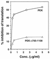
Addition of recombinant PEK to reticulocyte lysates reduced protein synthesis. Partially purified full-length PEK or truncated PEK-Δ785-1108 was added at the indicated concentrations to reticulocyte lysates. [35S]methionine was added to the cell-free translation system concomitant with recombinant PEK, and synthesized polypeptides were precipitated with tricarboxylic acid. The incorporation of [35S]methionine into the protein samples was measured by scintillation counting. The effect of PEK on protein synthesis in the cell-free system is expressed as a percentage of inhibition.

Similar articles

Cited by

References

    1. Abastado J P, Miller P F, Jackson B M, Hinnebusch A G. Suppression of ribosomal reinitiation at upstream open reading frames in amino acid-starved cells forms the basis of GCN4 translational control. Mol Cell Biol. 1991;11:486–496. - PMC - PubMed
    1. Barber G N, Jagus R, Meurs E F, Hovanessian A G, Katze M G. Molecular mechanisms responsible for malignant transformation by regulatory and catalytic domain variants of the interferon-induced enzyme RNA-dependent protein kinase. J Biol Chem. 1995;270:17423–17428. - PubMed
    1. Bonner-Weir S, Smith F E. Islets of langerhans: morphology and its implications. In: Kahn C R, Weir G C, editors. Joslin’s diabetes mellitus. 13th ed. Piladelphia, Pa: Lea & Febiger; 1994. pp. 15–28.
    1. Cesareni G, Murray J. Plasmid vectors carrying the replication origin of filamentous single-stranded phages. In: Setlow J K, Hollaender A, editors. Genetic engineering: principles and methods. Vol. 9. New York, N.Y: Plenum Press; 1987. pp. 135–154.
    1. Chen J J, London I M. Regulation of protein synthesis by heme-regulated eIF-2 alpha kinase. Trends Biochem Sci. 1995;20:105–108. - PubMed

Publication types

MeSH terms

Associated data