. 1998 Nov 2;143(3):837-47.
doi: 10.1083/jcb.143.3.837.

CD44 occupancy prevents macrophage multinucleation

Affiliations

CD44 occupancy prevents macrophage multinucleation

H Sterling et al. J Cell Biol. .

Abstract

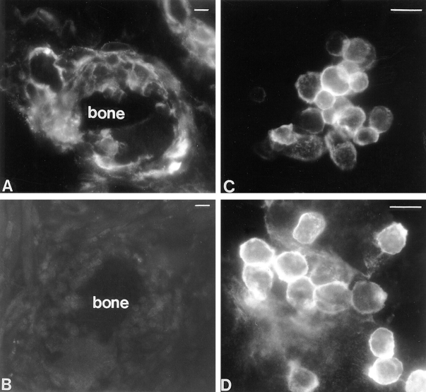
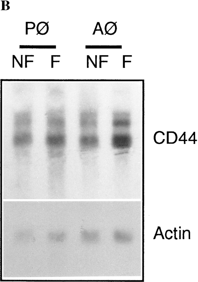
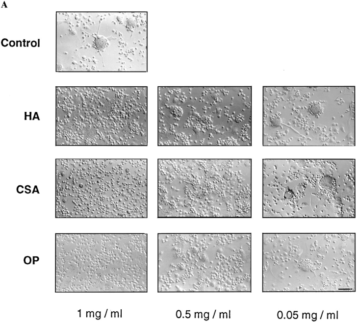
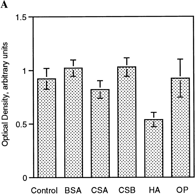
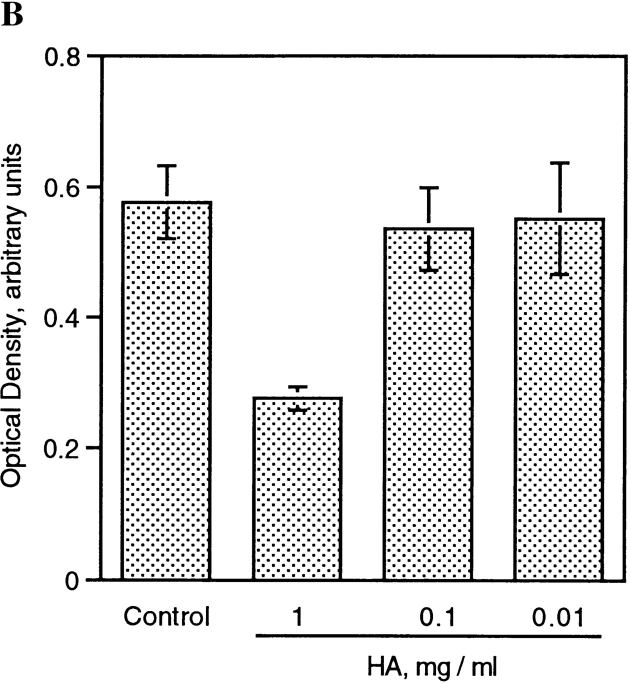
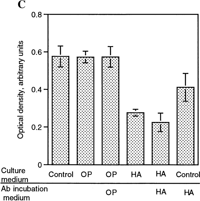
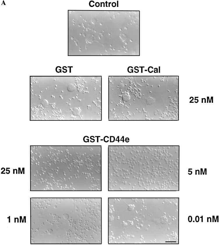
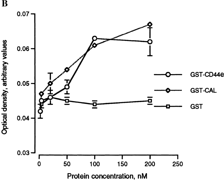
Cells of the mononuclear phagocyte lineage have the capability to adhere to and fuse with each other and to differentiate into osteoclasts and giant cells. To investigate the macrophage adhesion/fusion mechanism, we focused our attention on CD44, a surface glycoprotein known to play a role in hematopoietic cell-cell adhesion. We report that CD44 expression by macrophages is highly and transiently induced by fusogenic conditions both in vitro and in vivo. We show that CD44 ligands, hyaluronic acid, chondroitin sulfates, and osteopontin prevent macrophage multinucleation. In addition, we report that the recombinant extracellular domain of CD44 binds fusing macrophages and prevents multinucleation in vitro. These data suggest that CD44 may control the mononucleated status of macrophages in tissues by virtue of mediating cell-cell interaction.


Figures

Figure 1
Figure 1
High expression of CD44 on fusing macrophages in vivo (A) and in vitro (C and D). Multinucleation was induced in vivo by implanting rats intramuscularly with syngeneic bone particles that were recovered 10 d later and processed for immunolocalization. Frozen sections from bone implants were incubated with either mAb anti-rat CD44 (A) or mouse IgG1 (B) followed by goat anti–mouse IgG F(ab′)2 fragments conjugated to Cy3. Multinucleation was induced in vitro by plating alveolar macrophages on glass coverslips and culturing them under fusogenic conditions for either 1 (C) or 3 d (D). Cells were subjected to immunocytochemistry using anti-rat CD44 followed by goat anti–mouse IgG F(ab′)2 fragments conjugated to Cy3. Note the differences in fluorescence intensity between fusing cells. Bars, 20 μm.
Figure 2
Figure 2
Kinetics of CD44 expression in macrophages induced to fuse in vitro (A). Rat alveolar macrophages were isolated, plated at 5 × 10⁶ cells/ml in quadruplicate wells, using 96-well dishes (5 × 10⁴ cells/well), and cultured in fusogenic conditions for the indicated times. CD44 expression on paraformaldehyde fixed cells was determined by ELISA using mAb mouse anti-rat CD44 followed by goat anti–mouse IgG-conjugated to horseradish peroxidase (A and B). Mouse mAbs anti-rat CD4 and MHCII were used as controls. Northern blot analysis of CD44 (B). Each lane contained ∼8 μg of total RNA from freshly isolated as well as alveolar and peritoneal macrophages that had been cultured for 72 h under fusogenic conditions. Hybridization was performed using 32P-labeled PCR generated CD44 and β-actin probes.
Figure 2
Figure 2
Kinetics of CD44 expression in macrophages induced to fuse in vitro (A). Rat alveolar macrophages were isolated, plated at 5 × 10⁶ cells/ml in quadruplicate wells, using 96-well dishes (5 × 10⁴ cells/well), and cultured in fusogenic conditions for the indicated times. CD44 expression on paraformaldehyde fixed cells was determined by ELISA using mAb mouse anti-rat CD44 followed by goat anti–mouse IgG-conjugated to horseradish peroxidase (A and B). Mouse mAbs anti-rat CD4 and MHCII were used as controls. Northern blot analysis of CD44 (B). Each lane contained ∼8 μg of total RNA from freshly isolated as well as alveolar and peritoneal macrophages that had been cultured for 72 h under fusogenic conditions. Hybridization was performed using 32P-labeled PCR generated CD44 and β-actin probes.
Figure 3
Figure 3
CD44 ligands inhibit macrophage adhesion/ fusion. (A) Rat alveolar macrophages were isolated, plated at 5 × 10⁶ cells/ml in quadruplicate wells, using 96-well dishes (5 × 10⁴ cells/ well), and cultured in fusogenic conditions for the indicated times, in the absence or presence of hyaluronic acid (HA), chondroitin sulfate A (CSA), or osteopontin (OP) at the indicated concentrations. (B) Alveolar macrophages were cultured as in A, but in dishes coated with either HA (1 mg/ml) or CSA (1 mg/ml). Bars, 100 μm.
Figure 3
Figure 3
CD44 ligands inhibit macrophage adhesion/ fusion. (A) Rat alveolar macrophages were isolated, plated at 5 × 10⁶ cells/ml in quadruplicate wells, using 96-well dishes (5 × 10⁴ cells/ well), and cultured in fusogenic conditions for the indicated times, in the absence or presence of hyaluronic acid (HA), chondroitin sulfate A (CSA), or osteopontin (OP) at the indicated concentrations. (B) Alveolar macrophages were cultured as in A, but in dishes coated with either HA (1 mg/ml) or CSA (1 mg/ml). Bars, 100 μm.
Figure 4
Figure 4
HA decreases CD44 surface expression. (A) Rat alveolar macrophages were isolated, plated at 5 × 10⁶ cells/ml in quadruplicate wells, using 96-well dishes (5 × 10⁴ cells/well), and cultured in fusogenic conditions for 2 d, in the absence or presence of 1 mg/ml BSA, chondroitin sulfate A (CSA) or B (CSB), hyaluronic acid (HA), or 1 μM osteopontin (OP). CD44 expression was determined by ELISA using mAb mouse anti–rat CD44 followed by goat anti–mouse IgG-conjugated to horseradish peroxidase. (B) Cells were cultured as in A in the absence or presence of decreasing concentrations of HA and CD44 expression was determined by ELISA. (C) Cells were cultured as in A in the absence or presence of either OP (1 μM) or HA (1 mg/ml) for 3 d. CD44 expression was determined by ELISA in which mAb anti-CD44 incubation buffer was supplemented or not with either OP (1 μM) or HA (1 mg/ml).
Figure 4
Figure 4
HA decreases CD44 surface expression. (A) Rat alveolar macrophages were isolated, plated at 5 × 10⁶ cells/ml in quadruplicate wells, using 96-well dishes (5 × 10⁴ cells/well), and cultured in fusogenic conditions for 2 d, in the absence or presence of 1 mg/ml BSA, chondroitin sulfate A (CSA) or B (CSB), hyaluronic acid (HA), or 1 μM osteopontin (OP). CD44 expression was determined by ELISA using mAb mouse anti–rat CD44 followed by goat anti–mouse IgG-conjugated to horseradish peroxidase. (B) Cells were cultured as in A in the absence or presence of decreasing concentrations of HA and CD44 expression was determined by ELISA. (C) Cells were cultured as in A in the absence or presence of either OP (1 μM) or HA (1 mg/ml) for 3 d. CD44 expression was determined by ELISA in which mAb anti-CD44 incubation buffer was supplemented or not with either OP (1 μM) or HA (1 mg/ml).
Figure 5
Figure 5
Recombinant extracellular CD44 fusion protein (GST-CD44e) inhibits macrophage fusion. (A) Rat alveolar macrophages were isolated, plated at 5 × 10⁶ cells/ml in quadruplicate wells, using 96-well dishes (5 × 10⁴ cells/well), and cultured in fusogenic conditions for 3 d, in the presence of increasing concentrations of GST–CD44e, GST–Cal, and GST. (B) Cells were cultured as in A for 17 h and then incubated for 3 h at 4°C with GST–CD44e. Binding was determined by ELISA using anti-GST antibody coupled to horseradish peroxidase as described in Materials and Methods. Specific binding was determined by subtracting nonspecific binding from total binding for each concentration of GST–CD44e. Bar, 100 μM.
Figure 5
Figure 5
Recombinant extracellular CD44 fusion protein (GST-CD44e) inhibits macrophage fusion. (A) Rat alveolar macrophages were isolated, plated at 5 × 10⁶ cells/ml in quadruplicate wells, using 96-well dishes (5 × 10⁴ cells/well), and cultured in fusogenic conditions for 3 d, in the presence of increasing concentrations of GST–CD44e, GST–Cal, and GST. (B) Cells were cultured as in A for 17 h and then incubated for 3 h at 4°C with GST–CD44e. Binding was determined by ELISA using anti-GST antibody coupled to horseradish peroxidase as described in Materials and Methods. Specific binding was determined by subtracting nonspecific binding from total binding for each concentration of GST–CD44e. Bar, 100 μM.

Similar articles

Cited by

References

    1. Alkhatib G, Combadiere C, Broder C, Feng Y, Kennedy P, Murphy P, Berger E. CC CKR5: A RANTES, MIP-1α, MIP-1β receptor as a fusion cofactor for macrophage-tropic HIV-1. Science. 1996;272:1955–1958. - PubMed
    1. Belitsos PC, Hildreth JE, August JT. Homotypic cell aggregation induced by anti-CD44 (Pgp-1) monoclonal antibodies and related to CD44 (Pgp-1) expression. J Immunology. 1990;144:1661–1670. - PubMed
    1. Blobel CP, Wolfsberg TG, Turk CW, Miles DG, Primakoff P, White JM. A potential fusion peptide and an integrin ligand domain in a protein active in sperm-egg fusion. Nature. 1992;356:248–252. - PubMed
    1. Cherr GN, Yudin AI, Katz DF. Organization of the hamster cumulus extracellular matrix A hyaluronate glycoprotein gel which modulates sperm access to the oocyte. Dev Growth Differ. 1990;32:353–366. - PubMed
    1. Chiozzi P, Sanz J, Ferrari D, Falzoni S, Aleotti A, Buell G, Collo G, Di Virgilio F. Spontaneous cell fusion in macrophage cultures expressing high levels of the P2Z/P2X7receptor. J Cell Biol. 1997;138:697–706. - PMC - PubMed

Publication types