. 1998 Nov 10;95(23):13859-64.
doi: 10.1073/pnas.95.23.13859.

NF-kappaB activation provides the potential link between inflammation and hyperplasia in the arthritic joint

Affiliations

NF-kappaB activation provides the potential link between inflammation and hyperplasia in the arthritic joint

A V Miagkov et al. Proc Natl Acad Sci U S A. .

Abstract

The transcription factor NF-kappaB is a pivotal regulator of inflammatory responses. While the activation of NF-kappaB in the arthritic joint has been associated with rheumatoid arthritis (RA), its significance is poorly understood. Here, we examine the role of NF-kappaB in animal models of RA. We demonstrate that in vitro, NF-kappaB controlled expression of numerous inflammatory molecules in synoviocytes and protected cells against tumor necrosis factor alpha (TNFalpha) and Fas ligand (FasL) cytotoxicity. Similar to that observed in human RA, NF-kappaB was found to be activated in the synovium of rats with streptococcal cell wall (SCW)-induced arthritis. In vivo suppression of NF-kappaB by either proteasomal inhibitors or intraarticular adenoviral gene transfer of super-repressor IkappaBalpha profoundly enhanced apoptosis in the synovium of rats with SCW- and pristane-induced arthritis. This indicated that the activation of NF-kappaB protected the cells in the synovium against apoptosis and thus provided the potential link between inflammation and hyperplasia. Intraarticular administration of NF-kB decoys prevented the recurrence of SCW arthritis in treated joints. Unexpectedly, the severity of arthritis also was inhibited significantly in the contralateral, untreated joints, indicating beneficial systemic effects of local suppression of NF-kappaB. These results establish a mechanism regulating apoptosis in the arthritic joint and indicate the feasibility of therapeutic approaches to RA based on the specific suppression of NF-kappaB.


Figures
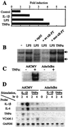
Figure 1
Figure 1
Suppression of NF-κB inhibits inflammatory responses in primary rat synovial fibroblasts. (A) Induction of NF-κB-mediated transcription in vitro. For luciferase assay, cells were transfected with a 3x wt κBLUC reporter vector followed by an overnight incubation in a low serum (0.5% FBS) medium and stimulated for 3 hr with IL-1β (10 ng/ml), lipopolysaccharide (10 μg/ml), and TNFα (10 ng/ml). The specificity of NF-κB activation was determined by assessing the expression of a 3x mut κBLUC vector (data not shown). (B) Induction of NF-κB DNA-binding activity. For EMSA, quiescent synoviocytes were stimulated for 1 hr with lipopolysaccharide (10 μg/ml) and TNFα (10 ng/ml). The nuclear extracts were preincubated with a radiolabeled κB probe and resolved on PAGE. The specificity of NF-κB binding was assessed by including an excessive amount (100:1) of cold wt and mut NF-κB decoys into the binding reactions. Both major NF-κB bands (arrows) contained RelA subunits, as determined by the supershift by a RelA (p65) Ab (data not shown). (C) AdsrIκBα expression inhibits NF-κB activation in vitro. Cells were transduced with indicated Ad vectors and 2 days later stimulated with TNFα (100 ng/ml) for 30 min. NF-κB DNA-binding activity was determined by EMSA as in B. (D) Suppression of NF-κB inhibits inflammatory responses in vitro. For Northern blotting, cells were transduced as indicated in the legend to C and stimulated with IL-1β (10 ng/ml) and TNFα (10 ng/ml). Total RNA was analyzed by sequentially hybridizing the blot with indicated radiolabeled probes.
Figure 2
Figure 2
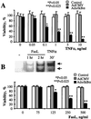
Suppression of NF-κB potentiates the TNFα- and FasL-induced cytotoxicity in rat synovial fibroblasts. (A) The expression of srIκBα potentiates the cytotoxicity of TNFα. Cells were transduced in duplicate with the AdsrIκBα vector and, 2 days later, stimulated with indicated concentrations of TNFα for an additional 24 hr. The cell viability was determined by using the MTT assay. (Bars = SD.) The significance of difference was calculated by using unpaired two-tailed Student’s t test. Data are representative of two experiments. (B) Activation of NF-κB by FasL inhibits the cytotoxicity of FasL. (Upper) Induction of NF-κB DNA-binding activity by FasL. For EMSA, cells were stimulated with FasL (100 ng/ml), and NF-κB DNA-binding activity was determined by EMSA as described in the legend to Fig. 1B. The nuclear extracts of TNFα-stimulated cells (see Fig. 1C) were used as a positive control. (Lower) srIκBα expression potentiates cytotoxicity of FasL. Cells were transduced in duplicate with the AdsrIκBα vector and, 2 days later, stimulated with the indicated concentrations of FasL for an additional 24 hr. Control, untransduced cells. The cell viability was determined by using the MTT assay. The significance of difference was calculated by using unpaired two-tailed Student’s t test. (Bars = SD.) Data are representative of three experiments.
Figure 3
Figure 3
Induction of the recurrence of SCW arthritis is associated with activation of NF-κB in the synovium. Activated NF-κB was detected by HPR immunostaining by using primary mAb against RelA NLS (21). (a) Normal synovium. Original magnification, ×200. (b) Inflamed synovium at day 1 after reactivation of SCW arthritis. Original magnification, ×200. (c) Same as b; original magnification, ×400. (d) Control immunostaining of the inflamed synovium (day 1 after reactivation) by using an mAb preabsorbed with a RelA NLS peptide. Original magnification, ×200. Data are representative of two experiments.
Figure 4
Figure 4
Inhibitors of NF-κB accelerate apoptosis in the inflamed rat synovium. (A) Proteasomal inhibitor MG132 induces apoptosis in the SCW arthritic joint. At day 1 after reactivation of the recurrence of SCW arthritis, ankle joints were injected either with MG132 dissolved in 10% DMSO (5 μg/joint) (a and c) or with the vehicle (b). One day later, synovial tissue was explanted and apoptosis was assessed by using TUNEL assay. Some of apoptotic cells are indicated by the arrowheads. Original magnification, ×100. (B) In vivo adenovirus-mediated gene transfer of srIκBα into the SCW arthritic joints. One day after reactivation of SCW arthritis, ankle joints were injected with the AdsrIκBα vector. Two days later, synovial tissue was explanted and srIκBα expression was assessed by HRP immunostaining for human IκBα (b). The specificity detection was confirmed by immunostaining the AdCMV-transduced joints (a). Data are representative of two experiments. Original magnification, ×200. (C) In vivo gene transfer of srIκBα induces apoptosis in the SCW arthritic joint. At day 1 after reactivation of SCW arthritis, ankle joints were injected either with the AdsrIκBα (a) or with the AdCMV (b) vectors. One day later, synovial tissue was explanted and apoptosis was assessed by using TUNEL assay. Some of apoptotic cells are indicated by the arrowheads. Note nuclear fragmentation in apoptotic cells. Data are representative of two experiments. Original magnification, ×1,000. (D) Quantitative assessment of apoptosis in SCW arthritic joints. A summary of the experiments is described in the legends to A and C. Each bar represents the average percentage of apoptotic cells counted in randomly chosen fields. (Bars = SD.) The frequency of apoptosis was counted at a low magnification in the indicated numbers of randomly chosen fields and compared with that in untransduced SCW arthritic rat synovium (the first bar in the row). MG 132, 12 fields in the explants of four joints; vehicle, 12 fields in the explants of four joints; AdsrIκBa, nine fields in the explants of three joints; AdCMV, nine fields in the explants of three joints. The significance of the difference between treated and untreated SCW joints was calculated by using unpaired two-tailed Student’s t test. (E) Time course of AdGFP (Upper) and AdsrIκBα (Lower) expression in rats with pristane-induced arthritis. Rats with established pristane arthritis received AdsrIκBα or AdGFP vectors into both ankle joints. Expression of the transgene was analyzed by Western blotting. (Upper) Immunodetection of Ad GFP in AdGFP-transduced joints. GFP, the arrow indicates the position of the band of recombinant GFP protein (not shown). (Lower) Immunodetection of srIκBα in AdsrIκBα-transduced joints. Pos., a positive control (in vitro AdsrIκBα-transduced cells); Neg., a negative control (in vitro AdCMV-transduced cells); srIκBα, the arrow indicates the position of the band of the srIκBα protein; n.s., a nonspecific band unrelated to human or rat IκBα, as determined by immunodetection with a peptide-blocked primary Ab (data not shown).
Figure 5
Figure 5
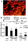
I.a. administration of NF-κB decoys inhibit the recurrence of SCW arthritis. (A) Distribution of fluorescein-labeled ODNs in the synovium. Ankle joints of rats with established SCW arthritis were injected with liposomal complexes of fluorescein-labeled NF-κB decoys. One day later, synovial explants were isolated, sectioned, counterstained with propidium iodide, and analyzed under a fluorescent microscope equipped with a fluorescein filter set. Note the bright-yellow fluorescent nuclei and red fluorescence of cytoplasmic RNA. Original magnification, ×200. (B and C) Therapeutic protocol. (B) Schedule of injections. Rats were injected into both ankle joints with PG-APS. Four weeks later, at Day 0, animals were divided into three groups and monoarticularly injected with liposomal complexes of NF-κB decoys (10 μg ODNs/joint). In the experimental group, animals were injected with wt-κB decoys, and two control groups received mut κB-PT decoys and saline. The contralateral ankle joints were injected with saline. At Day 1, reactivation of arthritis was induced by i.v. injection of PG-APS. (C) The severity of recurrent SCW arthritis in treated rats. The average mean values of joint swelling were calculated in each group as dn = <DnD1>, where Dn and D1 are joint diameters at day n and day 1, respectively. After reactivation, the maximal joint swelling in all groups was registered at day 4. ODNs, liposome-treated joints; CL, contralateral joints; N, number of joints per group. The significance of difference with the control, saline-treated group was calculated by using unpaired two-tailed Student’s t test. (Bars = SEM.) Results of a single experiment are shown. The therapeutic effect of wt NF-κB decoys was reproduced in an identical experiment.
Figure 6
Figure 6
NF-κB in the regulation of inflammation and apoptosis in the arthritic joint.

Similar articles

Cited by

References

    1. Firestein G S. Arthritis Rheum. 1996;39:1781–1790. - PubMed
    1. Zvaifler N J, Tsai V, Alsalameh S, von Kempis J, Firestein G S, Lotz M. Am J Pathol. 1997;150:1125–1138. - PMC - PubMed
    1. Mountz J D, Wu J, Cheng J, Zhou T. Arthritis Rheum. 1994;37:1415–1420. - PubMed
    1. Firestein G S, Nguyen K, Aupperle K R, Yeo M, Boyle D L, Zvaifler N J. Am J Pathol. 1996;149:2143–2151. - PMC - PubMed
    1. Nishioka K, Hasunuma T, Kato T, Sumida T, Kobata T. Arthritis Rheum. 1998;41:1–9. - PubMed

Publication types