. 1998 Dec;72(12):9441-52.
doi: 10.1128/JVI.72.12.9441-9452.1998.

Differential tropism and replication kinetics of human immunodeficiency virus type 1 isolates in thymocytes: coreceptor expression allows viral entry, but productive infection of distinct subsets is determined at the postentry level

Affiliations

Differential tropism and replication kinetics of human immunodeficiency virus type 1 isolates in thymocytes: coreceptor expression allows viral entry, but productive infection of distinct subsets is determined at the postentry level

L Pedroza-Martins et al. J Virol. 1998 Dec.

Abstract

Human thymocytes are readily infected with human immunodeficiency virus type 1 (HIV-1) in vivo and in vitro. In this study, we found that the kinetics of replication and cytopathic effects of two molecular isolates, NL4-3 and JR-CSF, in postnatal thymocytes are best explained by the distribution of chemokine receptors used for viral entry. CXCR4 was expressed at high levels on most thymocytes, whereas CCR5 expression was restricted to only 0.1 to 2% of thymocytes. The difference in the amount of proviral DNA detected after infection of fresh thymocytes with NL4-3 or JR-CSF correlated with the levels of CXCR4 and CCR5 surface expression. Anti-CCR5 blocking studies showed that low levels of CCR5 were necessary and sufficient for JR-CSF entry in thymocytes. Interleukin-2 (IL-2), IL-4, and IL-7, cytokines normally present in the thymus, influenced the expression of CXCR4 and CCR5 on thymocytes and thus increased the infectivity and spread of both NL4-3 and JR-CSF in culture. NL4-3 was produced by both immature and mature thymocytes, whereas JR-CSF production was restricted to the mature CD1(-)/CD69(+) population. Although CXCR4 and CCR5 distribution readily explained viral entry in mature CD69(+) and immature CD69(-) cells, and correlated with proviral DNA distribution, we found that viral production was favored in CD69(+) cells. Therefore, while expression of CD4 and appropriate coreceptors are essential determinants of viral entry, factors related to activation and stage-specific maturation contribute to HIV-1 replication in thymocyte subsets. These results have direct implications for HIV-1 pathogenesis in pediatric patients.


Figures

FIG. 1
FIG. 1
Distribution of chemokine receptor expression on freshly isolated human thymocytes. Thymocytes were isolated by nylon wool separation and phenotyped with CD3-PE and nonlabeled antibodies to the chemokine receptors CXCR-4 (antibody 12G5) and CCR5 (antibody 2D7), followed by goat anti-mouse IgG-FITC (GAM-FITC). Appropriate isotype control antibodies (IgG2a and IgG1) followed by goat anti-mouse IgG-FITC were used to set the cursors. The percentage of cells staining with the isotype control antibodies followed by goat anti-mouse IgG-FITC was 0%. Dead cells were excluded from the analysis by using 7-AAD.
FIG. 2
FIG. 2
Effects of cytokines on chemokine receptor expression. Thymocytes were cultured for 2 weeks in serum-free medium alone or supplemented with IL-2 plus IL-4 or IL-4 plus IL-7. Before culture (day 0) and on days 7 and 15, cells were removed for immunophenotyping to examine expression of chemokine receptors by flow cytometry. Appropriate isotype control antibodies and single-color staining with CD3-FITC were used to set the cursors defining the CD3+/high population (A and B). Dead cells were excluded from the analysis by using 7-AAD. (A and B) CXCR4 and CCR5 expression on thymocyte subsets was determined by using the antibodies CD3-FITC, CXCR4-PE (12G5), and CCR5-PE (2D7). (C) In a different experiment, immunophenotyping was performed on day 12 of culture to identify the distribution of CCR5 on thymocyte subsets that respond to IL-2 plus IL-4. Thymocytes were stained with CD3-PE, CD4-PE, or CD1-PE in combination with nonlabeled antibody to CCR5 (2D7) and then with goat anti-mouse IgG-FITC (GAM-FITC). The percentage of cells staining with the IgG1 isotype control antibody for CCR5 followed by goat anti-mouse IgG-FITC was 0%.
FIG. 2
FIG. 2
Effects of cytokines on chemokine receptor expression. Thymocytes were cultured for 2 weeks in serum-free medium alone or supplemented with IL-2 plus IL-4 or IL-4 plus IL-7. Before culture (day 0) and on days 7 and 15, cells were removed for immunophenotyping to examine expression of chemokine receptors by flow cytometry. Appropriate isotype control antibodies and single-color staining with CD3-FITC were used to set the cursors defining the CD3+/high population (A and B). Dead cells were excluded from the analysis by using 7-AAD. (A and B) CXCR4 and CCR5 expression on thymocyte subsets was determined by using the antibodies CD3-FITC, CXCR4-PE (12G5), and CCR5-PE (2D7). (C) In a different experiment, immunophenotyping was performed on day 12 of culture to identify the distribution of CCR5 on thymocyte subsets that respond to IL-2 plus IL-4. Thymocytes were stained with CD3-PE, CD4-PE, or CD1-PE in combination with nonlabeled antibody to CCR5 (2D7) and then with goat anti-mouse IgG-FITC (GAM-FITC). The percentage of cells staining with the IgG1 isotype control antibody for CCR5 followed by goat anti-mouse IgG-FITC was 0%.
FIG. 3
FIG. 3
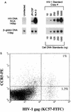
CXCR4 and CCR5 expression levels correlate with the amount of NL4-3 and JR-CSF proviral DNA after infection. Thymocytes were infected with JR-CSF (200 IU/104 cells) or NL-4-3 (10 IU/104 cells) and cultured with IL-2 plus IL-4. CCR5 expression on day 0 is shown in Fig. 1. (A) Twenty-four hours postinfection, 106 cells were removed and analyzed by using primers (R/U5) specific for the LTR region of HIV-1 to detect the presence of proviral DNA. To normalize for the amount of cellular DNA, PCR was performed in parallel for sequences in the β-globin gene. (B) JR-CSF-infected thymocytes were cultured with IL-2 plus IL-4. At 13 days postinfection, cells were subjected to surface staining with CCR5-PE/CD3-PerCP followed by intracellular staining with KC57-FITC.
FIG. 4
FIG. 4
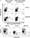
The antibody (2D7) to CCR5 is able to block productive infection of thymocytes by JR-CSF. Thymocytes were preincubated in the presence or absence of antibody to CCR5 (2D7) or CXCR4 (12G5) for 2 h before infection with JR-CSF (30 IU/104 cells). The antibodies were present during the infection and throughout the culture with IL-2 plus IL-4. HIV replication was detected by measuring p24 antigen in the culture supernatants on days 8, 15, and 22 postinfection. Preincubation conditions: no antibody (gray bars), 1 μg of 2D7 (vertically striped bars), 5 μg of 2D7 (diagonally striped bars), 10 μg of 12G5 (horizontally striped bars), 1 μg of 2D7 plus 10 μg of 12G5 (checkered bars), 5 μg of 2D7 plus 10 μg of 12G5 (black bars), and 100 μg of CD4-IgG (white bars).
FIG. 5
FIG. 5
The antibody to CCR5 (2D7) specifically blocks expression of JR-CSF in thymocytes. Thymocytes were preincubated in the presence or absence of 5 μg of CCR5 antibody per ml for 2 h before infection with JR-CSF (30 IU/104 cells) or NL4-3 (1.5 IU/104 cells). The antibody was present during infection and throughout the culture with IL-2 plus IL-4. (A) At 2 weeks postinfection, cells were subjected to surface staining with CD3-PerCP followed by intracellular staining with KC57-FITC. (B) To determine the effect of CCR5 antibody on thymocytes, uninfected and infected cells were immunophenotyped with CD4-PE and CD8-PerCP and intracellularly stained with KC57-FITC 2 weeks postinfection. CD4-PE/CD8-PerCP expression is shown.
FIG. 6
FIG. 6
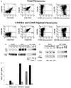
Infection of total thymocytes and CD69+ and CD69 thymocyte subsets by JR-CSF and NL4-3. CD69+ and CD69 subsets were obtained by using antibody-coated immunomagnetic beads from the CD45RA thymocytes and immunophenotyped to determine the purity of the isolation. The resulting CD45RA/CD69+ cells bound to beads, and CD45RA/CD69 cells were used for infection. (A) Immunophenotype of thymocytes before and after depletion of CD45RA- and CD69-expressing cells. (B) The total thymocyte population and the CD69+ and CD69 thymocyte subsets were infected with JR-CSF (100 IU/104 cells) or NL4-3 (10 IU/104 cells) and cultured for 2 weeks in the presence of IL-2 plus IL-4. Twenty-four hours postinfection 106 cells were removed and analyzed by using primers (R/U5) specific for the LTR region of HIV-1 to detect the presence of proviral DNA. To normalize for the amount of cellular DNA, PCR was performed in parallel for sequences in the β-globin gene. Heat-inactivated virus (−) were run in parallel with the live-virus-treated samples (+) as controls for DNA contamination from the inoculum. (C) PCR amplification of proviral DNA was performed on diluted DNA samples from the NL4-3-infected cells described above to detect the presence of fully reverse transcribed (RT) proviral DNA (LTR/gag) in parallel with the partially reverse transcribed (R/U5) proviral DNA. (D) HIV replication was detected by measuring p24 antigen in the culture supernatants of JR-CSF-infected CD69+ cells (vertically striped bars), JR-CSF-infected CD69 cells (white bars), NL4-3-infected CD69+ cells (black bars), and NL4-3-infected CD69 cells (gray bars) on days 1, 5, and 12 postinfection.

Similar articles

Cited by

References

    1. Adachi A, Gendelman H E, Koenig S, Folks T, Willey R, Rabson A, Martin M A. Production of acquired immunodeficiency syndrome-associated retrovirus in human and nonhuman cells transfected with an infectious molecular clone. J Virol. 1986;59:284–291. - PMC - PubMed
    1. Aldrovandi G M, Feuer G, Gao L, Jamieson B, Kristeva M, Chen I S Y, Zack J A. The SCID-hu mouse as a model for HIV-1 infection. Nature. 1993;363:732–736. - PubMed
    1. Alkhatib G, Combardiere C, Broder C C, Feng Y, Kennedy P E, Murphy P M, Berger E A. CC CKR5: a RANTES, MIP-1α, MIP-1β receptor as a fusion cofactor for macrophage-tropic HIV-1. Science. 1996;272:1955–1958. - PubMed
    1. Bazan H A, Alkhatib G, Broder C C, Berger E A. Patterns of CCR5, CXCR4, and CCR3 usage by envelope glycoproteins from human immunodeficiency virus type 1 primary isolates. J Virol. 1998;72:4485–4491. - PMC - PubMed
    1. Bjorndal A, Deng H, Jansson M, Fiore J R, Colognesi C, Karlsson A, Albert J, Scarlatti G, Littman D R, Fenyo E M. Coreceptor usage of primary human immunodeficiency virus type 1 isolates varies according to biological phenotype. J Virol. 1997;71:7478–7487. - PMC - PubMed

Publication types

MeSH terms