An inhibitor of HIV-1 protease modulates proteasome activity, antigen presentation, and T cell responses
- PMID: 9789051
- PMCID: PMC23730
- DOI: 10.1073/pnas.95.22.13120
An inhibitor of HIV-1 protease modulates proteasome activity, antigen presentation, and T cell responses
Abstract
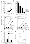
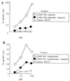
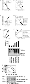
Inhibitors of the protease of HIV-1 have been used successfully for the treatment of HIV-1-infected patients and AIDS disease. We tested whether these protease inhibitory drugs exerted effects in addition to their antiviral activity. Here, we show in mice infected with lymphocytic choriomeningitis virus and treated with the HIV-1 protease inhibitor ritonavir a marked inhibition of antiviral cytotoxic T lymphocyte (CTL) activity and impaired major histocompatibility complex class I-restricted epitope presentation in the absence of direct effects on lymphocytic choriomeningitis virus replication. A potential molecular target was found: ritonavir selectively inhibited the chymotrypsin-like activity of the 20S proteasome. In view of the possible role of T cell-mediated immunopathology in AIDS pathogenesis, the two mechanisms of action (i.e., reduction of HIV replication and impairment of CTL responses) may complement each other beneficially. Thus, the surprising ability of ritonavir to block the presentation of antigen to CTLs may possibly contribute to therapy of HIV infections but potentially also to the therapy of virally induced immunopathology, autoimmune diseases, and transplantation reactions.
Figures


Similar articles
-
How an inhibitor of the HIV-I protease modulates proteasome activity.J Biol Chem. 1999 Dec 10;274(50):35734-40. doi: 10.1074/jbc.274.50.35734. J Biol Chem. 1999. PMID: 10585454
-
Effects of retroviral protease inhibitors on proteasome function and processing of HIV-derived MHC class I-restricted cytotoxic T lymphocyte epitopes.AIDS Res Hum Retroviruses. 2001 Jul 20;17(11):1063-6. doi: 10.1089/088922201300343744. AIDS Res Hum Retroviruses. 2001. PMID: 11485623 No abstract available.
-
DNA immunization: ubiquitination of a viral protein enhances cytotoxic T-lymphocyte induction and antiviral protection but abrogates antibody induction.J Virol. 1997 Nov;71(11):8497-503. doi: 10.1128/JVI.71.11.8497-8503.1997. J Virol. 1997. PMID: 9343207 Free PMC article.
-
T-cell-mediated immunopathology in viral infections.Transplant Rev. 1974;19(0):89-120. doi: 10.1111/j.1600-065x.1974.tb00129.x. Transplant Rev. 1974. PMID: 4601807 Review. No abstract available.
-
Biological role of major transplantation antigens in T cell self-recognition.Experientia. 1986 Sep 15;42(9):970-2. doi: 10.1007/BF01940698. Experientia. 1986. PMID: 3530799 Review.
Cited by
-
The HIV-1 protease inhibitor nelfinavir activates PP2 and inhibits MAPK signaling in macrophages: a pathway to reduce inflammation.J Leukoc Biol. 2012 Oct;92(4):795-805. doi: 10.1189/jlb.0911447. Epub 2012 Jul 11. J Leukoc Biol. 2012. PMID: 22786868 Free PMC article.
-
Intracellular HIV-1 Tat protein represses constitutive LMP2 transcription increasing proteasome activity by interfering with the binding of IRF-1 to STAT1.Biochem J. 2006 Jun 1;396(2):371-80. doi: 10.1042/BJ20051570. Biochem J. 2006. PMID: 16512786 Free PMC article.
-
Metabolic complications associated with HIV protease inhibitor therapy.Drugs. 2003;63(23):2555-74. doi: 10.2165/00003495-200363230-00001. Drugs. 2003. PMID: 14636077 Review.
-
Proteasome inhibition: a new strategy in cancer treatment.Invest New Drugs. 2000 May;18(2):109-21. doi: 10.1023/a:1006321828515. Invest New Drugs. 2000. PMID: 10857991 Review.
-
Proteasomes are a target of the anti-tumour drug vinblastine.Biochem J. 2001 Jun 15;356(Pt 3):835-41. doi: 10.1042/0264-6021:3560835. Biochem J. 2001. PMID: 11389692 Free PMC article.
References
-
- Richman D D. Science. 1996;272:1886–1888. - PubMed
-
- Danner S A, Carr A, Leonard J M, Lehman L M, Gudiol F, Gonzales J, Raventos A, Rubio R, Bouza E, Pintado V, et al. N Engl J Med. 1995;333:1528–1533. - PubMed
-
- Perrin L, Telenti A. Science. 1998;280:1871–1873. - PubMed
-
- Jacobson M A, Zegans M, Pavan P R, O’Donnell J J, Sattler F, Rao N, Owens S, Pollard R. Lancet. 1997;349:1443–1445. - PubMed
Publication types
MeSH terms
Substances
Grants and funding
LinkOut - more resources
Full Text Sources
Other Literature Sources

