The number of glutamate transporter subtype molecules at glutamatergic synapses: chemical and stereological quantification in young adult rat brain
- PMID: 9786982
- PMCID: PMC6793562
- DOI: 10.1523/JNEUROSCI.18-21-08751.1998
The number of glutamate transporter subtype molecules at glutamatergic synapses: chemical and stereological quantification in young adult rat brain
Abstract
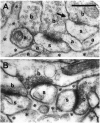
The role of transporters in shaping the glutamate concentration in the extracellular space after synaptic release is controversial because of their slow cycling and because diffusion alone gives a rapid removal. The transporter densities have been measured electrophysiologically, but these data are from immature brains and do not give precise information on the concentrations of the individual transporter subtypes. Here we show by quantitative immunoblotting that the numbers of the astroglial glutamate transporters GLAST (EAAT1) and GLT (EAAT2) are 3200 and 12,000 per micrometer3 tissue in the stratum radiatum of adult rat hippocampus (CA1) and 18,000 and 2800 in the cerebellar molecular layer, respectively. The total astroglial cell surface is 1.4 and 3.8 m2/cm3 in the two regions, respectively, implying average densities of GLAST and GLT molecules in the membranes around 2300 and 8500 micrometer-2 in the former and 4700 and 740 micrometer-2 in the latter region. The total concentration of glial glutamate transporters in both regions corresponds to three to five times the estimated number of glutamate molecules in one synaptic vesicle from each of all glutamatergic synapses. However, the role of glial glutamate transporters in limiting synaptic spillover is likely to vary between the two regions because of differences in the distribution of astroglia. Synapses are completely ensheathed and separated from each other by astroglia in the cerebellar molecular layer. In contrast, synapses in hippocampus (stratum radiatum) are only contacted by astroglia and are often found side by side without intervening glial processes.
Figures


Similar articles
-
The glutamate transporter EAAT4 in rat cerebellar Purkinje cells: a glutamate-gated chloride channel concentrated near the synapse in parts of the dendritic membrane facing astroglia.J Neurosci. 1998 May 15;18(10):3606-19. doi: 10.1523/JNEUROSCI.18-10-03606.1998. J Neurosci. 1998. PMID: 9570792 Free PMC article.
-
Glutamate transporters in glial plasma membranes: highly differentiated localizations revealed by quantitative ultrastructural immunocytochemistry.Neuron. 1995 Sep;15(3):711-20. doi: 10.1016/0896-6273(95)90158-2. Neuron. 1995. PMID: 7546749
-
Developmental expression of glutamate transporters and glutamate dehydrogenase in astrocytes of the postnatal rat hippocampus.Hippocampus. 2004;14(8):975-85. doi: 10.1002/hipo.20015. Hippocampus. 2004. PMID: 15390174
-
[Role of glutamate transporters in excitatory synapses in cerebellar Purkinje cells].Brain Nerve. 2007 Jul;59(7):669-76. Brain Nerve. 2007. PMID: 17663137 Review. Japanese.
-
Molecular organization of cerebellar glutamate synapses.Prog Brain Res. 1997;114:97-107. doi: 10.1016/s0079-6123(08)63360-9. Prog Brain Res. 1997. PMID: 9193140 Review.
Cited by
-
Glutamate Transporters/Na(+), K(+)-ATPase Involving in the Neuroprotective Effect as a Potential Regulatory Target of Glutamate Uptake.Mol Neurobiol. 2016 Mar;53(2):1124-1131. doi: 10.1007/s12035-014-9071-4. Epub 2015 Jan 14. Mol Neurobiol. 2016. PMID: 25586061 Review.
-
Aberrant protein S-nitrosylation contributes to hyperexcitability-induced synaptic damage in Alzheimer's disease: Mechanistic insights and potential therapies.Front Neural Circuits. 2023 Feb 2;17:1099467. doi: 10.3389/fncir.2023.1099467. eCollection 2023. Front Neural Circuits. 2023. PMID: 36817649 Free PMC article. Review.
-
Compromised glutamate transport in human glioma cells: reduction-mislocalization of sodium-dependent glutamate transporters and enhanced activity of cystine-glutamate exchange.J Neurosci. 1999 Dec 15;19(24):10767-77. doi: 10.1523/JNEUROSCI.19-24-10767.1999. J Neurosci. 1999. PMID: 10594060 Free PMC article.
-
Post-Traumatic Epilepsy and Comorbidities: Advanced Models, Molecular Mechanisms, Biomarkers, and Novel Therapeutic Interventions.Pharmacol Rev. 2022 Apr;74(2):387-438. doi: 10.1124/pharmrev.121.000375. Pharmacol Rev. 2022. PMID: 35302046 Free PMC article. Review.
-
The non-adrenergic imidazoline-1 receptor protein nischarin is a key regulator of astrocyte glutamate uptake.iScience. 2022 Mar 21;25(4):104127. doi: 10.1016/j.isci.2022.104127. eCollection 2022 Apr 15. iScience. 2022. PMID: 35434559 Free PMC article.
References
-
- Asztely F, Erdemli G, Kullmann DM. Extrasynaptic glutamate spillover in the hippocampus: dependence on temperature and the role of active glutamate uptake. Neuron. 1997;18:281–293. - PubMed
-
- Baddeley AJ, Gundersen HJG, Cruz-Orive LM. Estimation of surface area from vertical sections. J Microsc. 1986;142:259–276. - PubMed
-
- Barbour B, Häusser M. Intersynaptic diffusion of neurotransmitter. Trends Neurosci. 1997;20:377–384. - PubMed
Publication types
MeSH terms
Substances
LinkOut - more resources
Full Text Sources
Other Literature Sources
Molecular Biology Databases
Miscellaneous
