. 1998 Oct 1;12(19):2997-3007.
doi: 10.1101/gad.12.19.2997.

Senescence of human fibroblasts induced by oncogenic Raf

Affiliations

Senescence of human fibroblasts induced by oncogenic Raf

J Zhu et al. Genes Dev. .

Abstract

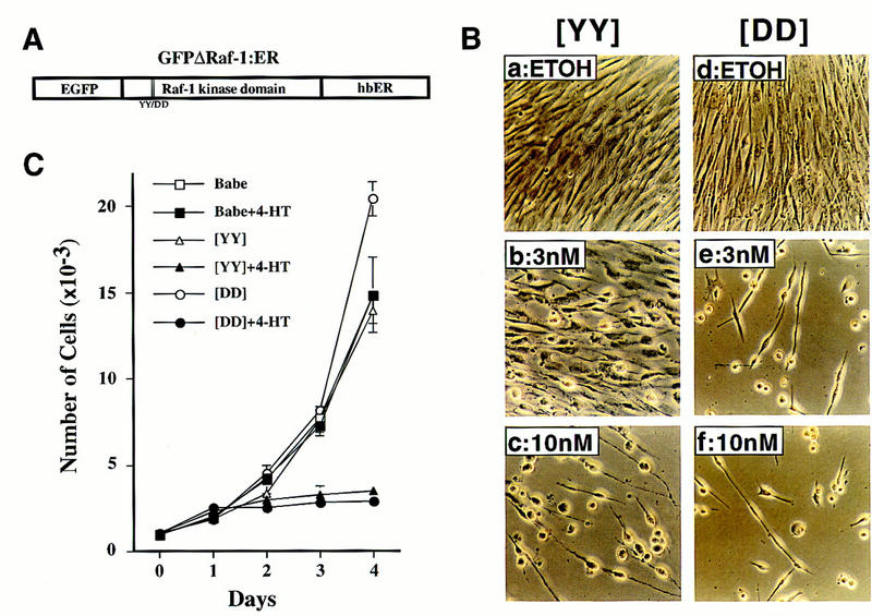
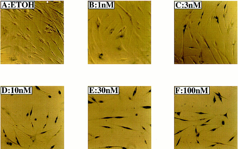
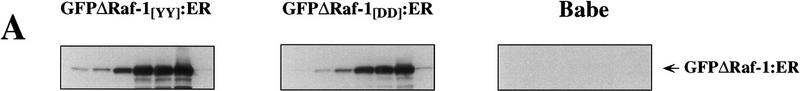
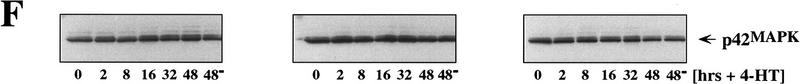
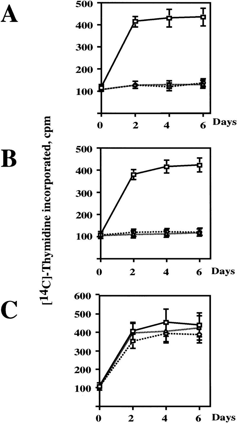
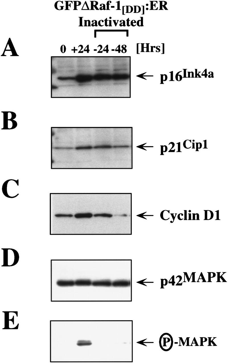
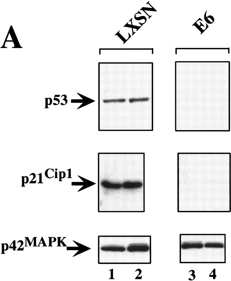
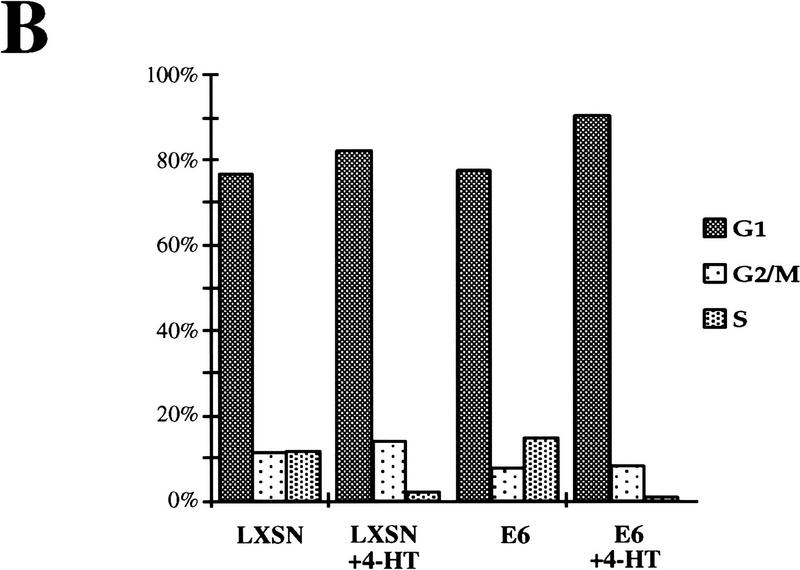
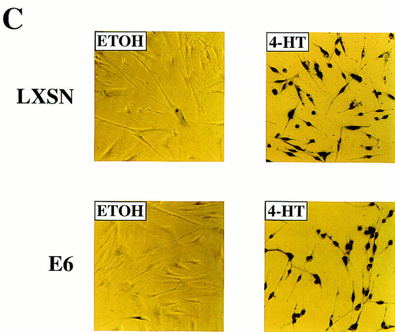
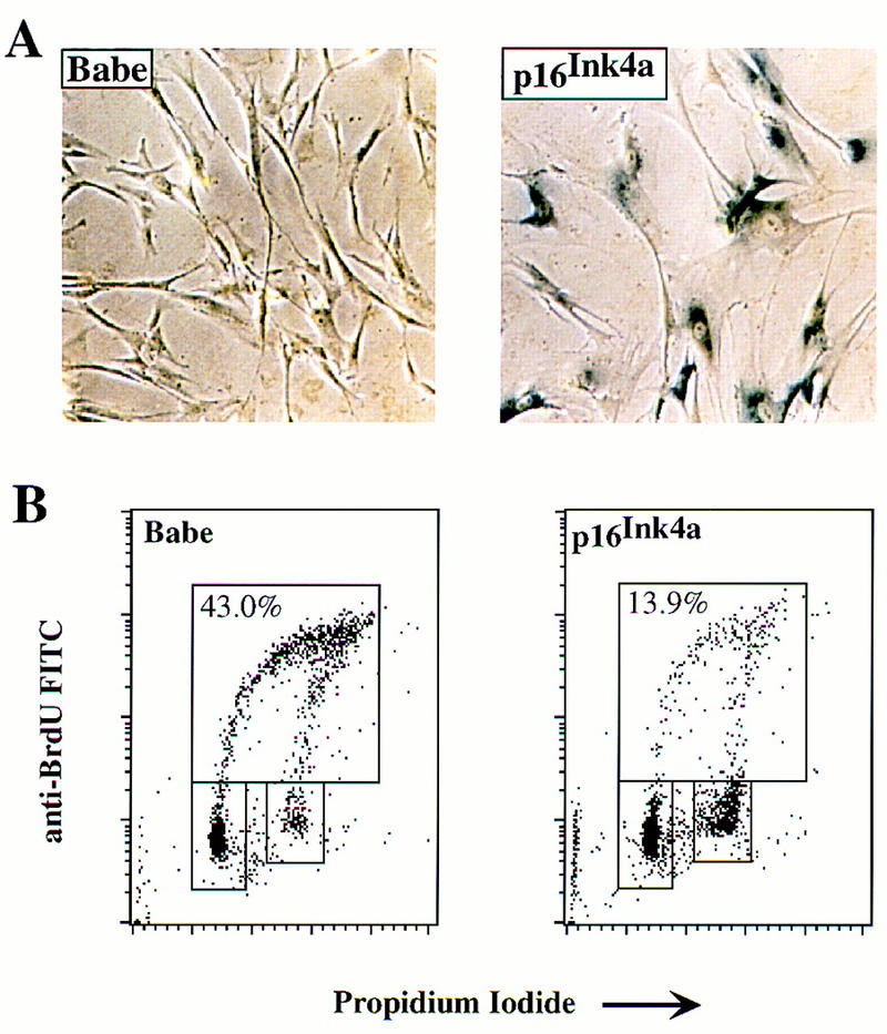
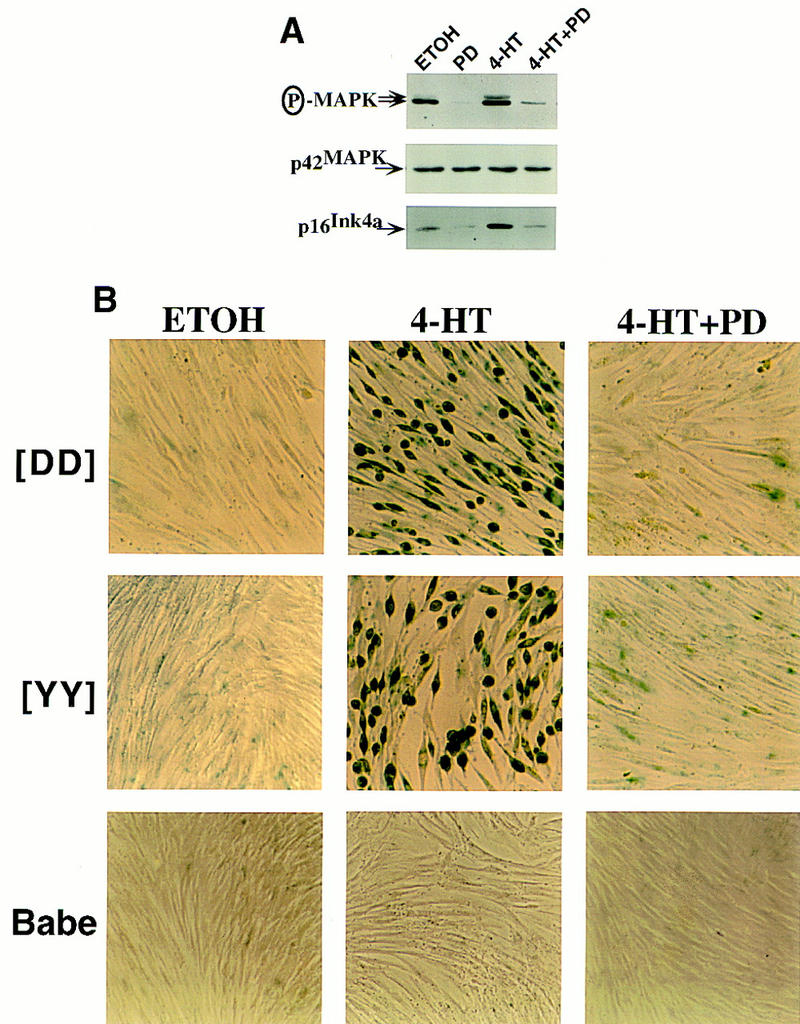
The oncogenes RAS and RAF came to view as agents of neoplastic transformation. However, in normal cells, these genes can have effects that run counter to oncogenic transformation, such as arrest of the cell division cycle, induction of cell differentiation, and apoptosis. Recent work has demonstrated that RAS elicits proliferative arrest and senescence in normal mouse and human fibroblasts. Because the Raf/MEK/MAP kinase signaling cascade is a key effector of signaling from Ras proteins, we examined the ability of conditionally active forms of Raf-1 to elicit cell cycle arrest and senescence in human cells. Activation of Raf-1 in nonimmortalized human lung fibroblasts (IMR-90) led to the prompt and irreversible arrest of cellular proliferation and the premature onset of senescence. Concomitant with the onset of cell cycle arrest, we observed the induction of the cyclin-dependent kinase (CDK) inhibitors p21(Cip1) and p16(Ink4a). Ablation of p53 and p21(Cip1) expression by use of the E6 oncoprotein of HPV16 demonstrated that expression of these proteins was not required for Raf-induced cell cycle arrest or senescence. Furthermore, cell cycle arrest and senescence were elicited in IMR-90 cells by the ectopic expression of p16(Ink4a) alone. Pharmacological inhibition of the Raf/MEK/MAP kinase cascade prevented Raf from inducing p16(Ink4a) and also prevented Raf-induced senescence. We conclude that the kinase cascade initiated by Raf can regulate the expression of p16(Ink4a) and the proliferative arrest and senescence that follows. Induction of senescence may provide a defense against neoplastic transformation when the MAP kinase signaling cascade is inappropriately active.


Figures

Figure 1
Figure 1
Activation of Raf kinase induces morphological alterations and arrests the proliferation of IMR-90 cells. (A) An inducible and tagged version of Raf. The illustrated construct was inserted into the replication-defective retrovirus described in Materials and Methods. (EGFP) Enhanced version of green fluorescent protein; (hbER) hormone-binding domain of the human estrogen receptor. The double vertical lines within the Raf-1 kinase domain and YY/DD below indicate the location of the tyrosine residues that were mutated to aspartic acid to generate the more active GFPΔRaf-1[DD]:ER as described in Materials and Methods and previously (Woods et al. 1997). (B) Morphological effects of Raf. IMR-90 cells expressing either the [YY] (a–c) or [DD] (d–e) forms of GFPΔRaf-1:ER were treated for 4 days with either ethanol as solvent control (ETOH) or different concentrations of 4-HT as indicated, then photographed with a phase-contrast microscope. (C) Proliferation of IMR-90 cells. Control (Babe) or GFPΔRaf-1:ER-expressing IMR-90 cells ([YY] or [DD]) were cultured in DMEM containing 15% vol/vol FCS in the absence or presence of 100 nm 4-HT as indicated. Cells were harvested at daily intervals and counted with a hemocytometer. Each data point represents the average of four independent measurements. A graph representative of multiple experiments is presented. (□) Babe; (█) Babe+4-HT; (▵) [YY]; (▴) [YY]+4-HT; (○) [DD]; (•) [DD]+4-HT.
Figure 2
Figure 2
Activity of senescence-associated, acidic β-galactosidase in IMR-90 cells. Actively proliferating IMR-90 cells expressing GFPΔRaf-1[YY]:ER were treated with either ethanol (ETOH) or with a range of 4-HT concentrations from 1–100 nm as indicated for 6 days, then stained to detect the activity of the senescence-associated, acidic β-galactosidase.
Figure 3
Figure 3
Effects of Raf on cell cycle regulators. Control (Babe) and GFPΔRaf-1:ER ([YY]- and [DD])-expressing IMR-90 cells were rendered quiescent for 24 hr, at which time 4-HT was added to a final concentration of 1 μm. Cell extracts were prepared at different times (2–48 hr) after the addition of 4-HT, and the expression of GFPΔRaf-1:ER (A), p16Ink4a (B), p21Cip1 (C), cyclin D1 (D), p27Kip1 (E), and p42 MAP kinase (F) was assessed by Western blotting with the appropriate antisera as described in Materials and Methods. As a control, an equivalent volume of ethanol was added to the cells for 48 hr (48).
Figure 3
Figure 3
Effects of Raf on cell cycle regulators. Control (Babe) and GFPΔRaf-1:ER ([YY]- and [DD])-expressing IMR-90 cells were rendered quiescent for 24 hr, at which time 4-HT was added to a final concentration of 1 μm. Cell extracts were prepared at different times (2–48 hr) after the addition of 4-HT, and the expression of GFPΔRaf-1:ER (A), p16Ink4a (B), p21Cip1 (C), cyclin D1 (D), p27Kip1 (E), and p42 MAP kinase (F) was assessed by Western blotting with the appropriate antisera as described in Materials and Methods. As a control, an equivalent volume of ethanol was added to the cells for 48 hr (48).
Figure 4
Figure 4
Raf-induced cell cycle arrest is irreversible. (A) GFPΔRaf-1[YY]:ER- and (B) GFPΔRaf-1[DD]:ER-expressing IMR-90 cells and (C) control cells (Babe) plated on Cytostar T plates were rendered quiescent in serum-free medium in the absence or presence of 1 μm 4-HT for 24 hr. At that time cells in the absence of 4-HT were stimulated with media containing 10% vol/vol FCS. Cells in the presence of 4-HT were either maintained in the continuous presence of 4-HT (4-HT continuous) or were washed extensively to remove the 4-HT (4-HT washed) prior to stimulation with media containing 10% vol/vol FCS. Cell proliferation was then measured over the following 6 days by the incorporation of methyl-[14C]thymidine into DNA. (□) 10% FCS; (▵) 4-HT continuous; (○) 4-HT washed.
Figure 5
Figure 5
Sustained expression of p16Ink4a following inactivation of GFPΔRaf-1:ER. IMR-90 cells expressing GFPΔRaf-1[DD]:ER were either untreated (0) or treated with 1 μm 4-HT for 24 hr (all other samples). After 24 hr in 4-HT, (+24) cells were washed extensively and then cultured in the absence of 4-HT for a further 24 (−24) or 48 hr (−48). Cell extracts were prepared, and the expression of p16Ink4a (A), p21Cip1 (B), cyclin D1 (C), and p42 MAP kinase (D) and the activity of p42/p44 MAP kinases (E) was assessed by Western blotting with the appropriate antisera as described in Materials and Methods.
Figure 6
Figure 6
Raf-induced cell cycle arrest and senescence of IMR-90 cells in the absence of detectable p53 and p21Cip1. (A) Effect of the E6 oncoprotein on p53 and p21Cip1 expression. IMR-90 cells expressing GFPΔRaf-1[YY]:ER cells were infected with retroviruses encoding resistance to neomycin alone (LXSN) or expressing the E6 protein of HPV16 and resistance to neomycin (E6), and pooled populations of drug-resistant clones were established. Cells were then treated for 24 hr with either ethanol (lanes 1,3) or 1 μm 4-HT (lanes 2,4). Cell lysates were prepared, and the expression of p53, p21Cip1, and p42 MAP kinase was assessed by Western blotting with the appropriate antisera. (B) Raf-induced cell cycle arrest is unaffected by expression of HPV-16 E6 protein. IMR-90 cell populations described above expressing GFPΔRaf-1[YY]:ER in the absence (LXSN) or presence of the HPV16 E6 oncoprotein (E6) were treated with either ethanol or 4-HT for 4 days. Cells were labeled with BrdU for 3 hr prior to harvest, fixed, and stained with a FITC-conjugated anti-BrdU antibody and propidium iodide to detect S-phase cells. Cells were analyzed by use of a Becton-Dickinson FACscan to determine the percentage of cells in the G1, S, and G2/M phases of the cell cycle. (C) Raf-induced SA–β-galactosidase activity. IMR-90 cell populations described above expressing GFPΔRaf-1[YY]:ER in the absence (LXSN) or presence of the HPV16 E6 oncoprotein (E6) cells were treated with either ethanol or 4-HT for 6 days at which time the activity of SA–β-galactosidase was assessed as described in Materials and Methods.
Figure 6
Figure 6
Raf-induced cell cycle arrest and senescence of IMR-90 cells in the absence of detectable p53 and p21Cip1. (A) Effect of the E6 oncoprotein on p53 and p21Cip1 expression. IMR-90 cells expressing GFPΔRaf-1[YY]:ER cells were infected with retroviruses encoding resistance to neomycin alone (LXSN) or expressing the E6 protein of HPV16 and resistance to neomycin (E6), and pooled populations of drug-resistant clones were established. Cells were then treated for 24 hr with either ethanol (lanes 1,3) or 1 μm 4-HT (lanes 2,4). Cell lysates were prepared, and the expression of p53, p21Cip1, and p42 MAP kinase was assessed by Western blotting with the appropriate antisera. (B) Raf-induced cell cycle arrest is unaffected by expression of HPV-16 E6 protein. IMR-90 cell populations described above expressing GFPΔRaf-1[YY]:ER in the absence (LXSN) or presence of the HPV16 E6 oncoprotein (E6) were treated with either ethanol or 4-HT for 4 days. Cells were labeled with BrdU for 3 hr prior to harvest, fixed, and stained with a FITC-conjugated anti-BrdU antibody and propidium iodide to detect S-phase cells. Cells were analyzed by use of a Becton-Dickinson FACscan to determine the percentage of cells in the G1, S, and G2/M phases of the cell cycle. (C) Raf-induced SA–β-galactosidase activity. IMR-90 cell populations described above expressing GFPΔRaf-1[YY]:ER in the absence (LXSN) or presence of the HPV16 E6 oncoprotein (E6) cells were treated with either ethanol or 4-HT for 6 days at which time the activity of SA–β-galactosidase was assessed as described in Materials and Methods.
Figure 7
Figure 7
Cell cycle arrest and induction of senescence by p16Ink4a. IMR-90 cells were infected with a retroviral vector expressing either the selectable marker for puromycin resistance alone (Babe) or the marker plus human p16Ink4a as indicated. The infected cells were then subjected to selection for 6 days. Surviving cells were analyzed for SA–β-galactosidase activity (A) or labeled with BrdU for analysis by FACscan for relative content of DNA (B). (B) Upper boxes delineate cells in S phase; lower boxes delineate cells in G0/G1 (left) or G2/M (right). Numbers in upper boxes indicate percentage of cells in S phase.
Figure 8
Figure 8
MEK activity is required for induction of p16Ink4a and senescence by Raf in IMR-90 cells. (A) Inhibition of MAP kinase activation and p16Ink4a expression by PD098059. Asynchronously growing IMR-90 cells expressing GFPΔRaf-1[YY]:ER were treated with either 10 nm 4-HT or an equivalent volume of ethanol (ETOH) for 4 days in the absence (DMSO) or presence of the MEK inhibitor PD098059 (PD, 25 μm) as indicated. The activity of p42/p44 MAP kinase and the expression of p42 MAP kinase and p16Ink4a were assessed by Western blotting with the appropriate antisera. (B) MEK activity is required for Raf-induced senescence in IMR-90 cells. Control (Babe) and GFPΔRaf-1:ER ([YY]- and [DD])-expressing IMR-90 cells were treated with various combinations of 4-HT (10 nm for [DD] and 3 nm for [YY]), PD (25 μm), or an equivalent volume of ethanol or DMSO for 6 days, and stained for SA–β-galactosidase activity.

Similar articles

Cited by

References

    1. Alcorta DA, Xiong Y, Phelps D, Hannon G, Beach D, Barrett JC. Involvement of the cyclin-dependent kinase inhibitor p16 (Ink4a) in replicative senescence of normal human fibroblasts. Proc Natl Acad Sci. 1996;93:13742–13747. - PMC - PubMed
    1. Alessi DR, Cuenda A, Cohen P, Dudley DT, Saltiel AR. PD 098059 is a specific inhibitor of the activation of mitogen-activated protein kinase kinase in vitro and in vivo. J Biol Chem. 1995;270:27489–27494. - PubMed
    1. Avruch J, Zhang XF, Kyriakis JM. Raf meets Ras: Completing the framework of a signal transduction pathway. Trends Biochem Sci. 1994;19:279–283. - PubMed
    1. Bodnar AG, Ouellette M, Frolkis M, Holt SE, Chiu CP, Morin GB, Harley CB, Shay JW, Lichtsteiner S, Wright WE. Extension of life-span by introduction of telomerase into normal human cells. Science. 1998;279:349–352. - PubMed
    1. Brown JP, Wei W, Sedivy JM. Bypass of senescence after disruption of p21CIP1/WAF1 gene in normal diploid human fibroblasts. Science. 1997;277:831–834. - PubMed

Publication types

MeSH terms