. 1998 Oct 5;143(1):207-15.
doi: 10.1083/jcb.143.1.207.

Regulated targeting of BAX to mitochondria

Affiliations

Regulated targeting of BAX to mitochondria

I S Goping et al. J Cell Biol. .

Abstract

The proapoptotic protein BAX contains a single predicted transmembrane domain at its COOH terminus. In unstimulated cells, BAX is located in the cytosol and in peripheral association with intracellular membranes including mitochondria, but inserts into mitochondrial membranes after a death signal. This failure to insert into mitochondrial membrane in the absence of a death signal correlates with repression of the transmembrane signal-anchor function of BAX by the NH2-terminal domain. Targeting can be instated by deleting the domain or by replacing the BAX transmembrane segment with that of BCL-2. In stimulated cells, the contribution of the NH2 terminus of BAX correlates with further exposure of this domain after membrane insertion of the protein. The peptidyl caspase inhibitor zVAD-fmk partly blocks the stimulated mitochondrial membrane insertion of BAX in vivo, which is consistent with the ability of apoptotic cell extracts to support mitochondrial targeting of BAX in vitro, dependent on activation of caspase(s). Taken together, our results suggest that regulated targeting of BAX to mitochondria in response to a death signal is mediated by discrete domains within the BAX polypeptide. The contribution of one or more caspases may reflect an initiation and/or amplification of this regulated targeting.


Figures

Figure 1
Figure 1
Subcellular distribution of BAX. BAX immunoreactivity localizes to mitochondria, perinuclear membrane, and cytosol in FL5.12 cells. Immunofluorescent confocal microscopy of FL5.12 cells using rabbit anti-BAX (P-19) antibody and goat Cy3 anti-rabbit Ig antibody shows both punctate and diffuse cytoplasmic immunoreactivity (A). MitoTracker Green FM shows exclusive punctate labeling (B). Dual exposure of BAX and MitoTracker labeling reveals colocalization of BAX with mitochondria (yellow) as well as nonmitochondrial BAX immunoreactivity (red; C). Conventional fluorescence detection of anti-BAX (P-19) immunoreactivity (Cy3) and Hoechst H33258 nuclear stain shows red cytoplasmic BAX immunoreactivity, blue nuclear Hoechst staining and pink perinuclear signal where the two reactivities overlap (D).
Figure 2
Figure 2
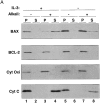
Mitochondrial BAX changes its alkali and protease sensitivity after a death stimulus. (A) BAX becomes alkali-resistant after a death stimulus. Heavy membranes enriched in mitochondria were prepared from FL5.12 cells cultured in IL-3 (lanes 1–4) or deprived of IL-3 for 12 h (lanes 5–8), incubated in isotonic buffer (lanes 1 and 2 and lanes 5 and 6), or in 0.1 M Na2CO3, pH 11.5 (lanes 3 and 4 and lanes 7 and 8), and centrifuged at 200,000 g for 45 min to yield supernatant (S) and pellet (P). The fractions were analyzed by immunoblot for BAX, BCL-2, cytochrome c oxidase subunit iv, and cytochrome c. (B) The NH2-terminus of BAX is further exposed after a death stimulus. The mitochondrial fraction prepared from FL5.12 cells in IL-3 (lanes 1 and 2 and lanes 6 and 7) or deprived of IL-3 for 12 h (lanes 3–5, and lanes 8–10) were incubated in isotonic buffer and treated with trypsin (30 μg/ml; lanes 2, 4, and 5) or proteinase K (100 μg/ml; prot. K, lanes 7, 9, and 10), for 20 min at 4°C. Trypsin was inactivated by adding a 30-fold weight excess of soybean trypsin inhibitor, and proteinase K was inactivated with phenylmethylsulfonylfluoride (1 mM). The fractions were analyzed by immunoblot with anti-BAX 651 antibody (amino acids 43–61; lanes 1–4 and lanes 6–9) or with anti-BAX N20 antibody (amino acids 11–30; Santa Cruz Biotechnology; lanes 5 and 10). (C) zVAD-fmk reduces mitochondrial membrane integration of BAX after IL-3 withdrawal. The mitochondrial fraction prepared from FL5.12 cells maintained in IL-3 (lanes 1, 2, 5, and 6) or deprived of IL-3 for 12 h (lanes 3, 4, 7, and 8) in the absence (lanes 1, 3, 5, and 7) or presence (lanes 2, 4, 6, and 8) of 50 μM zVAD-fmk for 12 h, were analyzed either directly (lanes 1–4) or after extraction with 0.1 M Na2CO3, pH 11.5 (lanes 5–8), as described in A.
Figure 2
Figure 2
Mitochondrial BAX changes its alkali and protease sensitivity after a death stimulus. (A) BAX becomes alkali-resistant after a death stimulus. Heavy membranes enriched in mitochondria were prepared from FL5.12 cells cultured in IL-3 (lanes 1–4) or deprived of IL-3 for 12 h (lanes 5–8), incubated in isotonic buffer (lanes 1 and 2 and lanes 5 and 6), or in 0.1 M Na2CO3, pH 11.5 (lanes 3 and 4 and lanes 7 and 8), and centrifuged at 200,000 g for 45 min to yield supernatant (S) and pellet (P). The fractions were analyzed by immunoblot for BAX, BCL-2, cytochrome c oxidase subunit iv, and cytochrome c. (B) The NH2-terminus of BAX is further exposed after a death stimulus. The mitochondrial fraction prepared from FL5.12 cells in IL-3 (lanes 1 and 2 and lanes 6 and 7) or deprived of IL-3 for 12 h (lanes 3–5, and lanes 8–10) were incubated in isotonic buffer and treated with trypsin (30 μg/ml; lanes 2, 4, and 5) or proteinase K (100 μg/ml; prot. K, lanes 7, 9, and 10), for 20 min at 4°C. Trypsin was inactivated by adding a 30-fold weight excess of soybean trypsin inhibitor, and proteinase K was inactivated with phenylmethylsulfonylfluoride (1 mM). The fractions were analyzed by immunoblot with anti-BAX 651 antibody (amino acids 43–61; lanes 1–4 and lanes 6–9) or with anti-BAX N20 antibody (amino acids 11–30; Santa Cruz Biotechnology; lanes 5 and 10). (C) zVAD-fmk reduces mitochondrial membrane integration of BAX after IL-3 withdrawal. The mitochondrial fraction prepared from FL5.12 cells maintained in IL-3 (lanes 1, 2, 5, and 6) or deprived of IL-3 for 12 h (lanes 3, 4, 7, and 8) in the absence (lanes 1, 3, 5, and 7) or presence (lanes 2, 4, 6, and 8) of 50 μM zVAD-fmk for 12 h, were analyzed either directly (lanes 1–4) or after extraction with 0.1 M Na2CO3, pH 11.5 (lanes 5–8), as described in A.
Figure 3
Figure 3
BAX constructs and their competence for mitochondrial targeting in vitro. ART domain (gray box); TM (black box); +, targeting-competent; −, targeting-incompetent. Amino acids 1–20 of BAX from the indicated species are shown. See text for additional details.
Figure 4
Figure 4
Apoptotic cell extract supports BAX targeting to mitochondria in vitro. (A) 35S-labeled transcription-translation products of BAX cDNA in rabbit reticulocyte lysate (BAX and BAXΔART) were incubated with purified intact mitochondria from rat heart in a standard protein import reaction supplemented with 1 mM dATP (lanes 3, 4, 6, and 7) or 1 mM dATP and 200 μg apoptotic cytosol protein (Extract; lanes 4 and 7) in a volume of 50 μl. At the end of the reaction, mitochondria were recovered by centrifugation, and the pellets were analyzed by SDS PAGE and fluorography either directly or after extraction with 0.1 M Na2CO3, pH 11.5 (Alkali; lanes 5–7). Lane 1, 10% of input translation product. (B) As in A except that import was conducted with BAXΔ1-19 cDNA transcription product (lane 2) at 4°C or 30°C as indicated, and was analyzed after extraction of the mitochondria with 0.1M Na2CO3, pH 11.5 (lanes 4 and 5). (C) 35S-labeled transcription-translation product of PARP cDNA in reticulocyte lysate (5% by volume) was incubated with (lanes 2 and 3) or without (lane 1) 100 μg apoptotic cytosol protein in a total volume of 20 μl, in the presence (lane 3) or absence (lanes 1 and 2) of 1 mM dATP, and equivalent portions were analyzed for the appearance of the 24-kD cleavage product of PARP (24K PARP) by 12% SDS PAGE and fluorography. (D) As in A, except that before import, mitochondria were treated with 0.4 mg/ml trypsin and 50-fold weight excess soybean trypsin inhibitor added either at the beginning (− Pre-Trypsin; columns 2 and 4) or at the end (+ Pre-Trypsin; columns 1 and 3) of the reaction. Mitochondria were collected by centrifugation and import conducted in the presence (columns 3 and 4) or absence (columns 1 and 2) of apoptotic cytosol (Extract), and the relative amount of alkali-insoluble full-length BAX was determined (maximum set at 100).
Figure 5
Figure 5
Failure of BAX to target mitochondria in vitro depends on the BAX TM. cDNAs encoding BAX (lanes 2–5), cytosolic dihydrofolate reductase fused at its COOH terminus to amino acids 169– 192 of BAX (DHFR-BAX™, lanes 7–10), and BAX in which the COOH-terminal 24 amino acids have been replaced by the corresponding domain of BCL-2 (amino acids 218–239) (BAX-BCL-2™, lanes 12–15) were transcribed and translated in reticulocyte lysate and the 35S-labeled, alkali-insoluble mitochondrial import products were analyzed by SDS PAGE and fluorography. Lanes 1, 6, and 11 represent 10% of input translation product, as indicated. The sequence (single letter code) of the COOH-terminal 24 amino acids of BAX and 22 amino acids of BCL-2 are shown; bold letters designate the predicted TMs.
Figure 6
Figure 6
The ability of apoptotic high-speed cytosol to support BAX targeting to mitochondria in vitro depends on cytochrome c, and is inhibited by zVAD-fmk. (A) The 35S-labeled transcription-translation products of BAX cDNA were subjected to mitochondrial import in vitro in the presence of 0 (lanes 2 and 6), 13 (lanes 3 and 7), 65 (lanes 4 and 8), or 260 (lanes 5 and 9) μg apoptotic high-speed cytrosolic protein (Extract) with (lanes 6–9) or without (lanes 2–5) added cytochrome c (Cyt C; 75 μg/ml), and the alkali-resistant products were visualized after SDS PAGE and fluorography. Lane 1, 10% of input translation product. (B; left) Cytochrome c was removed from apoptotic cell extract by immunoadsorption (Cyt C minus extract) using mouse monoclonal 2G8.B6 antibody (Mueller and Jemmerson, 1996) as described (Liu et al., 1996). Equivalent aliquots of the extract before (lane 1) and after (lane 2) immunoadsorption were analyzed by Western immunoblot using mouse monoclonal 7H8.2C12 antibody against cytochrome c (Liu et al., 1996), and were visualized by enhanced chemiluminescence. (Middle) Membrane insertion of BAX translation products, determined by resistance to extraction at pH 11.5, was conducted and analyzed as in A in the presence or absence of apoptotic cytosol, which had or had not been subjected to immunoadsorption with anti-cytochrome c or supplemented with added cytochrome c before the addition of dATP and incubation at 37°C, as indicated. (Right) The ability of this same apoptotic cytosol to generate the 24-kD apoptotic cleavage product of PARP was determined under the conditions indicated, as described in Fig. 4. (C) The ability of the apoptotic high-speed cytosol to support membrane insertion of BAX (alkaline-resistant product, lanes 2–4; left) and PARP cleavage (right) was assayed as in A in the presence or absence of 50 μM tetrapeptide zVAD-fmk that was delivered from a 100× concentrated stock dissolved in dimethylsulfoxide before adding dATP and incubating at 37°C. An equivalent volume of this solvent had no effect on BAX import (left, lane 3).
Figure 6
Figure 6
The ability of apoptotic high-speed cytosol to support BAX targeting to mitochondria in vitro depends on cytochrome c, and is inhibited by zVAD-fmk. (A) The 35S-labeled transcription-translation products of BAX cDNA were subjected to mitochondrial import in vitro in the presence of 0 (lanes 2 and 6), 13 (lanes 3 and 7), 65 (lanes 4 and 8), or 260 (lanes 5 and 9) μg apoptotic high-speed cytrosolic protein (Extract) with (lanes 6–9) or without (lanes 2–5) added cytochrome c (Cyt C; 75 μg/ml), and the alkali-resistant products were visualized after SDS PAGE and fluorography. Lane 1, 10% of input translation product. (B; left) Cytochrome c was removed from apoptotic cell extract by immunoadsorption (Cyt C minus extract) using mouse monoclonal 2G8.B6 antibody (Mueller and Jemmerson, 1996) as described (Liu et al., 1996). Equivalent aliquots of the extract before (lane 1) and after (lane 2) immunoadsorption were analyzed by Western immunoblot using mouse monoclonal 7H8.2C12 antibody against cytochrome c (Liu et al., 1996), and were visualized by enhanced chemiluminescence. (Middle) Membrane insertion of BAX translation products, determined by resistance to extraction at pH 11.5, was conducted and analyzed as in A in the presence or absence of apoptotic cytosol, which had or had not been subjected to immunoadsorption with anti-cytochrome c or supplemented with added cytochrome c before the addition of dATP and incubation at 37°C, as indicated. (Right) The ability of this same apoptotic cytosol to generate the 24-kD apoptotic cleavage product of PARP was determined under the conditions indicated, as described in Fig. 4. (C) The ability of the apoptotic high-speed cytosol to support membrane insertion of BAX (alkaline-resistant product, lanes 2–4; left) and PARP cleavage (right) was assayed as in A in the presence or absence of 50 μM tetrapeptide zVAD-fmk that was delivered from a 100× concentrated stock dissolved in dimethylsulfoxide before adding dATP and incubating at 37°C. An equivalent volume of this solvent had no effect on BAX import (left, lane 3).

Similar articles

Cited by

References

    1. Antonsson B, Conti F, Ciavatta A, Montessuit S, Lewis S, Martinou I, Bernasconi L, Bernard A, Mermod J-J, Mazzei G, et al. Inhibition of Bax channel-forming activity by BCL-2. Science. 1997;277:370–372. - PubMed
    1. Armstrong RC, Aja T, Xiang J, Gaur S, Krebs JF, Hoang K, Bai X, Korsmeyer SJ, Karanewsky DS, Fritz LC, Tomaselli KJ. Fas-induced activation of the cell death-related protease CPP32 is inhibited by BCL-2 and by ICE family protease inhibitors. J Biol Chem. 1996;271:16850–16855. - PubMed
    1. Boulakia CA, Chen G, Ng FWH, Teodoro JG, Branton PE, Nicholson DW, Poirier GG, Shore GC. BCL-2 and adenovirus E1B 19-kDa protein prevent E1A-induced processing of CPP32 and cleavage of poly(ADP-ribose) polymerase. Oncogene. 1996;12:529–535. - PubMed
    1. Cheng EH-Y, Levine B, Boise LH, Thompson CG, Hardwick JM. Bax-independent inhibition of apoptosis by BCL-XL . Nature. 1996;379:554–556. - PubMed
    1. Chinnaiyan AM, Orth K, O'Rourke K, Duan H, Poirier GG, Dixit VM. Molecular ordering of the cell death pathway. BCL-2 and BCL-XLfunction upstream of the CED-3-like apoptotic proteases. J Biol Chem. 1996;271:4573–4576. - PubMed

Publication types

MeSH terms

Substances