. 1998 Oct 5;143(1):81-94.
doi: 10.1083/jcb.143.1.81.

Sorting mechanisms regulating membrane protein traffic in the apical transcytotic pathway of polarized MDCK cells

Affiliations

Sorting mechanisms regulating membrane protein traffic in the apical transcytotic pathway of polarized MDCK cells

A Gibson et al. J Cell Biol. .

Abstract

The transcytotic pathway followed by the polymeric IgA receptor (pIgR) carrying its bound ligand (dIgA) from the basolateral to the apical surface of polarized MDCK cells has been mapped using morphological tracers. At 20 degreesC dIgA-pIgR internalize to interconnected groups of vacuoles and tubules that comprise the endosomal compartment and in which they codistribute with internalized transferrin receptors (TR) and epidermal growth factor receptors (EGFR). Upon transfer to 37 degreesC the endosome vacuoles develop long tubules that give rise to a distinctive population of 100-nm-diam cup-shaped vesicles containing pIgR. At the same time, the endosome gives rise to multivesicular endosomes (MVB) enriched in EGFR and to 60-nm-diam basolateral vesicles. The cup-shaped vesicles carry the dIgA/pIgR complexes to the apical surface where they exocytose. Using video microscopy and correlative electron microscopy to study cells grown thin and flat we show that endosome vacuoles tubulate in response to dIgA/pIgR but that the tubules contain TR as well as pIgR. However, we show that TR are removed from these dIgA-induced tubules via clathrin-coated buds and, as a result, the cup-shaped vesicles to which the tubules give rise become enriched in dIgA/pIgR. Taken together with the published information available on pIgR trafficking signals, our observations suggest that the steady-state concentrations of TR and unoccupied pIgR on the basolateral surface of polarized MDCK cells are maintained by a signal-dependent, clathrin-based sorting mechanism that operates along the length of the transcytotic pathway. We propose that the differential sorting of occupied receptors within the MDCK endosome is achieved by this clathrin-based mechanism continuously retrieving receptors like TR from the pathways that deliver pIgR to the apical surface and EGFR to the lysosome.


Figures
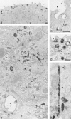
Figure 1
Figure 1
Transcytotic pathway of polarized PIR II MDCK cells outlined by dIgA-HRP internalized from the basolateral surface, 37°C. (a) Thick section showing dIgA-HRP loaded 100-nm-diam vesicles distributed below apical membrane. (The cup shape of these vesicles is more clearly shown at higher magnification in b). (b) Apical cytoplasm surrounding a centriole (C) and above Golgi stack (G). dIgA-HRP is distributed on the inner perimeter membranes of vacuoles (*), within MVB (MVB), throughout 100-nm-diam tubules (T) and in 100-nm-diam cup-shaped vesicles (csv). (c) High magnification showing apical vesicles containing transcytosed dIgA-HRP; arrowheads, fine filaments of DAB reaction product distributed on the apical surface; csv, cup-shaped vesicles. (d) As c, an undispersed core of reaction product (arrow) identifies a site of exocytosis. (e) 100-nm-diam tubule containing dIgA-HRP running in the long axis of the cell; arrowheads, position of microtubule. Bars, 0.2 μm.
Figure 2
Figure 2
Changes in proportions of dIgA-HRP containing vacuoles, tubules, and cup-shaped vesicles in polarized, filter-grown cells on transfer to 37°C. Polarized, filter-grown cells were loaded with dIgA-HRP at 20°C and then transferred to 37°C for 0, 15, and 30 min. With time at 37°C the proportion of vacuoles decreases, the proportion of tubules increases and then decreases, and the proportion of cup-shaped vesicles increases. The increase in cup-shaped vesicles is twice as rapid in the second 15-min interval. The data were obtained by estimating the surface area of all dIgA-HRP–containing elements within equivalent areas of cytoplasm in cells incubated for the 0–30-min time intervals and then assigning them to four groups; elements of less than 60-nm- diam, vesicles of 100-nm-diam (i.e., cup-shaped vesicles), tubules of 100-nm-diam, and elements of 0.1–0.3-μm-diam (vacuoles). Elements of less than 60 nm in diameter comprised 74% of the total DAB-positive structures.
Figure 3
Figure 3
Release of dIgA and degradation of EGF in polarized PIR II cells. Monolayers were preloaded basolaterally at 20°C with 125I-dIgA (solid symbols) or 125I-EGF (open symbols), chased at 37°C and apical (circles) and basolateral (squares) media collected. For EGF, only TCA-precipitable protein released is shown.
Figure 4
Figure 4
The distribution of fluorescent dIgA and Tf in pIgR/TR-expressing cells grown on glass coverslips. (a) Cells loaded with dIgA-FITC at 20°C for 30 min. Brightly fluorescing vacuoles of sorting endosome complexes are distributed throughout the cytoplasm. (b) As in a, then transferred to 37°C for 10 min. Long, dIgA-FITC–containing tubules have formed. (c) As in a, then transferred to 37°C for 30 min. Tubules replaced by fine punctate fluorescence distributed throughout peripheral cytoplasm. Arrow, pericentriolar concentration of dIgA fluorescence lying below the nucleus. (d) Cells preincubated with nocodazole 30 min at 5°C then treated as in c. Tubules do not form and the number of sorting endosome vacuoles is only slightly reduced compared with the 20°C condition shown in a. The vacuoles remain brightly fluorescent for dIgA and should be compared with the structures shown by electron microscopy in Fig. 6 d. (e) Cells loaded with Tf-HRP at 20°C for 30 min and then transferred to 37°C for 10 min in the presence of dIgA. Tubules are displayed. (f ) Cells treated as in e but no dIgA added on transfer to 37°C. No tubules form. Bars, 10 μm.
Figure 5
Figure 5
Video microscopy of endosomes at 20° and 37°C. (a) Stills taken from video in which cells grown flat were incubated with dIgA-FITC at 20°C for 60 min. Over the 10-min period the cluster of brightly fluorescent vacuoles continually changes shape and position but remains grouped together. (b) As in a but cells transferred to 37°C. During this 5-min interval, fine tubules (arrows) grow rapidly from the vacuole. (The full sequences of these videos can be seen on http://www.ucl.ac.uk/lmcb/gibson.htm). Bars, 5 μm.
Figure 6
Figure 6
Colocalization of pIgR, TR, and EGFR tracers in clusters of endosome vacuoles and tubules in cells growing on solid substrata. (a) Vacuoles and tubules containing internalized dIgA-HRP and 10-nm gold/anti-TR (arrowheads). The cytoplasmic domains of pIgR are labeled with 5-nm gold and show them to be distributed throughout the dIgA-HRP–loaded vacuoles and tubules. Vesicles surrounding the Golgi (G) are unlabeled. Cells were preloaded with dIgA-HRP and 10-nm gold/anti-TR at 20°C for 60 min. Internalized HRP cross-linked with DAB/H2O2, and cells permeabilized with digitonin before being labeled with 5-nm gold/anti-pIgR. (b) Vacuoles and tubules containing internalized Tf-HRP and 10-nm gold/dIgA. The cytoplasmic domains of TR are labeled with 5-nm gold. HRP-loaded vacuoles and most tubules that label with 5-nm gold/anti-TR also contain internalized pIgR tracer (arrowheads). Cells were prepared as in a. (c) Cells loaded with 10-nm gold/dIgA and 5-nm gold/anti-EGFR in presence of 200 ng/ml EGF at 20°C for 30 min. Sorting endosome vacuole (V) and associated tubules (arrows) contain both tracers. (d) Without microtubules, tubulation does not occur at 37°C and vacuoles remain loaded with dIgA-HRP/pIgR. These compact groups of vacuoles correspond to the strongly fluorescent spots seen in Fig 4 d. Cells were loaded with dIgA-HRP for 60 min at 20°C, incubated with nocodazole at 5°C for 30 min, and then incubated at 37°C for 40 min. Internalized dIgA-HRP cross-linked with DAB/H2O2, and cells permeabilized with digitonin before being labeled with 10-nm gold/anti-pIgR and 5-nm gold/anti-TR (arrowheads). Bars, 0.1 μm.
Figure 9
Figure 9
Distribution of pIg R and TR and their relationship to clathrin coats on vacuoles, fragmenting tubules, and on cup-shaped vesicles. (a) Distribution of AP 1 gold label on coated buds forming on an endosome vacuole (arrowheads) that is also giving rise to a 100-nm-diam dIgA-induced tubule (arrows). Immunogold labeling was as described in Futter et al., 1998. Cells were incubated with dIgA-HRP, Tf-HRP at 20°C for 60 min and then transferred to 37°C for 10 min. (b) Cytoplasmic domains of pIgR labeled with 5-nm gold on a fragment of a dIgA-induced tubule containing internalized 10-nm gold/anti-TR (arrowhead). Arrows indicate the distributions of clathrin lattices. Cells were incubated with dIgA-HRP, Tf-HRP at 20°C for 60 min, incubated with B3/25-gold at 5°C for 30 min, and then transferred to 37°C for 5 min. (c) Preparation as in b showing tubule fragment on which cytoplasmic domains of pIgR are labeled with 5-nm gold. Receptors within clathrin-coated domains (arrows) remain unlabeled but the presence of B3/25 gold (arrowheads) shows they contain TR. (d) Cup-shaped vesicles (csv) developing from tubule fragments, cytoplasmic domains of pIgR (10 nm), and TR (5 nm) labeled with gold. Arrows, clathrin lattices on tubule fragments (t); G, Golgi cisternae. Cells were preloaded at 20°C with dIgA-HRP and Tf-HRP and then transferred to 37°C for 15 min. The internalized HRP tracers have been cross-linked with DAB/H2O2. Cells were then permeabilized with digitonin and double labeled with gold antibody conjugates. (e) Distribution of pIgR (10-nm gold) and TR (5-nm gold) on 100-nm-diam vesicles (cup-shape not shown in this section) and 60-nm-diam vesicles. Membrane boundaries are visible, indicating that the section plane is close to equatorial in both vesicles. Both vesicles label for pIgR and TR but quantitation of these vesicle populations shows pIgR are more concentrated (60:40) in the larger vesicle and TR more concentrated (70:30) in the smaller. Bars, 0.1 μm.
Figure 7
Figure 7
(a) Whole mount showing distribution of TR and pIgR in a complex of endosome vacuoles and tubules loaded with Tf-HRP at 20°C. (This complex corresponds to the structures seen in section in Fig. 6, a–c). The interconnected vacuolar and tubular elements of a sorting endosome complex have been preserved by cross-linking their content of Tf-HRP and dIgA-HRP. Internalized 10-nm-diam gold complexes show linearly arrayed pIgR within short tubules (arrowheads) and 5-nm gold particles identify the cytoplasmic domains of TR. Arrows, free vesicles. Both pIgR and TR are distributed throughout the complex. Cells preloaded with TF-HRP, dIgA-HRP and 10-nm dIgA-gold at 20°C for 30 min, were cross-linked with DAB/H2O2, extracted with TX100, and then TR cytoplasmic domains were localized with TR antibody (H68.4) followed by 5-nm immunogold. Bar, 0.2 μm. (b) Whole mount showing tubulation induced by dIgA. Low magnification view showing dIgA-induced tubules extending throughout the cytoplasm (Fig. 7 a) with vacuoles (arrows) distributed at intervals along their length. Cell preloaded with dIgA-HRP and TF-HRP at 20°C for 30 min and transferred to 37°C for 15 min before being cross-linked with DAB/H2O2, extracted with TX100, and then labeled with antibody gold complexes (not evident in this micrograph, but see Fig. 8 a). n, nucleus. Bar, 1 μm.
Figure 8
Figure 8
dIgA-induced tubules extending from clusters of endosome vacuoles. (a) Whole mount showing group of vacuoles including MVBs (arrows) with 100-nm-diam tubule extension (T). Gold particles show pIgR(10 nm) and TR(5 nm) distributed randomly throughout vacuoles and tubule. Inset, segment of tubule enlarged to display TR (arrowheads) distributed amongst pIgR. Cells were preloaded with dIgA-HRP and TF-HRP at 20°C for 30 min and transferred to 37°C for 10 min before being cross-linked with DAB/H2O2, extracted with TX100, and then labeled with antibody gold complexes. (b) A group of vacuoles (V) and tubular extensions (T) similar to the interconnected complex shown in a but displayed in a conventional thin section. Large arrow, bulbous tips of tubules with invaginations similar to those seen in cup-shaped vesicles; small arrows, 60-nm-diam–coated vesicle. Cells preloaded at 20°C with dIgA-HRP and then transferred to 37°C for 10 min. (c) IgA-induced tubule varying in diameter from 50 to 120 nm. Arrows, coated buds. Cells were preloaded at 20°C with dIgA-HRP and then transferred to 37°C for 10 min. Bars, 0.1 μm.

Similar articles

Cited by

References

    1. Apodaca G, Katz LA, Mostov KE. Receptor-mediated transcytosis of IgA in MDCK cells is via apical recycling endosomes. J Cell Biol. 1994;125:67–86. - PMC - PubMed
    1. Aroeti G, Kosen PA, Kuntz ID, Cohen FE, Mostov KE. Mutational and secondary structural analysis of the basolateral sorting signal of the polymeric immunoglobulin receptor. J Cell Biol. 1993;123:1149–1160. - PMC - PubMed
    1. Barroso M, Sztul EJ. Basolateral to apical transcytosis in polarized cells is indirect and involves BFA and trimeric G protein sensitive passage through the apical endosome. J Cell Biol. 1994;124:83–100. - PMC - PubMed
    1. Bates P, Young JAT, Varmus HE. A receptor for subgroup A Rous sarcoma virus is related to the low density lipoprotein receptor. Cell. 1993;74:1043–1051. - PubMed
    1. Bomsel M, Mostov K. Sorting of plasma membrane proteins in epithelial cells. Curr Opin Cell Biol. 1991;3:647–653. - PubMed

Publication types