. 1998 Sep 21;142(6):1413-27.
doi: 10.1083/jcb.142.6.1413.

Annexin XIIIb associates with lipid microdomains to function in apical delivery

Affiliations

Annexin XIIIb associates with lipid microdomains to function in apical delivery

F Lafont et al. J Cell Biol. .

Abstract

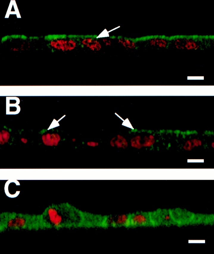
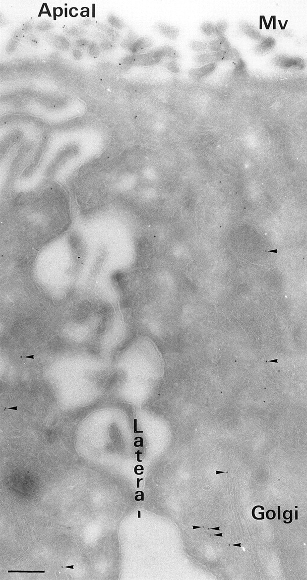
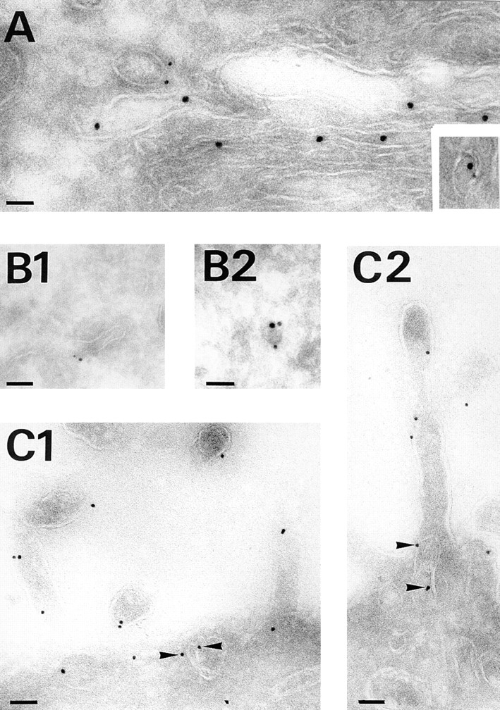
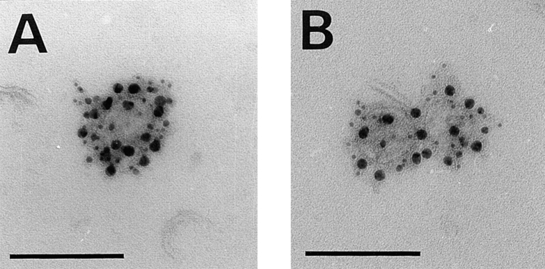
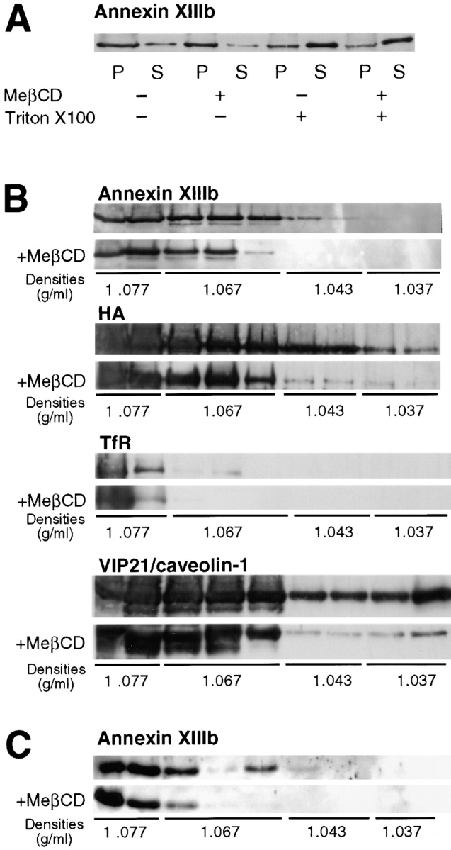
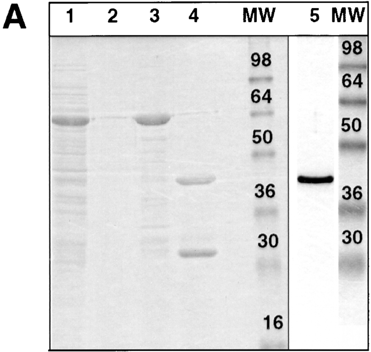
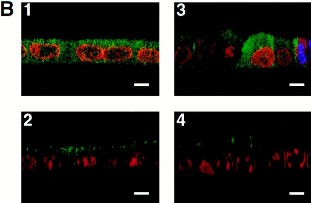
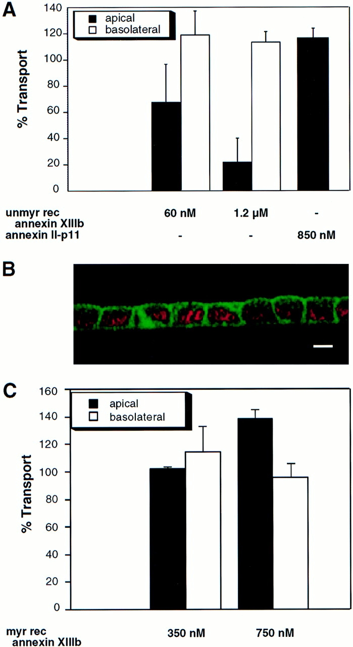
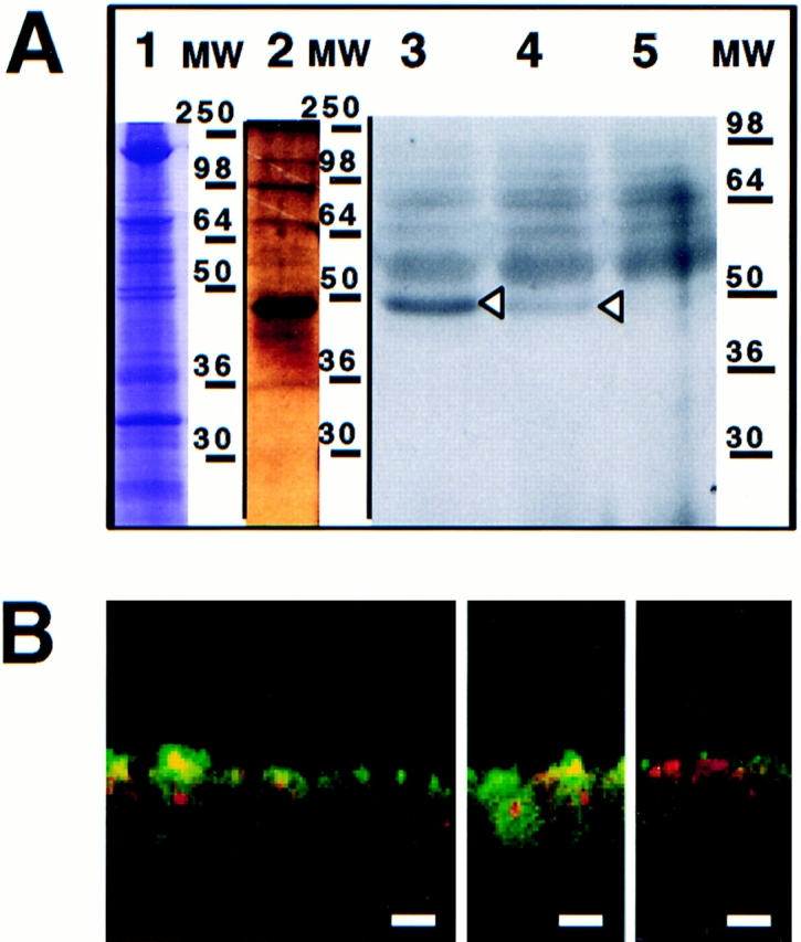
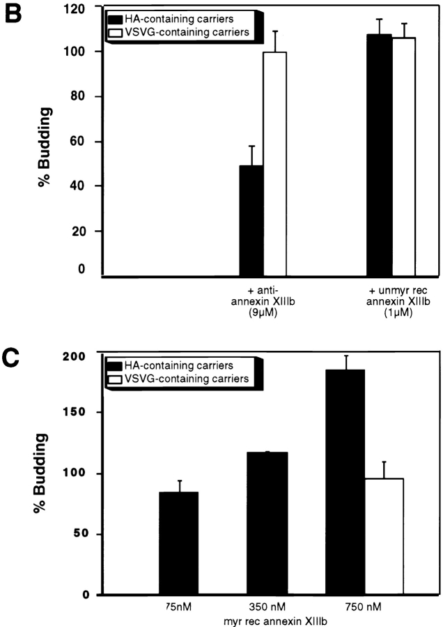
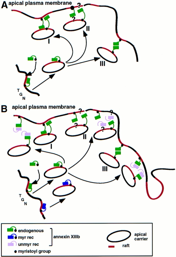
A member of the annexin XIII sub-family, annexin XIIIb, has been implicated in the apical exocytosis of epithelial kidney cells. Annexins are phospholipid-binding proteins that have been suggested to be involved in membrane trafficking events although their actual physiological function remains open. Unlike the other annexins, annexin XIIIs are myristoylated. Here, we show by immunoelectron microscopy that annexin XIIIb is localized to the trans-Golgi network (TGN), vesicular carriers and the apical cell surface. Polarized apical sorting involves clustering of apical proteins into dynamic sphingolipid-cholesterol rafts. We now provide evidence for the raft association of annexin XIIIb. Using in vitro assays and either myristoylated or unmyristoylated recombinant annexin XIIIb, we demonstrate that annexin XIIIb in its native myristoylated form stimulates specifically apical transport whereas the unmyristoylated form inhibits this route. Moreover, we show that formation of apical carriers from the TGN is inhibited by an anti-annexin XIIIb antibody whereas it is stimulated by myristoylated recombinant annexin XIIIb. These results suggest that annexin XIIIb directly participates in apical delivery.


Figures

Figure 1
Figure 1
Survey of annexin XIIIb during MDCK cell polarization. Filter-grown MDCK cells were immunostained with anti-annexin XIIIb or anti-annexin II antibodies (green) and nuclei were labeled with propidium iodide (red). Samples were viewed by confocal microscopy. Confocal (x, z) scans are presented. (A) Fully polarized cell processed for annexin XIIIb immunostaining. (B and C) Cells fixed after 24 h in culture before immunolabeling with the anti-annexin XIIIb (B) or anti-annexin II (C) antibodies. Notice that some cells in B (arrows) display specific apical surface labeling like that observed for the entire monolayer when the cells are fully polarized (A, arrow). Bar, 10 μm.
Figure 2
Figure 2
Overview of the ultrastructural distribution of annexin XIIIb in polarized MDCK cells. Polarized filter-grown MDCK cells were processed for immunoelectron microscopy with the anti-annexin XIIIb antibody. Arrowheads point out gold particles in the cytoplasm distributed in close vicinity of membranes. Bar, 400 nm.
Figure 3
Figure 3
Annexin XIIIb distributes along the apical pathway. Double immunostaining was performed on influenza virus infected polarized filter-grown MDCK cells expressing the VSV G-tagged sialyltransferase before processing for electron microscopy. (A) Sialyltransferase detected with anti-VSV G antibody (15 nm gold) and anti-annexin XIIIb (6 nm gold) labeling at the Golgi level after a TGN block at 20°C for 75 min. The inset illustrates colocalization of both sialyltransferase and annexin XIIIb in the same vesicular profile found nearby the Golgi stacks. (B1 and B2) Examples of colocalization of annexin XIIIb (6 nm gold) and HA (15 nm gold) in tubulovesicular structures. (C1 and C2). Annexin XIIIb staining in tubulovesicular structures underneath the apical surface (arrowheads) and in microvilli. Bars: (B1) 120 nm; (B2) 100 nm; and (C1 and C2) 120 nm.
Figure 4
Figure 4
Distribution of annexin XIIIb on exocytic TGN-derived carriers. (A) Immunostaining of HA (15 nm gold), annexin XIIIb (10 nm gold) and VIP21/caveolin-1 (5 nm gold) on TGN-derived carriers obtained from cells whose apical membrane have been ripped off (see Materials and Methods). (B) Immunostaining of VSV G (15 nm gold), annexin XIIIb (10 nm gold) and VIP21/ caveolin-1 (5 nm gold) on TGN-derived carriers. Bar, 100 nm. Note the enrichment of gold particles associated with annexin XIIIb on HA-containing carriers versus the amount observed on VSV G-containing carriers and see Table II. Bar, 100 nm.
Figure 5
Figure 5
Annexin XIIIb is raft associated. Filter-grown MDCK cells were infected with influenza virus. Cells treated with mevalonate and lovastatin were extracted with MeβCD. Then, cells were submitted to a TGN block and chased at 37°C before lysis. (A) Lysed cells were either extracted with Triton X-100 or not before centrifugation. Percentages of insoluble (P) and soluble (S) annexin XIIIb were analyzed by Western blotting. Similar result were obtained in four independent experiments. (B) Lysed cells were extracted with Triton X-100 and membranes were floated in an OptiPrep™ step gradient. Western blotting was performed on methanol-chloroform precipitated fractions. The first two fractions correspond to the volume of the sample submitted to floatation. (C) TGN-derived carriers from influenza virus infected cells were extracted with Triton X-100 and membranes were floated in an Optiprep™ step gradient. Western blotting was performed on methanol-chloroform precipitated fractions.
Figure 6
Figure 6
Purified unmyristoylated recombinant annexin XIIIb is efficiently reaching the intracellular compartment in permeabilized cells. (A) Purification of recombinant annexin XIIIb expressed in E. coli. Lane 1, E. coli lysate; lane 2, last wash, and lane 3, elution from glutathione column. Lane 4, Thrombin cleavage before run on Mono-Q column. Lane 5, Purified recombinant annexin XIIIb after Mono-Q column elution. Lanes 1–4, Coomassie blue staining. Lane 5, Silver staining. (B) Confocal (x,z) scans of cells either basolaterally (1, 2) or apically (3, 4) SLO-permeabilized. Recombinant annexin XIIIb (1.2 μM) was added to samples 1 and 3. Nuclei were labeled with propidium iodide (red) and annexin XIIIb and visualized using an anti-annexin XIIIb antibody (green). Note that the recombinant unmyristoylated protein is present in both the apical and basolateral compartments (B1 and B3). Bar, 10 μm.
Figure 6
Figure 6
Purified unmyristoylated recombinant annexin XIIIb is efficiently reaching the intracellular compartment in permeabilized cells. (A) Purification of recombinant annexin XIIIb expressed in E. coli. Lane 1, E. coli lysate; lane 2, last wash, and lane 3, elution from glutathione column. Lane 4, Thrombin cleavage before run on Mono-Q column. Lane 5, Purified recombinant annexin XIIIb after Mono-Q column elution. Lanes 1–4, Coomassie blue staining. Lane 5, Silver staining. (B) Confocal (x,z) scans of cells either basolaterally (1, 2) or apically (3, 4) SLO-permeabilized. Recombinant annexin XIIIb (1.2 μM) was added to samples 1 and 3. Nuclei were labeled with propidium iodide (red) and annexin XIIIb and visualized using an anti-annexin XIIIb antibody (green). Note that the recombinant unmyristoylated protein is present in both the apical and basolateral compartments (B1 and B3). Bar, 10 μm.
Figure 7
Figure 7
Exocytic apical delivery is specifically sensitive to unmyristoylated and myristoylated recombinant annexin XIIIb. (A) Dose-dependent effect of unmyristoylated recombinant annexin XIIIb (unmyr rec annexin XIIIb) expressed in E. coli in the in vitro transport assay with HA and VSV G as reporter proteins for the apical and basolateral exocytic pathway, respectively. Annexin II–p11 heterotetramer does not impair the apical transport of HA. (B) Confocal (x, z) scan of polarized MDCK cells double stained for nuclei (propidium iodide in red) and for annexin II (green). (C) Dose-dependent effect of myristoylated recombinant annexin XIIIb (myr rec annexin XIIIb) expressed in Drosophila Schneider SL3 cells in the in vitro transport assay with HA and VSV G as reporter proteins for the apical and basolateral exocytic pathway, respectively. Bar, 20 μm.
Figure 8
Figure 8
Purified myristoylated recombinant annexin XIIIb is reaching the apical compartment of MDCK cells. (A) Purification of annexin XIIIb from Drosophila Schneider cells. Lane 1, Coomassie blue staining of the total lysate. Lane 2, Silver staining of eluted purified recombinant annexin XIIIb (see Materials and Methods). Arrowheads indicate the myristoylated annexin XIIIb. Note the appearance of dimers at 90 kD (each monomer is formed by the fusion of annexin XIIIb and histidine plus FLAG tags). Lanes 3–5, Autoradiography of annexin XIIIb expressing clones (lanes 3 and 4 clones BM17 and BM18, respectively) and of control cells (lane 5) after 3H-myristate labeling. (B) Distribution of the myristoylated recombinant annexin XIIIb in basolaterally SLO-permeabilized MDCK cells. The myristoylated recombinant protein was added from the basolateral side. Confocal (x, z) scans of cells double stained for nuclei (propidium iodide in red) and for the myristoylated recombinant annexin XIIIb with the anti-Flag antibody (green). Note that although in some cases the myristoylated recombinant protein was observed in both apical and basolateral compartments, the recombinant protein was mainly found in the apical compartment. Bar, 20 μm.
Figure 9
Figure 9
Effects of anti-annexin XIIIb antibody, unmyristoylated and myristoylated recombinant annexin XIIIb on TGN-derived carriers release. (A) Confocal (x, z) scans of polarized MDCK cells double stained for nuclei (propidium iodide in red) and annexin V (green). (B) Anti-annexin XIIIb antibody can inhibit the budding of HA-containing carriers while it is not interfering with the budding of VSV G-containing carriers. Unmyristoylated recombinant annexin XIIIb (unmyr rec annexin XIIIb) is not affecting the release from the TGN of HA- or VSV G-containing carriers. The matched controls, i.e., anti-annexin V antibody and the annexin II–p11 complex have no effect on the budding of TGN-derived carriers (see text). (C) Myristoylated recombinant annexin XIIIb (myr rec annexin XIIIb) can stimulate in a dose- dependent manner the release of HA-containing carriers from the TGN while leaving the release of VSV G-containing carriers unaffected. Bar, 10 μm.
Figure 9
Figure 9
Effects of anti-annexin XIIIb antibody, unmyristoylated and myristoylated recombinant annexin XIIIb on TGN-derived carriers release. (A) Confocal (x, z) scans of polarized MDCK cells double stained for nuclei (propidium iodide in red) and annexin V (green). (B) Anti-annexin XIIIb antibody can inhibit the budding of HA-containing carriers while it is not interfering with the budding of VSV G-containing carriers. Unmyristoylated recombinant annexin XIIIb (unmyr rec annexin XIIIb) is not affecting the release from the TGN of HA- or VSV G-containing carriers. The matched controls, i.e., anti-annexin V antibody and the annexin II–p11 complex have no effect on the budding of TGN-derived carriers (see text). (C) Myristoylated recombinant annexin XIIIb (myr rec annexin XIIIb) can stimulate in a dose- dependent manner the release of HA-containing carriers from the TGN while leaving the release of VSV G-containing carriers unaffected. Bar, 10 μm.
Figure 10
Figure 10
Model for the participation of annexin XIIIb and for the role played by the recombinant proteins in the apical exocytic pathway. (A) The raft-associated annexin XIIIb in the TGN level promotes assembly of apical carriers and participates in the docking and/or fusion steps at the plasma membrane. The raft depicted in red lines should be considered as membrane microdomains whose lipids are enriched in sphingolipid and cholesterol. In the last steps of exocytic transport annexin XIIIb could function either by binding to a membrane receptor (lipid domain I and/or proteins II), or by oligomerization (III). (B) Influence of unmyristoylated (unmy rec) and myristoylated (myr rec) recombinant annexin XIIIb on the apical exocytic pathway. The unmyristoylated protein inhibits the docking and/or fusion steps by interfering with the binding of the endogenous annexin XIIIb to its membrane ligand: lipid microdomains (I), protein complex, including putative cytosolic proteins (II), or itself, oligomerization (III). The myristoylated recombinant protein acts as the endogenous protein and increases the efficiency of the apical transport.

Similar articles

Cited by

References

    1. Ahmed SN, Brown DA, London E. On the origin of sphingolipid/ cholesterol-rich detergent-insoluble cell membranes: physiological concentrations of cholesterol and sphingolipid induce formation of a detergent-insoluble, liquid-ordered lipid phase in model. Biochemistry. 1997;36:10944–10953. - PubMed
    1. Ali SM, Geisow MJ, Burgoyne RD. A role for calpactin in calcium-dependent exocytosis in adrenal chromaffin cells. Nature. 1989;340:313–315. - PubMed
    1. Balcarova-Ständer J, Pfeiffer SE, Fuller SD, Simons K. Development of cell surface polarity in the epithelial Madin-Darby canine kidney (MDCK) cell line. EMBO (Eur Mol Biol Organ) J. 1984;3:2687–2694. - PMC - PubMed
    1. Beckers CJM, Keller DS, Balch WE. Semi-intact cells permeable to macromolecules: use in reconstitution of protein transport from the endoplasmic reticulum to the golgi complex. Cell. 1987;50:523–534. - PubMed
    1. Bennett M, Wandinger-Ness A, Simons K. Release of putative exocytic transport vesicles from perforated MDCK cells. EMBO (Eur Mol Biol Organ) J. 1988;7:4075–4085. - PMC - PubMed

Publication types