. 1998 Sep 15;18(18):7402-10.
doi: 10.1523/JNEUROSCI.18-18-07402.1998.

A protein kinase, PKN, accumulates in Alzheimer neurofibrillary tangles and associated endoplasmic reticulum-derived vesicles and phosphorylates tau protein

Affiliations

A protein kinase, PKN, accumulates in Alzheimer neurofibrillary tangles and associated endoplasmic reticulum-derived vesicles and phosphorylates tau protein

T Kawamata et al. J Neurosci. .

Abstract

A possible role for a protein kinase, PKN, a fatty acid-activated serine/threonine kinase with a catalytic domain homologous to the protein kinase C family and a direct target for Rho, was investigated in the pathology of Alzheimer's disease (AD) using a sensitive immunocytochemistry on postmortem human brain tissues and a kinase assay for human tau protein. The present study provides evidences by light, electron, and confocal laser microscopy that in control human brains, PKN is enriched in neurons, where the kinase is concentrated in a subset of endoplasmic reticulum (ER) and ER-derived vesicles localized to the apical compartment of juxtanuclear cytoplasm, as well as late endosomes, multivesicular bodies, Golgi bodies, secretary vesicles, and nuclei. In AD-affected neurons, PKN was redistributed to the cortical cytoplasm and neurites and was closely associated with neurofibrillary tangles (NFTs) and their major constituent, abnormally modified tau. PKN was also found in degenerative neurites within senile plaques. In addition, we report that human tau protein is directly phosphorylated by PKN both in vitro and in vivo. Thus, our results suggest a specific role for PKN in NFT formation and neurodegeneration in AD damaged neurons.


Figures
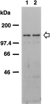
Fig. 1.
Fig. 1.
PKN expression in human brain. Total homogenates (50 μg of protein) of human brain tissue from the temporal or occipital cortices of control (lane 1) and AD cases (lane 2) were subjected to SDS-PAGE followed by immunoblotting. Immunostaining was performed with the antisera αC6 against PKN. The positions of marker proteins are indicated inkDa, and the position of PKN is indicated by anarrow.
Fig. 2.
Fig. 2.
PKN distribution in human brain tissues.A, In a representative neuron from the angular cortex of a control case, PKN was concentrated in many small or large vesicles accumulating in the proximal dendrites and cell bodies, especially the nucleus and perinuclear region. The axon (arrowhead) was weakly stained. Some immunoreactive dots looked like synaptic boutons (arrows) on dendrites. B, Dendrites, axons, and the cytoplasm just under cell membranes were strongly immunostained in a surviving neuron in AD angular gyrus, whereas PKN immunoreactivity was slightly decreased in or around the nucleus. Axonal immunoreactivity (arrowhead) was greater than that in control neuron. C, In the molecular layer (Figure legend continues) of the dentate gyrus from AD hippocampus, some apical dendrites (arrowheads) positive for PKN made synapses (small arrows) on immunoreactive varicose fibers (asterisk). Many varicose and thin fibers (long arrows) were also stained for PKN, which was accumulated in the dystrophic axon (thick arrow). D, A senile plaque (SP) (demarcated by arrowheads) was weakly stained for PKN, but PKN was highly enriched in the degenerative neurites (thick arrows). Faintly stained cells with glial profiles surrounded the SP (curved arrow).E, Intracellular NFTs (thick arrows) were strongly immunolabeled for PKN, as were degenerative neurites and neuropil threads in the subiculum of AD hippocampus. Extracellular NFTs were not labeled (thin arrows). A andB are at the same magnification. Scale bars:A, 25 μm; C, D, 50 μm;E, 100 μm.
Fig. 3.
Fig. 3.
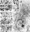
Subcellular localization of PKN in control and AD neurons. A, In control neuron, the antibody to PKN labeled nucleoplasm weakly (arrowheads) at high magnification. The nuclear envelope (double arrowheads), endoplasmic reticulum (thick arrow), multivesicular bodies (arrows), microtubules (asterisk), and small vesicles were moderately to intensely immunostained. Mitochondrial labeling (M) was not consistent: the mitochondria (M) inB was not immunoreactive for PKN. B,cis-/medial/trans-Golgi compartments (G) and trans-Golgi network, including secretory vesicles (arrowheads), were all strongly immunopositive. PKN was also found on many ribosomes. Note that a late endosome (arrow), fusing to primary lysosome (L), was immunolabeled for PKN. Lipofuscins were negative. C, In an asymmetric synapse, many synaptic vesicles in a presynaptic terminal (asterisk) were weakly immunoreactive; a spine apparatus (arrow) in the postsynapse was strongly stained for PKN. D, At lower magnification, many fine or coarse granular structures at the apical part of the perinuclear compartment (asterisk) and an intracellular NFT (black triangle) were intensely immunolabeled in a hippocampal pyramidal neuron. Top inset, Some of the abnormal filaments were markedly immunopositive for PKN (arrowheads), as were numerous vesicles 40–80 nm in diameter (arrows) surrounding the filamentous structures in a NFT. Bottom inset, Such filaments were composed of two types of tubules: straight tubules (arrowheads) and twisted tubules with a periodic constriction (small arrows) occurring every ∼80 nm, corresponding to paired helical filaments. Scale bars:A, 400 nm; B, C, 300 nm;D, 5 μm; top and bottom insets of D, 200 nm.
Fig. 4.
Fig. 4.
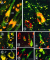
Colocalization of PKN with specific organelles and AD pathology in neurons. A, B, Double-labeling immunofluorescence microscopy demonstrating colocalization of PKN (red) and BiP (Grp-78, green). Numerous BiP-positive ER were scattered in cell bodies and apical dendrites of pyramidal neurons from a control hippocampus (A,green). A subset of these vesicles residing in the juxtanucleus and apical part of cell body and the proximal portion of apical dendrite was stained simultaneously for PKN and BiP (yellow, arrow inA). In tangle-bearing neurons in AD hippocampus, such doubly labeled vesicles were translocated and associated with intracellular tangles (arrows in B). Nuclear translocation of PKN was occasionally seen in AD degenerating neurons (arrowheads in B).C, Double immunolabeling for PKN (red) and cathepsin D, a candidate tau protease or APP secretase (green). Many small vesicles were doubly stained in the cytoplasm, especially in the perinuclear compartment of pyramidal neurons from AD hippocampus. Note that vesicles reactive for cathepsin D alone were near the cytoplasmic membrane (arrows), consistent with the report that cathepsin D is in early to late endosomes as well as in lysosomes in AD brain (Cataldo et al., 1997). D–H, Double immunofluorescent labeling of tangles for PKN (red) and NFT constituents (green). The AT8 monoclonal antibody recognized phosphoepitopes on tau in neuropil threads, degenerative neurites, and intracellular NFTs (D, green), which were decorated with many vesicles labeled for PKN (D, red). PKN and AT8 phosphoepitope were colocalized in filamentous structures within the cell soma (arrow) or within degenerative neurites (arrowheads). A similar pattern of filamentous structures within cell soma or degenerative neurites was seen in the staining with AT180 or AT270. PKN was localized in small vesicles within neurons overexpressing tau-1-positive nonphosphorylated tau (E, green). In F, PKN-containing vesicles (red) were seen within a vulnerable neuron overexpressing the Alz50 epitope (green). Some PKN-positive vesicles were seen very close to tau having an abnormal conformation (arrow in F). Few PKN-positive vesicles (G, red) were found on ubiquitinated intracellular NFTs (G, green). Extracellular NFTs labeled for complement protein C4d (H, green) were not associated with vesicles containing PKN (H, red). Scale bar: 20 μm in all panels.
Fig. 5.
Fig. 5.
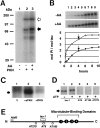
Phosphorylation of human tau protein by PKN. A, Phosphorylation was detected using autoradiography and SDS-PAGE. The white arrowheadindicates the position of autophosphorylation of recombinant PKN. One hundred nanograms of recombinant human tau were incubated with assay mixture without (lane 1) or with (lanes 2 and 3) recombinant PKN in the absence (lanes 1 and 2) or presence (lane 3) of 40 μm arachidonic acid. The reaction was terminated after 5 min, followed by separation on SDS-PAGE. Theblack arrowhead indicates the position of GST-tau fusion protein. Note the upper band of tau in lane 3, indicating molecular weight shift on more phosphorylation than inlane 2. B, Time course of tau phosphorylation by PKN. One hundred nanograms of GST-tau protein were incubated with recombinant PKN in the absence (closed circles or −AA) or presence (open circles or +AA) of arachidonic acid for 5 min (lane 1), 10 min (lane 2), 30 min (lane 3), 1 hr (lane 4), 2 hr (lane 5), 4 hr (lane 6), 6 hr (lane 7), 8 hr (lane 8), and 10 hr (lane 10). Mol of Pi incorporated into a mol of tau was calculated from the radioactivity quantified with an image analyzer.Arrows indicate the time point when 50 ng of additional PKN was applied in the assay mixture. C, In vivo phosphorylation of tau in human neuroblastoma SK-N-MC cells transfected with vector only or PKN transgenes. After immunoprecipitation with tau-1 antibody, intense phosphorylation signal was seen in the cells expressing active PKN (+aPKN, lane 2), but not in those expressing no (V, lane 1) or inactive PKN (+iPKN, lane 3). D, Immunoblot analysis with tau-1 (lanes 1 and2), AT8 (lane 3), AT180 (lane 4), and AT270 (lane 5) antibodies of recombinant tau phosphorylated by PKN in the absence (lane 1) or presence (lanes 2–5) of ATP. Note that tau-1 recognizes PKN-phosphorylated tau (lane 2), which does not react with AT8, AT180, or AT270 (lanes 3–5).E, Diagram of human tau isoform used. The four repeats of microtubule-binding domain in the C terminus are numbered 1–4 (closed circle). Epitopes of a couple of phosphorylation-dependent antibodies (open circles), a dephosphorylation-dependent antibody (open rectangle), and a conformation-dependent antibody (solid line) are depicted with their names initalic.

Similar articles

Cited by

References

    1. Alafuzoff I, Iqbal K, Friden H, Adolfsson R, Winblad B. Histopathological criteria for progressive dementia disorders: clinico-pathological correlation and classification by multivariate data analysis. Acta Neuropathol (Berl) 1987;74:209–225. - PubMed
    1. Amano M, Mukai H, Ono Y, Chihara K, Matsui T, Hamajima Y, Okawa K, Iwamatsu A, Kaibuchi K. Identification of a putative target for Rho as the serine-threonine kinase protein kinase N. Science. 1996;271:648–650. - PubMed
    1. Bancher C, Grundke-Iqbal I, Iqbal K, Fried VA, Smith HT, Wisniewski HM. Abnormal phosphorylation of tau preceded ubiquitination in neurofibrillary pathology of Alzheimer disease. Brain Res. 1991;539:11–18. - PubMed
    1. Billingsley ML, Kincaid RL. Regulated phosphorylation and dephosphorylation of tau protein: effects on microtubule interaction, intracellular trafficking and neurodegeneration. Biochem J. 1997;323:577–591. - PMC - PubMed
    1. Carmel G, Mager EM, Binder LI, Kuret J. The structural basis of monoclonal antibody Alz50’s selectivity for Alzheimer’s disease pathology. J Biol Chem. 1996;271:32789–32795. - PubMed

MeSH terms