. 1998 Aug 15;12(16):2598-609.
doi: 10.1101/gad.12.16.2598.

DNA-binding polarity of human replication protein A positions nucleases in nucleotide excision repair

Affiliations

DNA-binding polarity of human replication protein A positions nucleases in nucleotide excision repair

W L de Laat et al. Genes Dev. .

Abstract

The human single-stranded DNA-binding replication A protein (RPA) is involved in various DNA-processing events. By comparing the affinity of hRPA for artificial DNA hairpin structures with 3'- or 5'-protruding single-stranded arms, we found that hRPA binds ssDNA with a defined polarity; a strong ssDNA interaction domain of hRPA is positioned at the 5' side of its binding region, a weak ssDNA-binding domain resides at the 3' side. Polarity appears crucial for positioning of the excision repair nucleases XPG and ERCC1-XPF on the DNA. With the 3'-oriented side of hRPA facing a duplex ssDNA junction, hRPA interacts with and stimulates ERCC1-XPF, whereas the 5'-oriented side of hRPA at a DNA junction allows stable binding of XPG to hRPA. Our data pinpoint hRPA to the undamaged strand during nucleotide excision repair. Polarity of hRPA on ssDNA is likely to contribute to the directionality of other hRPA-dependent processes as well.


Figures

Figure 1
Figure 1
RPA preferentially binds to 3′-protruding single-stranded arms. (A–F) Gel-retardation assays showing the binding characteristics of hRPA to 3′-protruding 19-d(T) substrate (A), 5′-protruding 19-d(T) substrate (B), 3′-protruding 10-d(T) substrate (C), 5′-protruding 10-d(T) substrate (D), 3′-protruding 28-d(T) substrate (E), 5′-protruding 28-d(T) substrate (F). (Di and tri) Dimeric and trimeric hRPA complexes bound to DNA, respectively. No glutaraldehyde was added to any of the reactions. (G) Graphic presentation showing DNA-binding efficiency of hRPA (in percentage of bound substrate at 10 nm hRPA, which is the hRPA concentration that is referred to in the text as the approximately equimolar concentration) vs. the size of the single-stranded overhang. The DNA-binding efficiencies presented here are not meant to give a quantative measure of hRPA’s affinity for the different substrates. (H) Coomassie-stained protein gel of purified hRPA.
Figure 2
Figure 2
hRPA does not significantly unwind hairpin structures. Cleavage pattern of HaeIII in 3′- and 5′-protruding 19 nucleotide substrates is not altered when substrates are preincubated with hRPA (cf. lanes 3–5 with lane 2, and lanes 8–10 with lane 7, respectively). Note that, independent of the presence of hRPA, HaeIII is hardly able to cleave the nonprotruding strand of the 3′-protruding substrate (lanes 2–5). The dissimilar migration pattern of the released dinucleotides in lanes 2–5 vs. lanes 7–10 we attribute to different nucleotide compositions (CC vs. GG).
Figure 3
Figure 3
hRPA inhibits ERCC1–XPF endonuclease activity on 3′-protruding singles-stranded-arms. (A) Silver-stained protein gel of purified ERCC1–XPF. (B) Schematic presentation of ERCC1–XPF and hRPA acting on a 3′-protruding substrate. Asterisk indicates position of radioactive label. The 3′- and 5′-oriented side of hRPA, representing the weak and the strong ssDNA-binding side of hRPA, respectively, are indicated. (C,D) Denaturing polyacrylamide gels analyzing DNA incision products. (C) ERCC1–XPF nuclease assays on 3′-protruding 28-d(T) substrates, with increasing amounts of hRPA (lanes 3–6) and E. coli SSB protein (lanes 9–12). (D) ERCC1–XPF nuclease assays on 3′-protruding 19-d(T) substrates, with increasing amounts of hRPA (lanes 3–6) and E. coli SSB protein (lanes 9–12). Note that E. coli SSB protein concentration is given in nanomoles of tetramer per liter. The minor incision products visible in C and D do not correspond to known duplex single-stranded DNA junctions and probably arise from weak cutting activity near uncharacterized secondary structures in the DNA hairpin substrates.
Figure 4
Figure 4
hRPA stimulates ERCC1–XPF endonuclease activity on 5′-protruding single-stranded arms. (A) Schematic presentation of ERCC1–XPF and hRPA acting on a 5′-protruding substrate. Asterisk indicates position of radioactive label. The 3′- and 5′-oriented side of hRPA, representing the weak and the strong ssDNA-binding side of hRPA, respectively, are indicated. (B) ERCC1–XPF nuclease assays on 5′-protruding 28-d(T) substrates, with increasing amounts of hRPA (lanes 3–6) and E. coli SSB protein (lanes 9–12). (C) ERCC1–XPF nuclease assays on 5′-protruding 19-d(T) substrates, with increasing amounts of hRPA (lanes 3–6) and E. coli SSB protein (lanes 9–12). E. coli SSB protein concentration is given in nanomoles of tetramer per liter. (D) In C, lanes 1–5, but with ERCC1–XPF preparation purified from E. coli, to demonstrate that the effects of hRPA on ERCC1–XPF are not influenced by contaminants in the protein prep. Minor incision products visible in B–D do not correspond to known duplex ssDNA junctions and probably arise from weak cutting activity near uncharacterized secondary structures in the DNA hairpin substrates. Note that the ERCC1–XPF preparation obtained from E. coli (D) is more active, but yields similar incision products.
Figure 5
Figure 5
Polarity of hRPA on ssDNA has minor implications for XPG endonuclease activity. (A) Coomassie-stained protein gel of purified XPG. (B) Schematic presentation of XPG and hRPA acting on a 3′-protruding substrate (top) and a 5′-protruding substrate (bottom). Asterisks indicate positions of radioactive label. The 3′-oriented and 5′-oriented side of hRPA, representing the weak and the strong ssDNA-binding side of hRPA, respectively, are indicated. (C–D) XPG nuclease assays on C 3′-protruding 28-d(T) substrates, −/+ hRPA; (D) 5′-protruding 28-d(T) substrates, −/+ hRPA.
Figure 6
Figure 6
hRPA–ERCC1–XPF complex formation occurs specifically on 5′-protruding substrates. (A–C) Gel-retardation assays; all incubations were performed in 5 mm CaCl2, in the presence of 0.05% glutaraldehyde. (A) hRPA–ERCC1–XPF complex formation on 5′-protruding 28-d(T) substrates. For clarity, only 5 mm hRPA was used; higher concentrations of hRPA induced some hRPA dimer formation on these substrates, which migrated similar to the ternary hRPA–ERCC1–XPF complexes. Ternary complex formation was also observed at 10 mm hRPA (data not shown). (B) No complex formation between E. coli SSB and ERCC1–XPF on 5′-protruding 28-d(T) substrates. (C) No hRPA–ERCC1-XPF complex formation on 3′-protruding 28-d(T) substrates.
Figure 7
Figure 7
hRPA–XPG complex formation occurs specifically on 3′-protruding substrates. (A–C) Gel-retardation assays; all incubations were performed in 0.75 mm MnCl2, in the presence of 0.05% glutaraldehyde. (A) hRPA–XPG complex formation on 3′-protruding 28-d(T) substrates. Note that at 10 mm hRPA some hRPA dimer formation, (di) occurs (lane 2). (B) No complex formation between E. coli SSB and XPG on 3′-protruding 28-d(T) substrates. (C) No hRPA–XPG complex formation on 5′-protruding 28-d(T) substrates.
Figure 8
Figure 8
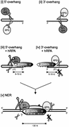
Model of hRPA binding to the undamaged strand during NER. (i,ii) ERCC1–XPF and XPG cleavage of 5′- and 3′-protruding substrates, respectively. (iii) On short 5′-protruding arms, hRPA weakly interacts with the single-stranded stretch, as bordering duplex DNA physically hinders stable binding of the strong DNA-interaction domain at the 5′-oriented side of hRPA. The 3′-oriented side of hRPA positions and strongly stimulates ERCC1–XPF-mediated incisions in the non-hRPA-bound strand, whereas it does not interact with XPG and slightly inhibits its activity. (iv) On short 3′-protruding arms, the strong binding domain of hRPA stably interacts with the single-stranded portion. This 5′-oriented side of hRPA positions XPG-mediated incisions (and slightly inhibits XPG-activity; see text), and completely blocks ERCC1–XPF-mediated incisions. (v) Extrapolation to nucleotide excision repair. On the formation of an opened DNA intermediate, which requires the activities of XPA, XPC–HHR23B, TFIIH, and hRPA (Evans et al. 1997b; Mu et al. 1997), an hRPA monomer is bound to the undamaged DNA strand and positions both nucleases onto the damaged strand; bound as such, it is able to stimulate strongly ERCC1–XPF incisions in the damaged strand and completely block ERCC1–XPF incisions in the undamaged strand. For clarity, XPA, XPC–HHR23B, and TFIIH are not depicted.

Similar articles

Cited by

References

    1. Baumann P, West SC. The human rad51 protein: Polarity of strand transfer and stimulation by hRP-A. EMBO J. 1997;16:5198–5206. - PMC - PubMed
    1. Benson F, Baumann P, West SC. Synergistic actions of Rad51 and Rad52 in recombination and DNA repair. Nature. 1998;391:401–404. - PubMed
    1. Bessho T, Sancar A, Thompson LH, Thelen MP. Reconstitution of human excision nuclease with recombinant XPF-ERCC1complex. J Biol Chem. 1997;272:3833–3837. - PubMed
    1. Blackwell LJ, Borowiec JA. Human replication protein A binds single-stranded DNA in two distinct complexes. Mol Cell Biol. 1994;14:3993–4001. - PMC - PubMed
    1. Blackwell LJ, Borowiec JA, Masrangelo IA. Single-stranded-DNA binding alters human replication protein A structure and facilitates interaction with DNA-dependent protein kinase. Mol Cell Biol. 1996;16:4798–4807. - PMC - PubMed

Publication types

LinkOut - more resources