. 1998 Aug;153(2):457-67.
doi: 10.1016/S0002-9440(10)65589-7.

Expression of autoactivated stromelysin-1 in mammary glands of transgenic mice leads to a reactive stroma during early development

Affiliations

Expression of autoactivated stromelysin-1 in mammary glands of transgenic mice leads to a reactive stroma during early development

N Thomasset et al. Am J Pathol. 1998 Aug.

Abstract

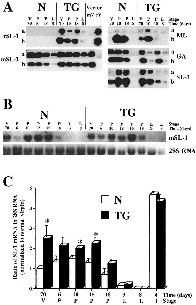
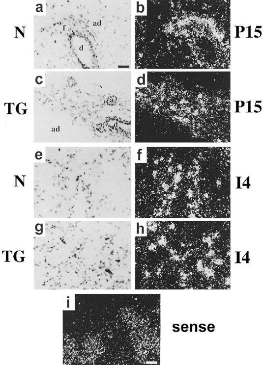
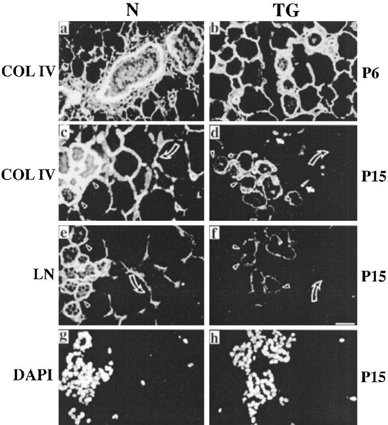
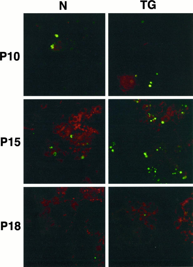
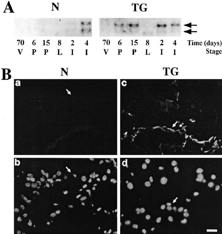
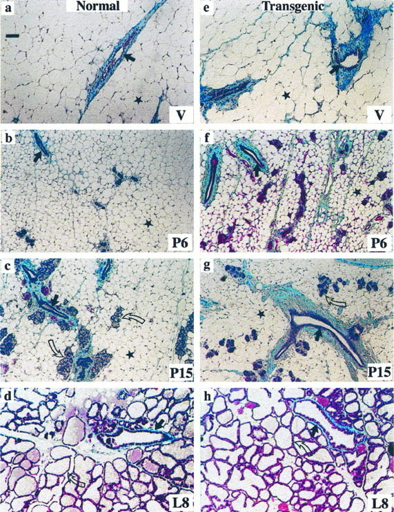
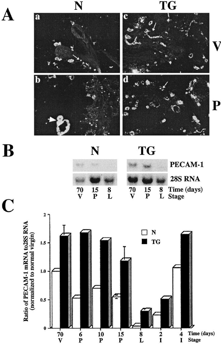
Extracellular matrix and extracellular matrix-degrading matrix metalloproteinases play a key role in interactions between the epithelium and the mesenchyme during mammary gland development and disease. In patients with breast cancer, the mammary mesenchyme undergoes a stromal reaction, the etiology of which is unknown. We previously showed that targeting of an autoactivating mutant of the matrix metalloproteinase stromelysin-1 to mammary epithelia of transgenic mice resulted in reduced mammary function during pregnancy and development of preneoplastic and neoplastic lesions. Here we examine the cascade of alterations before breast tumor formation in the mammary gland stroma once the expression of the stromelysin-1 transgene commences. Beginning in postpubertal virgin animals, low levels of transgene expression in mammary epithelia led to increased expression of endogenous stromelysin-1 in stromal fibroblasts and up-regulation of other matrix metalloproteinases, without basement membrane disruption. These changes were accompanied by the progressive development of a compensatory reactive stroma, characterized by increased collagen content and vascularization in glands from virgin mice. This remodeling of the gland affected epithelial-mesenchymal communication as indicated by inappropriate expression of tenascin-C starting by day 6 of pregnancy. This, together with increased transgene expression, led to basement membrane disruption starting by day 15 of pregnancy. We propose that the highly reactive stroma provides a prelude to breast epithelial tumors observed in these animals.


Figures

Figure 1.
Figure 1.
MMP expression during mammary gland development of normal and transgenic mice. A: Reverse transcription-PCR analysis of expression of the rat SL-1 (rSL-1) transgene, endogenous SL-1 (mSL-1), matrilysin (ML), gelatinase A (GA), and SL-3 (SL-3) in mammary glands in 70-day virgin (V 70), 10-day pregnant (P 10), 18-day pregnant (P 18), and 8-day lactating (L 8) normal (N) and transgenic (TG) mice. For each amplification product, the top row of each panel (a) shows negative images of ethidium bromide-stained PCR products after 40 cycles of amplification, and the bottom row of each panel (b) shows the Southern blot analysis with digoxygenin-labeled internal oliogodeoxynucleotides to confirm the identity of PCR products. The specificity of the primers for mouse and rat SL-1 was verified using vectors containing the appropriate mouse (mV) or rat (rV) cDNA. Note that rat SL-1 mRNA is expressed only in mammary glands of transgenic but not normal mice as expected, whereas endogenous mouse SL-1 mRNA is expressed in both normal and transgenic mice at all stages of development. B: Northern blot analysis of expression of endogenous SL-1 mRNA. Total RNA was extracted from two glands of 70-day virgin (V 70), 6-day pregnant (P 6), 10-day pregnant (P 10), 12-day pregnant (P 12), 15-day pregnant (P15), 18-day pregnant (P 18), 3-day lactating (L 3) and 8-day lactating (L 8) normal (N) and transgenic (TG) mice. Blots were hybridized with a cDNA probe specific for mouse SL-1 (mSL-1), stripped, and reprobed with a cDNA probe complementary to 28S ribosomal RNA (28S RNA). C: Northern hybridization signals were quantitated by scanning densitometry and normalized to signals obtained from blots reprobed with a cDNA complementary to 28S RNA. The value obtained at 8-day lactation for normal (open bars; N) and transgenic (solid bars; TG) mice was established as the baseline. For comparison of different blots, mRNA levels at various stages of development are expressed as a ratio relative to mRNA present at the normal glands of virgin animals, which was set to 1. Data are shown as means, with error bars representing SEM for four different sets of virgin, pregnant, and lactating glands. *Significant differences (P < 0.05) as determined by the Mann-Whitney U test.
Figure 2.
Figure 2.
Localization of endogenous SL-1 mRNA in mid-pregnant and involuting glands. In situ hybridization was performed with an antisense (a to h) or sense (i) mouse SL-1 probes in 15-day pregnant (P15) (a to d and i) and 4-day involuting (I4) glands (e to h) from normal (N) (a, b, e, and f) and transgenic (TG) (c, d, g, h, and i) mice; bright-field (a, c, e, and g) and dark-field (b, d, f, h, and i) microscopy images. Bar in a indicates 100 μm for (a to h); bar in i indicates 200 μm. a, alveolus; ad, adipocyte; f, fibroblasts; d, duct.
Figure 3.
Figure 3.
Immunolocalization of type IV collagen and laminin in mammary glands. Type IV collagen (COL IV) (a to d) and laminin (LN) (e and f) were localized by indirect immunofluorescence staining in 6-day pregnant (P6) (a and b) and 15-day pregnant (P15) (c to f) glands from normal (N) (a, c, and e) and transgenic (TG) (b, d, and f) mice; g and h show 4′,6′-diamidino-2-phenyl indole staining for nuclei of the same samples as e and f, respectively. Note the reduction in type IV collagen and laminin in the BM surrounding alveoli (arrowheads) and adipocytes (open curved arrows) in glands from 15-day pregnant transgenic mice compared with normal mice (compare c with d and e with f). In contrast, the staining of the BM surrounding blood vessels in d (filled white arrows) is not altered. Bar in f, 50 μm for all panels.
Figure 4.
Figure 4.
Apoptosis in normal and transgenic mammary glands. In situ detection of fragmented DNA detected by fluorescein isothiocyanate-digoxigenin nucleotide labeling of 3′-OH end (Apotag, Oncor) in 10-day (P10), 15-day (P15), and 18-day (P18) pregnant glands from normal (N) and transgenic (TG) mice. Note the increase of epithelial cells undergoing apoptosis in the 15-day pregnant gland from transgenic mice. The total number of cells per field was also greatly decreased in glands from late pregnant transgenic mice.
Figure 5.
Figure 5.
Expression of tenascin-C in mammary glands of normal and transgenic mice. A: Northern blot analysis for tenascin-C mRNA. Total RNA was extracted from mammary glands of 70-day virgin (70 V), 6-day pregnant (6 P), 15-day pregnant (15 P), 8-day lactating (8 L), 2-day involuting (2 I), and 4-day involuting (4 I) normal (N) and transgenic (TG) mice. Blots were hybridized with a cDNA probe for tenascin-C. Arrows indicate positions of the large and small tenascin-C transcripts. B: Immunolocalization of tenascin-C in mammary glands from 8-day lactating mice. Frozen tissue sections from mammary glands of normal (N) (a and b) and transgenic (TG) (c and d) mice were stained with a polyclonal antiserum to tenascin-C followed by a fluorescein-labeled conjugate (a and c) and counterstained with 4′,6′-diamidino-2-phenyl indole to visualize nuclei (b and d). Arrows indicate borders of duct. Bar in d, 25 μm for a to d. Note that tenascin-C was not detected in normal lactating mice (a) but was found in the proximity of the BM surrounding alveoli and ducts (arrow) in glands from transgenic mice.
Figure 6.
Figure 6.
Accumulation of collagen in mammary glands of transgenic mice. Gomori’s trichrome staining for total collagen (blue) in 70-day virgin (V) (a and e), 6-day pregnant (P6) (b and f), 15-day pregnant (P15) (c and g) and 8-day lactating (L8) (d and h) glands from normal (a to d) and transgenic (e to h) mice. Straight arrows indicate mammary ducts, curved arrows indicate alveolar structures; stars indicate adipocytes. Bar in a, 50 μm for a to h. Note that collagen deposition around mammary ducts is much more prominent in transgenic compared with normal mice (see also Table 1 ▶ ).
Figure 7.
Figure 7.
Vascularization of mammary glands in transgenic mice. A: Immunolocalization of vWF in blood vessels localized in alveoli (arrows) and in periductal stroma (arrowhead) of virgin (V) (a and c) and mid-pregnant (P) (b and d) mammary glands from normal (N) (a and b) and transgenic (TG) (c and d) mice. Note the increase in staining of vWF in glands from virgin and pregnant transgenic mice. B: Expression of the endothelial cell marker PECAM-1 in the glands of 70-day virgin (70 V), 15-day pregnant (15 P), and 8-day lactating (8 L) normal (N) and transgenic (TG) mice. Total RNA was analyzed by Northern blot analysis with a cDNA specific for PECAM-1 and a cDNA complementary to 28S ribosomal RNA (28S RNA). C: Quantification of PECAM-1 mRNA levels in mammary glands of 70-day virgin (70 V), 6-day pregnant (6 P), 10-day pregnant (10 P), 15-day pregnant (15 P), 8-day lactating (8 L), 2-day involuting (2 I), and 4-day involuting (4 I) normal (open bars; N) and transgenic (filled bars; TG) mice by scanning densitometry. The PECAM-1 levels were normalized by hybridization with a 28S RNA probe. For comparison of different blots, mRNA levels at various stages of development are expressed as a ratio relative to mRNA present at the normal virgin stage, which was set to 1. Error bars represent standard deviations from four independent experiments using glands from virgin 15-day pregnant and 8-day lactating mice. Values for 6 P, 10 P, 2 I, and 4 I represent the mean for two independent experiments.

Similar articles

Cited by

References

    1. Van den Hooff A: Stromal involvement in malignant growth. Adv Cancer Res 1988, 50:159-196 - PubMed
    1. Sakakura T: New aspects of stroma-parenchyma relations in mammary gland differentiation. Int Rev Cytol 1991, 125:165-202 - PubMed
    1. Bissell MJ, Hall HG: Form and function in the mammary gland: the role of extracellular matrix. Neville M Daniel C eds. The Mammary Gland Development, Regulation and Function. 1987, :pp 97-146 Plenum Publishing Corp, New York
    1. Adams JC, Watt FM: Regulation of development and differentiation by the extracellular matrix. Development 1993, 117:1183-1198 - PubMed
    1. Roskelley CD, Srebrow A, Bissell MJ: A hierarchy of ECM-mediated signaling regulates tissue-specific gene expression. Curr Opin Cell Biol 1995, 7:736-747 - PMC - PubMed

Publication types

MeSH terms