

A novel mechanism for the acquisition of virulence by a human influenza A virus
- PMID: 9707628
- PMCID: PMC21489
- DOI: 10.1073/pnas.95.17.10224
A novel mechanism for the acquisition of virulence by a human influenza A virus
Abstract
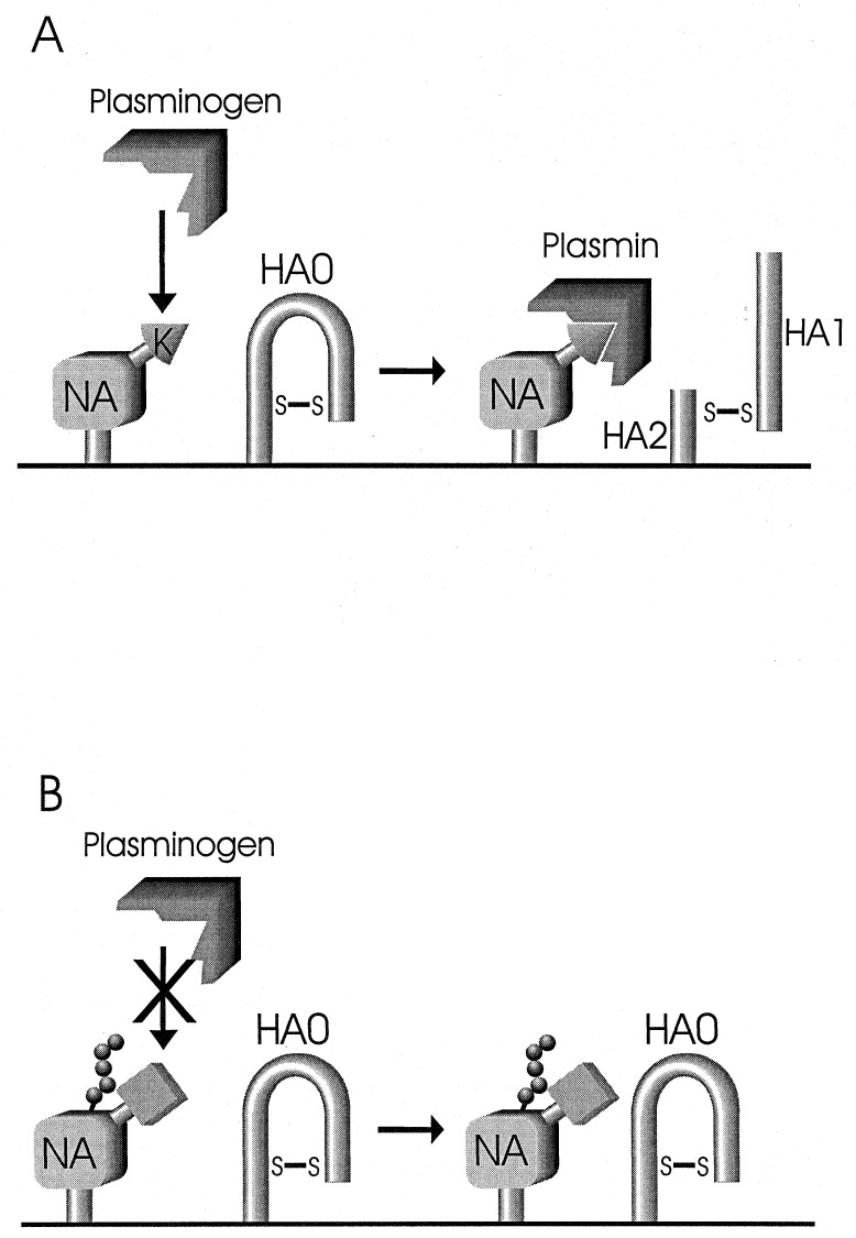
Cleavage of the hemagglutinin (HA) molecule by proteases is a prerequisite for the infectivity of influenza A viruses. Here, we describe a novel mechanism of HA cleavage for a descendant of the 1918 pandemic strain of human influenza virus. We demonstrate that neuraminidase, the second major protein on the virion surface, binds and sequesters plasminogen, leading to higher local concentrations of this ubiquitous protease precursor and thus to increased cleavage of the HA. The structural basis of this unusual function of the neuraminidase molecule appears to be the presence of a carboxyl-terminal lysine and the absence of an oligosaccharide side chain at position 146 (N2 numbering). These findings suggest a means by which influenza A viruses, and perhaps other viruses as well, could become highly pathogenic in humans.
Figures
Comment in
-
Influenza virus hemagglutinin cleavage into HA1, HA2: no laughing matter.Proc Natl Acad Sci U S A. 1998 Aug 18;95(17):9713-5. doi: 10.1073/pnas.95.17.9713. Proc Natl Acad Sci U S A. 1998. PMID: 9707539 Free PMC article. Review. No abstract available.
Similar articles
-
[Novel function of plasminogen-binding activity of the NA determines the pathogenicity of influenza A virus].Uirusu. 2004 Jun;54(1):83-91. doi: 10.2222/jsv.54.83. Uirusu. 2004. PMID: 15449908 Review. Japanese.
-
Assays for functional binding of plasminogen to viral proteins.Methods. 2000 Jun;21(2):159-63. doi: 10.1006/meth.2000.0987. Methods. 2000. PMID: 10816377
-
Alterations in hemagglutinin receptor-binding specificity accompany the emergence of highly pathogenic avian influenza viruses.J Virol. 2015 May;89(10):5395-405. doi: 10.1128/JVI.03304-14. Epub 2015 Mar 4. J Virol. 2015. PMID: 25741006 Free PMC article.
-
A Novel Neuraminidase-Dependent Hemagglutinin Cleavage Mechanism Enables the Systemic Spread of an H7N6 Avian Influenza Virus.mBio. 2019 Nov 5;10(6):e02369-19. doi: 10.1128/mBio.02369-19. mBio. 2019. PMID: 31690675 Free PMC article.
-
Competitive Cooperation of Hemagglutinin and Neuraminidase during Influenza A Virus Entry.Viruses. 2019 May 20;11(5):458. doi: 10.3390/v11050458. Viruses. 2019. PMID: 31137516 Free PMC article. Review.
Cited by
-
Subcellular localization and calcium and pH requirements for proteolytic processing of the Hendra virus fusion protein.J Virol. 2004 Sep;78(17):9154-63. doi: 10.1128/JVI.78.17.9154-9163.2004. J Virol. 2004. PMID: 15308711 Free PMC article.
-
Computational analysis and determination of a highly conserved surface exposed segment in H5N1 avian flu and H1N1 swine flu neuraminidase.BMC Struct Biol. 2010 Feb 22;10:6. doi: 10.1186/1472-6807-10-6. BMC Struct Biol. 2010. PMID: 20170556 Free PMC article.
-
Leptospira interrogans enolase is secreted extracellularly and interacts with plasminogen.PLoS One. 2013 Oct 18;8(10):e78150. doi: 10.1371/journal.pone.0078150. eCollection 2013. PLoS One. 2013. PMID: 24205133 Free PMC article.
-
Genetic diversity in the yellow head nidovirus complex.Virology. 2008 Oct 25;380(2):213-25. doi: 10.1016/j.virol.2008.07.005. Epub 2008 Sep 2. Virology. 2008. PMID: 18768192 Free PMC article.
-
H4N8 subtype avian influenza virus isolated from shorebirds contains a unique PB1 gene and causes severe respiratory disease in mice.Virology. 2012 Feb 5;423(1):77-88. doi: 10.1016/j.virol.2011.11.019. Epub 2011 Dec 20. Virology. 2012. PMID: 22192630 Free PMC article.
References
-
- Paulson J C. In: Interactions of Animal Viruses with Cell Surface Receptors. Connor M, editor. Orlando, FL: Academic; 1985. pp. 131–219.
-
- Klenk H-D, Garten W. Trends Microbiol. 1994;2:39–43. - PubMed
-
- Palese P, Tobita K, Ueda M, Compans R W. Virology. 1974;61:397–410. - PubMed
-
- Air G M, Laver W G. Proteins Struct Funct Genet. 1989;6:341–356. - PubMed
-
- Lazarowitz S G, Choppin P W. Virology. 1975;68:440–454. - PubMed
Publication types
MeSH terms
Substances
Grants and funding
LinkOut - more resources
Full Text Sources
Other Literature Sources
