Enhanced Galphaq signaling: a common pathway mediates cardiac hypertrophy and apoptotic heart failure
- PMID: 9707614
- PMCID: PMC21475
- DOI: 10.1073/pnas.95.17.10140
Enhanced Galphaq signaling: a common pathway mediates cardiac hypertrophy and apoptotic heart failure
Abstract
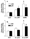
Receptor-mediated Gq signaling promotes hypertrophic growth of cultured neonatal rat cardiac myocytes and is postulated to transduce in vivo cardiac pressure overload hypertrophy. Although initially compensatory, hypertrophy can proceed by unknown mechanisms to cardiac failure. We used adenoviral infection and transgenic overexpression of the alpha subunit of Gq to autonomously activate Gq signaling in cardiomyocytes. In cultured cardiac myocytes, overexpression of wild-type Galphaq resulted in hypertrophic growth. Strikingly, expression of a constitutively activated mutant of Galphaq, which further increased Gq signaling, produced initial hypertrophy, which rapidly progressed to apoptotic cardiomyocyte death. This paradigm was recapitulated during pregnancy in Galphaq overexpressing mice and in transgenic mice expressing high levels of wild-type Galphaq. The consequence of cardiomyocyte apoptosis was a transition from compensated hypertrophy to a rapidly progressive and lethal cardiomyopathy. Progression from hypertrophy to apoptosis in vitro and in vivo was coincident with activation of p38 and Jun kinases. These data suggest a mechanism in which moderate levels of Gq signaling stimulate cardiac hypertrophy whereas high level Gq activation results in cardiomyocyte apoptosis. The identification of a single biochemical stimulus regulating cardiomyocyte growth and death suggests a plausible mechanism for the progression of compensated hypertrophy to decompensated heart failure.
Figures


Similar articles
-
Mitochondrial reprogramming induced by CaMKIIδ mediates hypertrophy decompensation.Circ Res. 2015 Feb 27;116(5):e28-39. doi: 10.1161/CIRCRESAHA.116.304682. Epub 2015 Jan 20. Circ Res. 2015. PMID: 25605649 Free PMC article.
-
Calcineurin-mediated hypertrophy protects cardiomyocytes from apoptosis in vitro and in vivo: An apoptosis-independent model of dilated heart failure.Circ Res. 2000 Feb 18;86(3):255-63. doi: 10.1161/01.res.86.3.255. Circ Res. 2000. PMID: 10679475
-
ROCK1 plays an essential role in the transition from cardiac hypertrophy to failure in mice.J Mol Cell Cardiol. 2010 Nov;49(5):819-28. doi: 10.1016/j.yjmcc.2010.08.008. Epub 2010 Aug 13. J Mol Cell Cardiol. 2010. PMID: 20709073 Free PMC article.
-
Gq signaling in cardiac adaptation and maladaptation.Trends Cardiovasc Med. 1999 Jan-Feb;9(1-2):26-34. doi: 10.1016/s1050-1738(99)00004-3. Trends Cardiovasc Med. 1999. PMID: 10189964 Review.
-
G-proteins in growth and apoptosis: lessons from the heart.Oncogene. 2001 Mar 26;20(13):1626-34. doi: 10.1038/sj.onc.1204275. Oncogene. 2001. PMID: 11313910 Review.
Cited by
-
Constitutive BDNF/TrkB signaling is required for normal cardiac contraction and relaxation.Proc Natl Acad Sci U S A. 2015 Feb 10;112(6):1880-5. doi: 10.1073/pnas.1417949112. Epub 2015 Jan 12. Proc Natl Acad Sci U S A. 2015. PMID: 25583515 Free PMC article.
-
The A-kinase anchoring protein (AKAP)-Lbc-signaling complex mediates alpha1 adrenergic receptor-induced cardiomyocyte hypertrophy.Proc Natl Acad Sci U S A. 2007 Jun 12;104(24):10140-5. doi: 10.1073/pnas.0701099104. Epub 2007 May 30. Proc Natl Acad Sci U S A. 2007. PMID: 17537920 Free PMC article.
-
Golgi localized β1-adrenergic receptors stimulate Golgi PI4P hydrolysis by PLCε to regulate cardiac hypertrophy.Elife. 2019 Aug 21;8:e48167. doi: 10.7554/eLife.48167. Elife. 2019. PMID: 31433293 Free PMC article.
-
Activation of Rho-associated coiled-coil protein kinase 1 (ROCK-1) by caspase-3 cleavage plays an essential role in cardiac myocyte apoptosis.Proc Natl Acad Sci U S A. 2006 Sep 26;103(39):14495-500. doi: 10.1073/pnas.0601911103. Epub 2006 Sep 18. Proc Natl Acad Sci U S A. 2006. PMID: 16983089 Free PMC article.
-
Transient cardiac expression of constitutively active Galphaq leads to hypertrophy and dilated cardiomyopathy by calcineurin-dependent and independent pathways.Proc Natl Acad Sci U S A. 1998 Nov 10;95(23):13893-8. doi: 10.1073/pnas.95.23.13893. Proc Natl Acad Sci U S A. 1998. PMID: 9811897 Free PMC article.
References
-
- Simon M I, Strathmann M P, Gautam N. Science. 1991;252:802–808. - PubMed
-
- Post G R, Brown J H. FASEB J. 1996;10:741–749. - PubMed
-
- Sadoshima J-I, Izumo S. Circ Res. 1993;73:413–423. - PubMed
-
- Shubeita H E, McDonough P M, Harris A N, Knowlton K U, Glembotski C C, Brown J H, Chien K R. J Biol Chem. 1990;265:20555–20562. - PubMed
-
- Knowlton K U, Michel M C, Itani M, Shubeita H E, Ishihara K, Brown J H, Chien K R. J Biol Chem. 1993;268:15374–15380. - PubMed
Publication types
MeSH terms
Substances
Grants and funding
LinkOut - more resources
Full Text Sources
Other Literature Sources
Medical
Molecular Biology Databases
Miscellaneous

