. 1998 Sep;72(9):7407-19.
doi: 10.1128/JVI.72.9.7407-7419.1998.

A C-terminal helicase domain of the human papillomavirus E1 protein binds E2 and the DNA polymerase alpha-primase p68 subunit

Affiliations

A C-terminal helicase domain of the human papillomavirus E1 protein binds E2 and the DNA polymerase alpha-primase p68 subunit

P J Masterson et al. J Virol. 1998 Sep.

Abstract

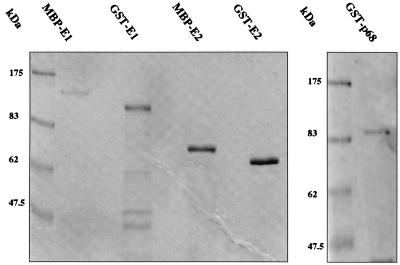
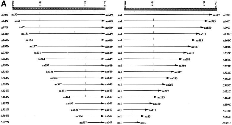
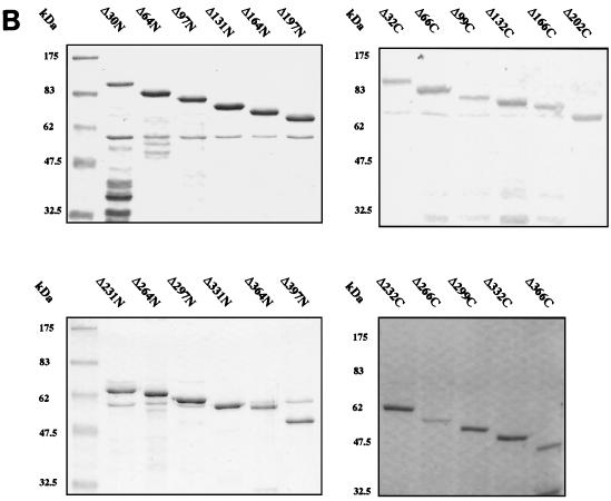
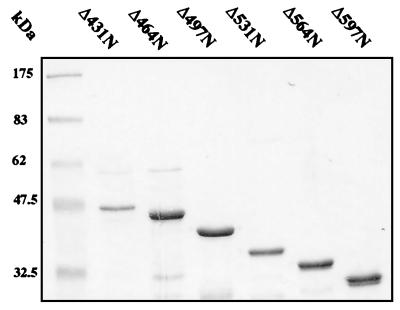
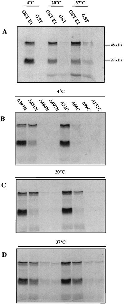
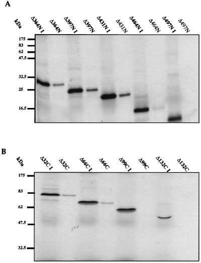
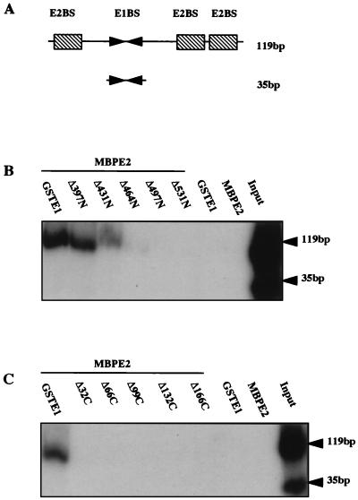
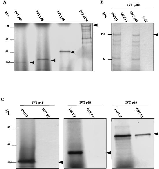
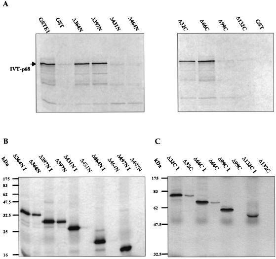
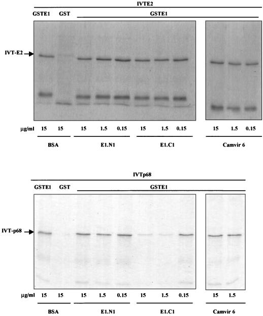
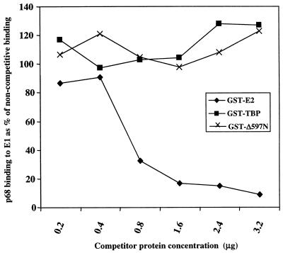
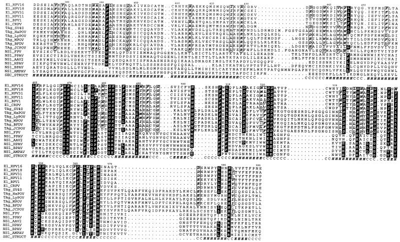
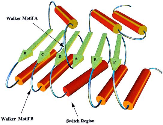
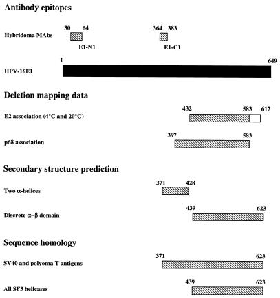
The human papillomavirus (HPV) E1 and E2 proteins bind cooperatively to the viral origin of replication (ori), forming an E1-E2-ori complex that is essential for initiation of DNA replication. All other replication proteins, including DNA polymerase alpha-primase (polalpha-primase), are derived from the host cell. We have carried out a detailed analysis of the interactions of HPV type 16 (HPV-16) E1 with E2, ori, and the four polalpha-primase subunits. Deletion analysis showed that a C-terminal region of E1 (amino acids [aa] 432 to 583 or 617) is required for E2 binding. HPV-16 E1 was unable to bind the ori in the absence of E2, but the same C-terminal domain of E1 was sufficient to tether E1 to the ori via E2. Of the polalpha-primase subunits, only p68 bound E1, and binding was competitive with E2. The E1 region required (aa 397 to 583) was the same as that required for E2 binding but additionally contained 34 N-terminal residues. In confirmation of these differences, we found that a monoclonal antibody, mapping adjacent to the N-terminal junction of the p68-binding region, blocked E1-p68 but not E1-E2 binding. Sequence alignments and secondary-structure prediction for HPV-16 E1 and other superfamily 3 (SF3) viral helicases closely parallel the mapping data in suggesting that aa 439 to 623 constitute a discrete helicase domain. Assuming a common nucleoside triphosphate-binding fold, we have generated a structural model of this domain based on the X-ray structures of the hepatitis C virus and Bacillus stearothermophilus (SF2) helicases. The modelling closely matches the deletion analysis in suggesting that this region of E1 is indeed a structural domain, and our results suggest that it is multifunctional and critical to several stages of HPV DNA replication.


Figures

FIG. 1
FIG. 1
Fusion proteins purified from E. coli. GST-E1, GST-E2, MBP-E1, MBP-E2, and GST-p68 were expressed and purified as described in Materials and Methods and then separated by SDS-PAGE and stained with Coomassie blue. The positions of molecular mass markers are indicated on the left of each gel.
FIG. 2
FIG. 2
(A) Schematic representation of the amino-terminal (N) and carboxyl-terminal (C) GST-E1 deletion series. Mutants are annotated according to the number of amino acids removed from the N or C terminus. Internal restriction sites utilized in construction are indicated where appropriate. (B) SDS-PAGE and Coomassie blue staining of glutathione-agarose-purified GST-E1 deletion proteins. The positions of molecular mass markers are indicated to the left of each gel.
FIG. 2
FIG. 2
(A) Schematic representation of the amino-terminal (N) and carboxyl-terminal (C) GST-E1 deletion series. Mutants are annotated according to the number of amino acids removed from the N or C terminus. Internal restriction sites utilized in construction are indicated where appropriate. (B) SDS-PAGE and Coomassie blue staining of glutathione-agarose-purified GST-E1 deletion proteins. The positions of molecular mass markers are indicated to the left of each gel.
FIG. 3
FIG. 3
Deletion analysis of GST-E1 binding to IVT E2. In vitro association of bead-bound GST-E1 or GST to radiolabelled IVT E2 was assayed as described in Materials and Methods. Autoradiographs show E2 protein that bound and was eluted from the beads. In addition to full-length E2 (48 kDa), a 27-kDa truncated form was also seen. (A) Binding of E2 to full-length GST-E1 and GST (as a control) at 4, 20, and 37°C. (B to D) Binding to selected GST-E1 N- and C-terminal deletion mutants that span the cutoff points for E2 binding at 4°C (B), 20°C (C), and 37°C (D). These results show that deletion of 432 N-terminal residues significantly affects binding at each assayed temperature while deletion of 66 C-terminal residues significantly affects binding at 4°C but not at the higher temperatures.
FIG. 4
FIG. 4
Deletion analysis of IVT E1 binding to GST-E2. In vitro association of bead-bound GST-E2 to radiolabelled IVT E1 N- and C-terminal deletion mutants was assayed as described in Materials and Methods. Autoradiographs show E1 protein that bound to and then was eluted from the beads. Input IVT protein is denoted I. (A) Binding of selected IVT E1 N-terminal deletion mutants to GST-E2 at 20°C. (B) Binding of selected IVT E1 C-terminal deletion mutants to GST-E2 at 20°C. This reciprocal binding experiment confirms the deletion analysis data obtained with IVT E2 and GST-E1 at 20°C. The positions of molecular mass markers are shown to the left of each gel.
FIG. 5
FIG. 5
Binding of E1 and E2 to the HPV-16 ori. McKay precipitation assays were performed as described in Materials and Methods. MBP-E2, in the presence of GST-E1 or GST-E1 N- or C-terminal deletion mutants, was incubated with two ori probes. Following incubation with glutathione-agarose beads and washes, bead-bound probes were recovered and analyzed by SDS-PAGE. (A) Schematic representation of HPV ori probes, the larger (119 bp) containing the entire ORI-binding E1BS and E2BS and the smaller (35 bp) containing only the E1BS. (B) Binding of selected GST-E1 N-terminal deletion mutants to the ori probes in the presence of MBP-E2. (C) Binding of selected GST-E1 C-terminal deletion mutants to the ori probes in the presence of MBP-E2. Input lanes show the ratios of the two probes.
FIG. 6
FIG. 6
Investigation of polα-primase subunit binding to E1. In vitro association of bead-bound GST-E1 to radiolabelled p180, p68, p58, or p48 subunits was assayed as described previously. (A) In vitro translation products of each polα-primase subunit. (B) Binding of p180 to full-length GST-E1, GST-p68, and GST, demonstrating clear interaction with p68 but no interaction with GST-E1 above background levels. (C) Binding of p48, p58, and p68 to full-length GST-E1, revealing strong interaction with only the p68 subunit. The positions of molecular mass markers are indicated. The arrowheads indicate full-length IVT protein products.
FIG. 7
FIG. 7
(A) Binding of p68 to GST-E1 and selected N- and C-terminal deletion mutants, demonstrating cutoff points for binding in each deletion series. (B and C) Reciprocal assays of binding of GST-p68 to IVT E1 N-terminal (B) and C-terminal (C) deletion mutants. The positions of molecular mass markers are indicated.
FIG. 8
FIG. 8
Antibody analysis of E1-E2 and E1-p68 interaction. The cross-blocking effect of two anti-E1 antibodies (E1-N1 and E1-C1) on the association of bead-bound GST-E1 with IVT E2 (A) and IVT p68 (B) was assayed as described in Materials and Methods. Antibodies were tested over a threefold range of concentrations. E1-N1 and Camvir 6 (anti-HPV 16L1) did not affect interaction of E1 with either E2 or p68, while E1-C1 significantly reduced only E1-p68 association.
FIG. 9
FIG. 9
Competitive binding of E2 and p68 to E1. Competition between IVT p68 and GST-E2 for binding to GST-E1 was assayed as described in Materials and Methods. The results demonstrate that GST-E2 and p68 compete for E1 binding with p68 in a dose-dependent manner, compared to GST-TBP and GST-E1Δ597N control proteins. Autoradiograms were scanned densitometrically to quantify the data.
FIG. 10
FIG. 10
Multiple-sequence alignment of the C-terminal regions (HPV-16 aa 371 to 591) of a number of SF3 helicases. E1, E1 protein; TAg, large T antigen; NS1, NS-1 protein; CRPV, cottontail rabbit PV; HaPOV, hamster polyomavirus; LyPOV, lymphotropic polyomavirus; MPOV, mouse polyomavirus; BFDV, budgerigar fledgling disease virus; JCPOV, polyomavirus JC; FPV, feline panleukopenia virus; PPAV, porcine parvovirus; AAV2, adeno-associated virus type 2; HPAV, human parvovirus; BPAV, bovine parvovirus; AMPAV, Aleutian mink disease parvovirus. Residues conserved across the complete alignment are displayed as white boldfaced letters on a black background, while residues conserved across E1 and T antigen sequences are boxed, italicized, and in boldface. A secondary-structure prediction for HPV-16 E1 is labelled SEC-STRUCT in the alignment. The prediction is a consensus of the predictions from three methods, DSC (23), PHDsec (42), and ZPRED (69). H, alpha helix; E, beta strand; C, other (no regular secondary structure).
FIG. 11
FIG. 11
A simplified model for the NTP-binding domain of E1 based on the X-ray crystallographic structures of the hepatitis C virus and B. stearothermophilus helicases. Note that this model is not to scale but is intended to display the order and relative positioning of structural elements of the domain.
FIG. 12
FIG. 12
Domains of E1 identified by deletion mapping and sequence analysis. Included are the following: epitopes of two novel HPV-16 E1 MAbs mapped by using the deletion series, deletion mapping data for E2 and p68 interaction sites (the larger E2 region is required at 4°C), secondary-structure prediction made by using multiple alignments as input, and sequence homology. Note that the deletion mapping coordinates given refer only to the outer boundaries for simplicity, so the true domains could be several residues shorter at either end. Note the close correspondence of deletion mapping and sequence analysis data, suggesting that the core helicase–E2-binding domain is aa 432 to 583 or 617 whereas an additional region (aa 397 to 432; equivalent to region A, conserved for papovavirus helicases only) is required for p68 binding.

Similar articles

Cited by

References

    1. Benson J D, Howley P M. Amino-terminal domains of the bovine papillomavirus type 1 E1 and E2 proteins participate in complex formation. J Virol. 1995;69:4364–4372. - PMC - PubMed
    1. Berg M, Stenlund A. Functional interactions between papillomavirus E1 and E2 proteins. J Virol. 1997;71:3853–3863. - PMC - PubMed
    1. Bird L E, Subramanya H S, Wigley D B. Helicases: a unifying structural theme? Curr Opin Struct Biol. 1998;8:14–19. - PubMed
    1. Blitz I L, Laimins L A. The 68-kilodalton E1 protein of bovine papillomavirus is a DNA binding phosphoprotein which associates with the E2 transcriptional activator in vitro. J Virol. 1991;65:649–656. - PMC - PubMed
    1. Bonne-Andrea C, Santucci S, Clertant P, Tillier F. Bovine papillomavirus E1 protein binds specifically DNA polymerase α but not replication protein A. J Virol. 1995;69:2341–2350. - PMC - PubMed

Publication types

MeSH terms

LinkOut - more resources